Abstract. CT urography (CTU) is an imaging method tailored specifically for the evaluation of the upper urothelial tract and urinary bladder. High diagnostic accuracy in detecting primary and recurrent urothelial tumours, their differentiation from benign lesions, anatomic variation and malformation characterization, and evaluation of important pathology in the surrounding tissues, renders CTU a valuable imaging study of the urothelial tract. Haematuria, initial staging and post treatment follow up of upper urinary tract and bladder malignancies are the most important indications for CTU. Triple phase and split bolus techniques are most commonly used CTU protocols that consist of a non-contrast, nephrographic, and excretory phase, obtained by three or two acquisitions. CTU scanning protocols are tailored to achieve adequate image quality with optimal opacification and distension of the urinary tract at the lowest achievable radiation dose within the range of 5-15 mSv, comparable to the doses of intravenous urography. Detailed patient history, clinical examination, and urine analysis are essential for proper patient selection and targeted CTU protocol, which are the most important tools for increasing diagnostic accuracy and lowering patient radiation dose of CT urography.
Key words: computed tomography; hematuria; urography; urologic neoplasms
Sažetak. CT urografija (CTU) je slikovna metoda specifično prilagođena oslikavanju i procjeni gornjeg mokraćnog sustava i mokraćnog mjehura. Visoka dijagnostička točnost u detekciji primarnih i recidivnih tumora urotela, razlikovanje malignih od benignih lezija, karakterizacija anatomskih varijacija i malformacija te procjena relevantnih patomorfoloških promjena u okolnim tkivima čine CT urografiju vrijednom metodom za oslikavanje mokraćnog sustava. Hematurija, inicijalna procjena proširenosti i praćenje tumora gornjeg mokraćnog sustava i mokraćnog mjehura najvažnije su indikacije za CTU. „Triple phase” i „split bolus” tehnike su najčešće korišteni CTU protokoli koje sačinjavaju nativna, nefrografska i ekskretorna faza, dobivene skeniranjem regije tri ili dva puta. CTU protokoli skeniranja su dizajnirani s ciljem ostvarivanja najbolje kvalitete slike te optimalne distenzije i opacifikacije mokraćnog sustava uz najmanju moguću dozu zračenja, u rasponu od 5 – 15 mSv, što je usporedivo s dozama zračenja intravenske urografije. Detaljna anamneza, klinički pregled i analiza urina ključni su za pravilan probir pacijenata i ciljani odabir CTU protokola, te su ujedno najučinkovitiji način poboljšavanja dijagnostičke točnosti i smanjivanja doze zračenja CT urografije.
Ključne riječi: hematurija; kompjutorizirana tomografija; urografija; urološke neoplazme
*Corresponding author:
Doris Dodig, MD Department of Radiology, Clinical Hospital Center Rijeka Krešimirova 42, 51 000 Rijeka
e-mail: [email protected] Department of Radiology, Clinical Hospital Center Rijeka, Rijeka
CT urography: principles and indications
CT urografija: načela i indikacije
INTRODUCTION
CT urography (CTU) is an imaging method tai-lored specifically for the evaluation of the upper urothelial tract and urinary bladder. High diag-nostic accuracy in detecting primary and recur-rent urothelial tumours, their differecur-rentiation from benign lesions, anatomic variation and mal-formation characterization, and evaluation of im-portant pathology in the surrounding tissues renders CTU a valuable imaging study of the urothelial tract. Relatively high radiation dose was the only downside of CTU technique. How-ever, multidetector CT technology advancement and protocol adjustment for the individual pa-tient, enables significant radiation dose reduction within ranges comparable to intravenous urogra-phy (IVU).
INDICATIONS
Haematuria
Haematuria can be caused by many benign and malignant conditions located anywhere within the urinary system and it is the most common symp-tom encountered in the clinical practice. CTU has completely replaced IVU, retrograde uretero-pyelography and ultrasonography (US) as the im-aging modality of choice in the high risk patient population who present with macrohaematuria, are older than 40 years and carry additional risk
factors for urothelial malignancy1-6. In this clinical
setting the benefit of high sensitivity, specificity and diagnostic accuracy of CTU for upper urinary tract carcinoma detection of 93.5-95.8%, 94.8-100%, and 94.2-99.6%, respectively, outweighs the cost of higher patient radiation exposure
com-pared to other imaging modalities4. Current
guide-lines by the American College of Radiology and the European Society of Urogenital Radiology recom-mend additional cystoscopy in the evaluation of high risk patients with macrohaematuria. Howev-er sevHowev-eral studies have shown that CTU is compa-rable to flexible cystoscopy in excluding and detecting bladder cancer with sensitivity, specifici-ty, positive and negative predictive values of 0.93,
0.99, 0.98, and 0.98, respectively5,7-9.
Low and medium risk patients presenting with painless microhaematuria require thorough
clini-cal assessment and follow up before proceeding with the appropriate imaging modality. The diag-nosis of microhaematuria needs to be correctly established by the findings of three or more red blood cells per high powered field on a properly collected urinary specimen. The most common causes such as vigorous exercise, menstruation, infection, and recent urologic procedure or trau-ma need to be excluded by detailed history and physical examination. When those are excluded, renal function testing, cystoscopy and CTU are
in-CT urography is the modality of choice for imaging the
pelvicalyceal system, ureters and urinary blader. It has
replaced other methods, particularly IVU. Haematuria
in selected patients, staging and follow-up of urothelial
malignancies, evaluation of congenital and post
trea-tment anatomy distortions, trauma, complicated
infec-tions, and 3D planning for difficult percutaneous
nephrolithotomy are indications for CTU.
dicated. The American Urological Association as-serts CTU over US and IVU as the imaging modality of choice for the evaluation of micro-haematuria based on the evidence that the risk of missing a significant pathology requiring treat-ment is significantly higher than the radiation
burden of an optimally performed CTU1. Patients
with negative initial work up and persistent mi-croscopic haematuria should undergo yearly urine analysis for two consecutive years. If urine analysis is negative, patients can be released from care. If there is persistent haematuria com-plete imaging follow up should be considered in
3-5 years upon clinicians decision1.
Upper urinary tract tumours
CTU has the highest diagnostic accuracy in the initial evaluation of upper urinary tract cancer (Figure 1). It is essential for TNM staging, detec-tion of synchronous or metachronous urinary tract tumours, and post treatment follow up. CTU enables detection of very small tumours (around 5 mm), and those that present only as urothelial wall thickening. Moreover, it is possible to evalu-ate periureteric spread, differentievalu-ate organ con-fined (stage T1 and T2) from locally invasive
(stage T3 and T4) disease, and asses nodal or
dis-tant metastases4,10. This information pertinent to
patient management and outcome are not ob-tainable by IVU. In addition, CTU has a high nega-tive predicnega-tive value (0.98) for bladder lesions which occur in 40% of patients with upper uri-nary tract tumours, possibly obviating the need for further invasive diagnostic procedures when no suspicious bladder lesions in patients with
up-per urinary tract cancer are detected9-11.
Urinary bladder tumours
Urinary bladder transitional cell carcinoma (TCC) has the highest recurrence rate for all tumours, and 2-4% of patients will have synchronous or metachronous tumour in the upper urinary tract.
Thus evaluation and surveillance of the whole urinary tract is mandatory initially and in the post
treatment follow up12-14. Cystoscopy and deep
bi-opsy are still gold standard in T staging of urinary bladder TCC, especially in superficial tumours. However, in addition to a good detection rate and positive predictive value for diagnosing pri-mary bladder tumours, CTU has the benefit of perivesical tumour spread assessment (sensitivi-ty and specifici(sensitivi-ty 89-92% and 95%-98%, respec-tively), accurate staging of T3 and T4 tumours, assessment of nodal and distant abdominopelvic metastases, detection of synchronous TCC in the upper urinary tract, and recurrence after transurethral resection (TUR) or radical
cystecto-my (Figure 2)14,15. A study by Kim et al. showed
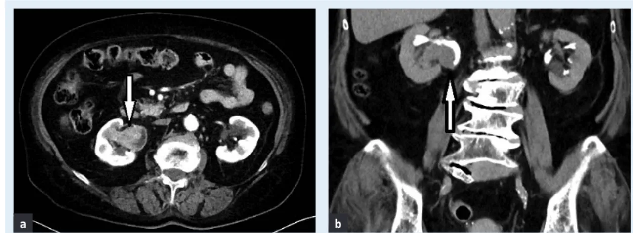
Figure 1 Transitional cell carcinoma of the pelvicalyceal system of the right kidney shown in a) nephrographic and b) excretory phase.
Figure 2 Sagittal reformatted images of a small (< 10 mm) urinary bladder tumour detected on CT urography in a) nephrographic phase as an enhancing lesion, and b) excretory phase as a contrast filling defect.
a b
that CTU has the accuracy of 91.7% in detection of recurrent TCC in the bladder after TUR, which was comparable to the reported 95% accuracy of
cys-toscopy11. CTU is essential after radical cystectomy
for invasive bladder TCC in assessing post-surgical complications and recurrent or metachronous dis-ease in a complicated setting of distorted anatomy of both genitourinary and gastrointestinal tracts
(Figure 3)16.
Other
Other important indications for CTU are congeni-tal conditions, hydronephrosis, iatrogenic or non-iatrogenic urinary tract trauma, 3D planning for difficult cases of percutaneous nephrolithotomy, and complex urinary tract infections, such as
tu-berculosis2,3,17.
MR urography combined with US and/or retro-grade pyelography is the alternative imaging
modality in patients with relative or absolute contra indications for CTU such as renal insuffi-ciency, contrast allergy, pregnancy, young age
and frequent examinations1.
Indications for CT urography are summarized in Table 1.
TECHNIQUES
Thin-slice multidetector CT imaging and intrave-nous contrast application are mandatory for eva-luating the urothelial tract. Thin-slices enable sufficient spatial resolution required for optimal detection and evaluation of the subtle urothelial tract pathology. Imaging in all three planes and 3D reformations help analyse normal anatomy, characterize and localize pathology, congenital malformations and complex post-surgical anato-mical relations of the urinary tract.
Triple phase and split bolus techniques
CTU scanning protocols are tailored to achieve op-timal opacification and distension of the urinary tract. Standard CTU protocols consist of three phases acquired in three or two aquisitions: a non-contrast, nephrographic and excretory phase. Non-contrast images are essential for detecting urinary tract stones, intralesional fat and calcifica-tion, and differentiation of hyperdense blood clots and non-enhancing benign lesions from malignant masses. Detection of enhancing malignant urothe-lial lesions or wall thickening that are more hyper-dense than the surrounding urine and conspicuity of kidney lesions are highest in the nephrographic phase (80-100 s after the start of contrast injec-tion). Homogenous opacification and adequate distension of the urinary tract in the excretory phase (5-12 minutes after contrast injection) al-Table 1 CT urography indications
CT urography indications Comment
Macrohaematuria Method of choice in patients over 40 years. Complementary to ultrasound and cystoscopy in younger patients.
Microhaematuria Complementary to ultrasound and cystoscopy after excluding infection, trauma, intensive exercise, mentrual bleeding and kidney disease. Detection, staging and surveillance of urothelial tumors Method of choice for upper urinary tract malignancies. Comparable to
cystoscopy for bladder carcinoma. Evaluation of disease spread in abdomen and pelvis.
Obstruction, trauma, congenital abnormalities, percutaneous nephrolithotomy, complex infections
CT urography in excretory phase is sufficient for most indications.
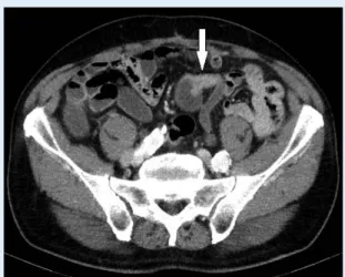
Figure 3 Recurrent transitional cell carcinoma at the anastomosis of the left ureter and neobladder after radical cystectomy seen on axial images as the enhancing wall thickening in the nephrographic phase.
lows detection of subtle filling defects represent-ing small lesions (5-15 mm) in the pelvicalyceal system, ureters or the urinary bladder, in addition to comprehensive overview of the anatomic
rela-tions18.
Triple phase and split bolus CTU technique are most commonly used, although there is no univer-sally accepted scanning protocol serving as the gold standard. In the triple phase technique the unenhanced scan of the abdomen and pelvis is fol-lowed by two separate scans in the nephrographic
and excretory phase after intravenous administra-tion of 100-150 ml of nonionic iodinated contrast agent at the rate of 2-3 ml/s. Split bolus technique is designed to image the urinary tract simultane-ously in the combined nephrographic and excreto-ry phase: after the unenhanced scan and the application of 30-50 ml of contrast agent at the rate of 2-3 ml/s, the second contrast bolus of 80-100 ml at the same rate is administered 5-12 min-utes after the first, followed by image acquisition
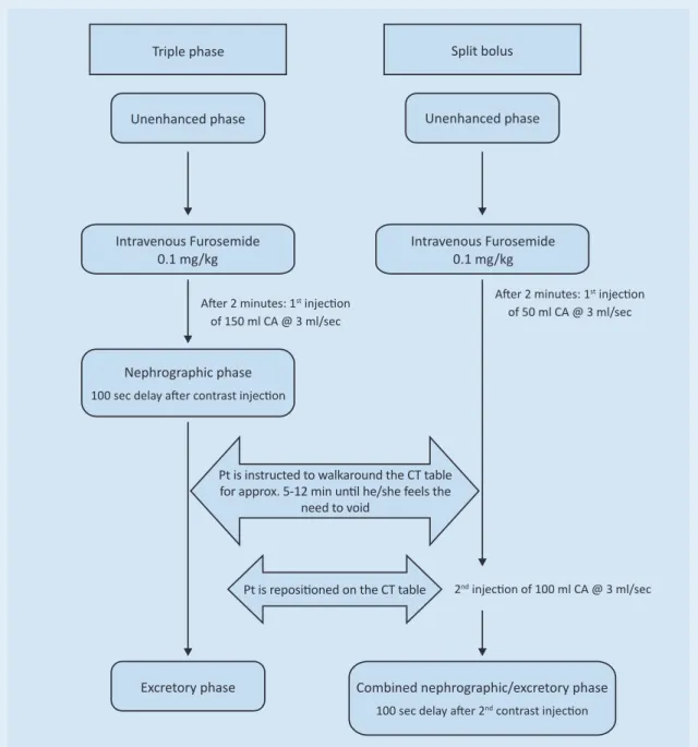
with a 100 s delay19-22. A graphic display of CTU
Figure 4 Diagram of the CT urography protocols used at our institution. Patients are instructed to empty their bladder 60 minutes before the examination and to drink 1000 ml of water over the next 20-30 minutes. CA contrast agent; Pt patient
Triple phase
Unenhanced phase
Intravenous Furosemide 0.1 mg/kg
Nephrographic phase
100 sec delay after contrast injection
Intravenous Furosemide 0.1 mg/kg Unenhanced phase
Split bolus
After 2 minutes: 1st injection
of 150 ml CA @ 3 ml/sec
After 2 minutes: 1st injection
of 50 ml CA @ 3 ml/sec
2nd injection of 100 ml CA @ 3 ml/sec
Pt is repositioned on the CT table
Excretory phase Combined nephrographic/excretory phase
100 sec delay after 2nd contrast injection
Pt is instructed to walkaround the CT table for approx. 5-12 min until he/she feels the
protocols used at our institution is shown in Figure 4. The advantage of split bolus technique is radia-tion dose reducradia-tion by 30-50% compared to the triple phase protocol, which is an important
con-sideration when imaging younger patients23.
De-tection of small lesions, especially in the distal ureter, in the combined nephrographic and excre-tory phase may theoretically be lower due to sub-optimal distension and lower contrast resolution between the hyperdense enhancing lesion and the surrounding opacified urine. However, studies did not show significantly inferior diagnostic accu-racy of split bolus compared to the triple phase
technique22,23.
Promising imaging techniques
Few studies have shown that urothelial tumours opacify the strongest 60-80 s after intravenous contrast application, suggesting that portal ve-nous (PV) phase increases the diagnostic accura-cy of CTU by significantly improving bladder tumour detection compared to the
nephrograph-ic phase24,25. Dual energy CT technology has the
advantage of computing virtual non-enhanced from the contrast enhanced images, thus reduc-ing the number of image acquisitions required for performing three phase or split bolus CTU. However, further research is needed to establish the correlation between attenuation values of
virtual and real non-enhanced images26,27.
Manoeuvres for achieving optimal urinary tract distension and opacification
To achieve optimal urinary tract distension and opacification many additional manoeuvres and strategies have been employed, such as abdomi-nal compression, supine and prone positioning of the patient on the CT table, the “log rolling” tech-nique, oral hydration, intravenous saline infusion, application of intravenous diuretics and busco-pane. Oral hydration with 750-1000 ml of water over 60 minutes before the examination and intra-venous injection of 0.1 mg/kg (maximal dose of 10 mg) of furosemide 2-3 minutes prior to contrast injection have been proven superior to other
tech-niques28-30. Optimally timed and sufficient oral
hy-dration is a simple method of inducing diuresis, contrast excretion and distension of the pelvicalyc-eal system, ureters and the urinary bladder, which
is important for lesion detection in all phases29.
Additional furosemide ensures continuous diure-sis and homogenous opacification of the urinary tract resulting in lower attenuation values of the excreted urine. This diminishes possible streak ar-tefacts from the overly dense urine and increases contrast resolution between the enhancing intra-luminal lesions and the surrounding opacified urine, which is especially important for lesion de-tection in the combined nephropyelographic
High diagnostic accuracy for urinary tract pathology
de-tection and characterization on CTU overcomes the
cost of radiation burden to the patient. Technology
ad-vances and protocol adjustments enable significant
ra-diation dose reduction. However, diligent patient
selection remains the best method for maintaining the
cost benefit ratio in favor of the patient.
phase in the split bolus protocol28,30. Attention
should be paid not to administer furosemide to patients with known allergy to furosemide or oth-er sulfa drugs, patients with systolic blood pres-sure under 90 mmHg, acute renal colic and urinary tract obstruction.
RADIATION DOSE REDUCTION STRATEGIES
At the beginning of the CTU technique the re-ported radiation dose was in the range of 15-25 mSv for triple phase and 10 mSv for split bolus technique, which is about 1.5 times more than
the dose of IVU31. However, a significant dose
re-duction has been achieved by modifying scan-ning parameters and CTU protocols. Dahlman et al. reports dose reduction of a triple phase CTU protocol from 16.2 to 9.4 mSv by performing low dose unenhanced and excretory phase (100 kV,
reference dose of 20mAseff and 40 mAseff,
respec-tively)32. Fewer acquisitions in the split bolus
technique reduce the radiation dose by 30-50%. Recently introduced iterative reconstruction al-gorithms improved image quality and allowed dose reduction by additional 40-50% with report-ed radiation dose of 6. 1 mSv for a triple phase protocol, and possible split bolus study at 4.2 mSv33-35. Dual energy CT technology offers even
more dose reduction by omitting the hanced phase and computing the virtual unen-hanced images, however this technique is not yet established in the clinical setting and further re-search is needed.
CONCLUSION
CTU is the imaging modality of choice for evalu-ating pelvicalyceal system, ureters and urinary bladder and is replacing IVU. Haematuria, initial staging and post treatment follow up of upper urinary tract and bladder malignancies are the most important indications for CTU. High dia-gnostic accuracy of CTU outweighs the radiation burden of this imaging technique in selected pa-tient groups. Detailed papa-tient history, clinical examination, and urine analysis are essential for proper patient selection and targeted tailoring of CTU protocol, and therefore remain the most important tools for patient radiation dose re-duction.
Conflicts of interest statement: The authors report no conflicts of interest.
REfERENCES
1. Davis R, Jones JS, Barocas DA, Castle EP, Lang EK, Levei-llee RJ et al. Diagnosis, evaluation and follow-up of asymptomatic microhematuria (AMH) in adults: AUA guideline. J Urol 2012;188 (6 Suppl):2473-81.
2. Nolte-Ernsting C, Cowan N. Understanding multislice CT urography techniques: Many roads lead to Rome. Eur Radiol 2006;16:2670-86.
3. Potenta SE, D’Agostino R, Sternberg KM, Tatsumi K, Pe-russe K. CT Urography for Evaluation of the Ureter. Ra-diographics 2015;35:709-26.
4. Jinzaki M, Kikuchi E, Akita H, Sugiura H, Shinmoto H, Oya M. Role of computed tomography urography in the clinical evaluation of upper tract urothelial carcinoma. Int J Urol 2016;23:284-98.
5. Luyao S, Raman SS, Beland MD, Coursey Moreno C, Gol-dfarb S, Harvin HJ et al. ACR Appropriateness Criteria® Hematuria. American College of Radiology [Internet]. [cited 2017 Jan 10]. Available from: https://acsearch. acr. org/docs/69490/Narrative/.
6. Cowan NC. CT urography for hematuria. Nat Rev Urol 2012;9:218-26.
7. Sadow CA, Silverman SG, O’Leary MP, Signorovitch JE. Bladder cancer detection with CT urography in an aca-demic medical center. Radiology 2008;249:195-202. 8. Van Der Molen AJ, Cowan NC, Mueller-Lisse UG,
Nol-te-Ernsting CC, Takahashi S, Cohan RH et al. CT uro-graphy: definition, indications and techniques. A guideline for clinical practice. Eur Radiol 2008;18: 4-17.
9. Turney BW, Willatt JM, Nixon D, Crew JP, Cowan NC. Computed tomography urography for diagnosing blad-der cancer. BJU 2006;98:345-8.
10. Vikram R, Sandler CM, Ng CS. Imaging and staging of transitional cell carcinoma: part 2, upper urinary tract. AJR Am J Roentgenol 2009;192:1488-93.
11. Kim JY, Kim SH, Lee HJ, Kim MJ, Kim YH, Cho SH. MDCT urography for detecting recurrence after transurethral resection of bladder cancer: comparison of nephro-graphic phase with pyelonephro-graphic phase. AJR Am J Roen-tgenol 2014;203:1021-7.
12. Fleshner N, Kondylis F. Demographics and epidemiology of urothelial cancer of urinary bladder. In: Droller MJ (ed). Urothelial tumors. Hamilton, ON: Decker, 2004:1-16. 13. Elmajian DA. Transitional cell carcinoma of the ureter
and renal pelvis. In: Nachtsheim D (ed). Urological on-cology. Austin, TX: Landes Bioscience, 2005:43-52. 14. Vikram R, Sandler CM, Ng CS. Imaging and staging of
transitional cell carcinoma: part 1, lower urinary tract. AJR Am J Roentgenol 2009;192:1481-7.
15. Kim JK, Park SY, Ahn HJ, Kim CS, Cho KS. Bladder cancer: analysis of multi-detector row helical CT enhancement pattern and accuracy in tumor detection and perivesi-cal staging. Radiology 2004;231:725-31.
16. Shinagare AB, Sadow CA, Silverman SG. Surveillance of patients with bladder cancer following cystectomy: yi-eld of CT urography. Abdom Imaging 2013;38:1415-21. 17. Hiorns MP. Imaging of the urinary tract: the role of CT
and MRI. Pediatr Nephrol 2011;26:59-68.
18. Silverman SG, Leyendecker JR, Amis ES, Jr. What is the current role of CT urography and MR urography in the evaluation of the urinary tract? Radiology 2009;250: 309-23.
19. Sanyal R, Deshmukh A, Singh Sheorain V, Taori K. CT urography: a comparison of strategies for upper urinary tract opacification. Eur Radiol 2007;17:1262-6. 20. Maher MM, Kalra MK, Rizzo S, Mueller PR, Saini S.
Multi-detector CT urography in imaging of the urinary tract in patients with hematuria. Korean J Radiol 2004;5:1-10. 21. Chow LC, Sommer FG. Multidetector CT urography with
abdominal compression and three-dimensional recon-struction. AJR Am J Roentgenol 2001;177:849-55. 22. Maheshwari E, O’Malley ME, Ghai S, Staunton M,
Ma-ssey C. Split-bolus MDCT urography: Upper tract opaci-fication and performance for upper tract tumors in patients with hematuria. AJR Am J Roentgenol 2010; 194:453-8.
23. Dillman JR, Caoili EM, Cohan RH, Ellis JH, Francis IR, Nan B et al. Comparison of urinary tract distension and opa-cification using single-bolus 3-phase vs split-bolus 2-phase multidetector row CT urography. J Comput Assist Tomogr 2007;31:750-7.
24. Kupershmidt M, Margolis M, Jang HJ, Massey C, Metser U. Evaluation of upper urinary tract tumors with portal venous phase MDCT: a case-control study. AJR Am J Ro-entgenol 2011;197:424-8.
25. Park SB, Kim JK, Lee HJ, Choi HJ, Cho KS. Hematuria: portal venous phase multi detector row CT of the blad-der--a prospective study. Radiology 2007;245:798-805. 26. Chen CY, Hsu JS, Jaw TS, Shih MC, Lee LJ, Tsai TH et al.
Split-Bolus Portal Venous Phase Dual-Energy CT Uro-graphy: Protocol Design, Image Quality, and Dose Re-duction. AJR Am J Roentgenol 2015;205:W492-501.
27. Chen CY, Tsai TH, Jaw TS, Lai ML, Chao MF, Liu GC et al. Diagnostic Performance of Split-Bolus Portal Venous Phase Dual-Energy CT Urography in Patients With He-maturia. AJR Am J Roentgenol 2016;206:1013-22. 28. Silverman SG, Akbar SA, Mortele KJ, Tuncali K, Bhagwat
JG, Seifter JL. Multi-detector row CT urography of normal urinary collecting system: furosemide versus saline as adjunct to contrast medium. Radiology 2006;240:749-55. 29. Curic J, Vukelic-Markovic M, Marusic P, Hrkac-Pustahija A, Brkljacic B. Influence of bladder distension on opaci-fication of urinary collecting system during CT urograp-hy. Eur Radiol 2008;18:1065-70.
30. Kemper J, Regier M, Stork A, Adam G, Nolte-Ernsting C. Improved visualization of the urinary tract in multidetec-tor CT urography (MDCTU): analysis of individual acquisiti-on delay and opacificatiacquisiti-on using furosemide and low-dose test images. J Comput Assist Tomogr 2006;30:751-7. 31. Stacul F, Rossi A, Cova MA. CT urography: the end of
IVU? Radiol Med 2008;113:658-69.
32. Dahlman P, van der Molen AJ, Magnusson M, Magnu-sson A. How much dose can be saved in three-phase CT urography? A combination of normal-dose cortico-medullary phase with low-dose unenhanced and excretory phases. AJR Am J Roentgenol 2012;199: 852-60.
33. Juri H, Tsuboyama T, Kumano S, Inada Y, Koyama M, Azuma H et al. Detection of bladder cancer: compari-son of low-dose scans with AIDR 3D and routine-dose scans with FBP on the excretory phase in CT urography. Br J Radiol 2016;89:20150495.
34. Bahn YE, Kim SH, Kim MJ, Kim CS, Kim YH, Cho SH. De-tection of Urothelial Carcinoma: Comparison of Redu-ced-Dose Iterative Reconstruction with Standard-Dose Filtered Back Projection. Radiology 2016;279:471-80. 35. van der Molen AJ, Miclea RL, Geleijns J, Joemai RM. A
Survey of Radiation Doses in CT Urography Before and After Implementation of Iterative Reconstruction. AJR Am J Roentgenol 2015;205:572-7.