Inhibitive Effect of Antibiotic-Loaded Beads to Cure Chronic Osteomyelitis in
Developing Country: Hand-made vs Commercial Beads
Hermawan N. Rasyid
1,2,4, Jim R. van Horn
3, Henny C. van der Mei
1, Soegijardjo Soegijoko
2,
Henk J. Busscher
1, and Daniëlle Neut
1,31 Department of Biomedical Engineering, University of Groningen, The Netherlands
2 Biomedical Engineering Program, School of Electrical Engineering and Informatics, Institut Teknologi Bandung, Indonesia 3 Department of Orthopedic Surgery, University Medical Center Groningen, The Netherlands
4 Department of Orthopedic and Traumatology, Medical Faculty Padjadjaran University/dr.Hasan Sadikin Hospital, Bandung, Indonesia
Abstract—Local antibiotic-loaded beads have been approved for standard treatment of orthopedic pathogens, especially chronic osteomyelitis. Septopal®, the only commercial local antibiotic bead available on the market, is antibiotic-loaded beads (fosfomycin-sodium) with Septopal® for ten bacterial strains of chronic osteomyelitis by diffusion technique at various indicated time points. The results showed that Septopal® could inhibit all bacterial strains except CNS 7334 and CNS 7391. Hand-made fosfomycin beads were only slightly effective in two of the ten bacterial strains is used (Staphylococcus aureus 7323 and CNS 7391). In conclusion, this study demonstrated that Septopal® can inhibit common orthopaedic pathogens better than hand-made beads in vitro
study, however,
fosfomycin
concentrations required to kill orthopedic implant related bacterial strains are high and its release from hand-made beads is too low to be considered efective when compared with Septopal beads.
Keywords— Local antibiotic-loaded beads, Chronic osteomyelitis, Septopal, Fosfomycin-sodium.
I.
I
NTRODUCTIONThe uses of polymethylmethacrylate (PMMA) for delivery of local antibiotics for the
treatment of have shown amounts of
the antibiotics antibiotics by diffusion to avascular areas of the bone and hypovascular areas of surrounding soft tissue that are inaccessible by systemic antibiotics and in many
circumstances of
organisms that are resistant to drug concentrations achieved systemically.5,6
The concept of antibiotic delivery by
incorporation of
gentamicin into acrylic bone cement was infections, especially chronic osteomyelitis, which is caused mostly by Gram positive bacteria such as Staphylococcus aureus.5
In developing
countries, such as Indonesia there are many poor people that always make use of the service of traditional medicine like bonesetter when dealing with open factures of the bone. In treating these fractures, initially they reduce the
fracture without
cleaning the wound, then directly covered the wound by herbals, then, fixed them using wood as a slab. After several months, the patient comes to the orthopedic clinic with persistent
sinus tract and active pus, meaning that chronic osteomyelitis occurred
Chronic
osteomyelitis in
developing countries is generally treated by clearing the cavity of infected materials, systemic administration of antibiotics and planting a chain of hand-made antibiotic-loaded beads. These beads are left in situ for about fourteen days, after which the cavity is filled with bone graft.
Antibiotic-loaded PMMA beads are commercially available under the name Septopal®, but the price of these beads is far too high for most patients. Fosfomycin is the
commonly used
antibiotic in these bone cement beads, because of its low costs, wide antibacterial spectrum, small molecular weight (138 Da) and its ability to remain stable up to the high temperatures as
reached during
polymerization of the bone cement.7
The treatment of
osteomyelitis in
Indonesia generally yields satisfactory clinical results, but it is unknown up to what extend the fosfomycin-loaded, hand-made beads contribute to these results. The aim of this study is to evaluate the in vitro antibacterial efficacy and kinetics of antibiotic release of several currently used, hand-made fosfomycin-loaded beads. All results will be compared with those of commercially available
gentamicin-loaded PMMA beads (Septopal®).
II.
M
ATERIALS AND METHODSA. Antibiotic-loaded beads preparation
A number of orthopedic surgeons in
Indonesia were
requested to describe their personal concepts developed to prepare antibiotic-loaded beads. In general, PMMA bone cements were used, in combination with a variety of different mixing techniques and sometimes templates. Fosfomycin-sodium was the general antibiotic included. Based on this inventory, six concepts were selected for further research and the participating surgeons were asked to submit an extensive protocol of their concept, as summarized in Table 1, together with samples of their beads.
Antibiotic-loaded PMMA beads are commercially available under the name Septopal® (Biomet Deutchland, Darmstadt, Germany). It consists of 30 beads threaded on made antibiotic-loaded beads. All beads contained fosfomycin and mixing was done by hand using a spatula.
B. Antibacterial efficacy
Beads#1 to #6 and Septopal® beads were
Presented at The International Conference of Biomed 2006 Kuala Lumpur [December 2006] Page 2
Beads Cementbase Fosfomycincontent [g] Shaping [cm]Size Area/volumeratio [cm-1]
#1 Zimmer 40 g 4 Template 1.7 1.8 #2 Simplex P
40 g 2 Handrolled 2.8 1.0 #3 Simplex P
40 g
2 Handrolled 1.6 1.8
#4 Simplex P 40 g
2 Handrolled 2 1.5
immersed in 10 ml of sterile phosphate buffer saline (PBS) and incubated at 370C. After
24 h of incubation 15 µl samples were taken and these were placed on bacterial streaked trypton soya brooth (TSB) agar plates. The zones of inhibition were
established by
measuring the diameters of the clear areas around one drop of elution fluid. Absence of an inhibition zone was taken as a sign that the antibiotic concentration was too low to inhibit bacterial growth. For this study we used ten clinical strains isolated from patients with an reported to be involved in the occurrence of osteomyelitis.8,9 Each
strain was diluted in 4 ml of 0.9% saline to concentration
approximately of 108
bacteria/ml. After overnight incubation at 370C on agar plates, the
determined by an E-test (AB Biodisk, Dälvagen, Sweden).
C. Kinetics of antibiotic release
kinetics of antibiotics release was established by taking four 15 µl elution fluid samples at the indicated time points and placing the droplets on the four quadrants of a bacterial streaked TSB agar plate. Kinetics of antibiotic release was only studied with strains
toward which
antibacterial efficacy could be established, as described above. After 24h incubation at 370C
the inhibition zone diameters were scored in all four quadrants.
D. Scanning electron microscopy
For scanning
electron microscopy (SEM), beads were exposed overnight to an osmiumtetroxide vapor. field emission scanning electron microscope type 6301F.
III.
R
ESULTSA. Minimal Inhibitory Concentration (MIC value) from 0.19 to over 1024 µg/ml for fosfomycin and 0.38 to over 256 µg/ml for gentamicin. Resistant sub-population could be observed within inhibition zones in both strains. The efficacy of fosfomycin is effective in S. aureus 7323, CNS 7391 and E.coli BS 6202 (MIC value 0.19, 0.50 and 1.5 µg/ml). Two bacterial strains were gentamicin resistant. Efficacy of gentamicin on the growth of 10 bacterial strains was evaluated, it has MIC value ranged aeruginosa 7348.
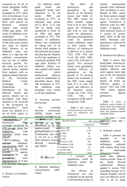
Table 2. MIC values of 10 bacterial strains used in this study for fosfomycin and gentamicin.
Bacterial strains MIC value (µg/ml) Fosfomycin
Micrococcus 7397 192
P. aeruginosa 5148 >1024
P. aeruginosa 7348 32
E. coli BS 6206 1.5*
Klebsiella 333257 >1024
* resistant sub-evaluated on the growth of 20 bacterial strains of
chronic osteomyelitis patients from Indonesia after incubation in vitro. Results of MIC values for these isolates ranged from 1.5 to over 1024 µg/ml. Fosfomycin is effective with low MIC values (1.5µg/ml) in three bacterial strains of S. aureus (S. aureus InB1; InP4; In B4 and InB7). Resistant sub-populations could not be observed in these inhibition zones.
B. Antibacterial efficacy Table 3 shows that hand-made fosfomycin-loaded beads were only slightly effective and never against more than two of the ten bacterial strains is included. Nearly all beads are effective against S. aureus 7323 and CNS elution fluids from hand-made fosfomycin-loaded PMMA beads and gentamicin-loaded Septopal® beads. millimeters achieved with elution fluid from
the hand-made
fosfomycin-loaded beads after different elution periods. None of the beads showed antibacterial efficacy against S. aureus 7323 extending beyond 24 h, whereas beads#1 and #3 were not effective at all. This is all in sharp
Presented at The International Conference of Biomed 2006 Kuala Lumpur [December 2006] Page 3
contrast with the
inhibition zones
achieved with elution fluid from Septopal®
beads, showing
sustained release against S. aureus 7323 for at least 144 h. Due to its gentamicin-resistance, no inhibition zones could be measured against CNS 7391.
Table 4. Diameters of clear zones of inhibition (mm).
- =no zone; *
resistant
sub-populations could be observed within the inhibition zones; sept=septopal
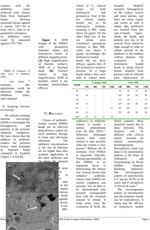
D. Scanning electron microscopy
We utilized scanning electron microscopy (SEM) to investigate the
homogenicity and
porosity of the polymer antibiotic complexes. We have shown that the addition of antibiotic renders the polymer texture more homogen in Septopal beads compared to beads#2 (Figure 1 A and B).
Figure 1: SEM images of the PMMA
with gentamicin
Septopal beads and fosfomycin beads in different magnifications.
(A) High magnification of fracture surface): SEM of a Septopal bead. (B) Fracture surface in high magnification: SEM of beads#2, which is the broadest antimicrobial efficacy.
IV.
D
ISCUSIONChains of antibiotic-loaded cement PMMA beads are an effective drug delivery system for local antibiotic therapy in bone and soft-tissue
infections. The
antibiotic concentrations at the site of infection are far higher than after systemic application of the same antibiotic and far above the minimal inhibitory
concentrations of most common pathogens.
The elution of fosfomycin-loaded beads were only effective in two of the ten bacterial strains tested, while the elution of Septopal® beads were effective against eight of the ten strains used. An E-test determined the MIC
values of the clinical strains for both
fosfomycin and
gentamicin. Four of the ten clinical strains turned out to be fosfomycin resistant, meaning a MIC value above 64 µg/ml. On the other hand two of the ten strains were considered gentamicin resistant as their MIC value was above 4 µg/ml. Accordingly, the fosfomycin-loaded beads did not show efficacy against four of the fosfomycin sensitive bacteria. In a large beads surface, they were able to release more
antibiotics as antibiotic release is mostly a surface phenomenon (van de Belt 2001).10
Moreover, prolonged release from bone cement is only possible when the cement is very porous.11 Release rate of
antibiotic from PMMA is important clinically. Porosity/permeability of the PMMA is an important factor in determining the release rate. Known factors that influence antibiotic release from PMMA are surface roughness and porosity. Van de Belt et al, demonstrated that porosity corresponded best with the eventual amount of release. It make sense since the evaluation of SEM, the
broadest beads#2
revealed homogenicity of the surface texture and more porous, and there are many craters and cracks as well. It means that antibiotics are leached from this type of beads. Space inside the beads and particle size has to be noticed. Pores should be large enough in order to release process of the antibiotic to the medium without obstacle. The
evaluation of
fosfomycin loaded beads is using clinical strains isolated from patients with an infected joint replacement in
Dutch patients, these situations mostly due to deep infection. This situation will be different with clinical strains focused on chronic osteomyelitis hematogenously. Retrospective study was done in 56 osteomyelitis patients at the Dept. of
Orthopedic and
Traumatology dr. Hasan Sadikin Hospital, Bandung, Indonesia. The microorganism pattern of osteomyelitis is S. aureus 26.9% of all cases, and P. aeruginosa 11.0% of all cases.12
The microorganism pattern of osteomyelitis and the treatment results can be contradictory. It seems that the efficacy of fosfomycin loaded
Beads S. aureus 7323 CNS 7391
24h 48h 72h 144h 24h 48h 72h 144h
#1 - - - - 20 20 15 20
#2 16 - - - 25* 20 20 20
#3 - - - - 15* 15 - 15
#4 11 - - - 20* 20 20 20
#5 7 - - 11 25* 20* - 20*
#6 13 - - - 25* 20 20 20
sept 16 12 11 14 - - -
-A
beads did not appear, but standard surgical
debridement and
systemic antibiotic administration should have an important role to treat this disease. We conclude from the presented observations that a mixture of fosfomycin and PMMA bone cement is not an effective one. Efficacy could only be seen in two of the ten bacterial strains. We assume that the hand-made beads were not porous enough as porosity of bone cement is the key to high antibiotic release. Therefore we have to try other procedures to increase the porosity, like adding glycine as in currently done in the Septopal® beads.13 Also
trying an other antibiotic is an option, together with changing the antibiotic carrier from PMMA bone cement to calcium phosphate.14
One big advantage of calcium phosphate above PMMA bone cement is that calcium
phosphate is
biodegradable, so no second operation is needed to remove the beads.
V.
C
ONCLUSIONSFosfomycin
concentrations required to kill orthopedic implant related bacterial strains are high and its release from hand-made beads is too low to be considered effective when compared with Septopal beads.
A
CKNOWLEDGEMENTSThe author would like to thank to Mr. Ietse Stokroos, Department of Cell Biology and Electron Microscopy for his help with the electron microscopy. This work is funded in part by Eric
Bleumink Fund,
University of
Groningen, The
Netherlands.
R
EFERENCES1.Buchholz, H.W., and Engelbrecht H. (1970) Uber die Depotwirkung einiger Antibiotica bei Vermischung mit dem Kunstharz Palacos. Chirurg 41:511-5.
2.Klemm, K. (1979) Gentamicin-PMMA-Kugeln in der Behandlung abszedierender Knochen und Weichteilinfektionen. Zentral bl. Chirurg 104:934-42. 3.Brinker, M.R.(2001) Antibiotic beads in general principles of trauma, in: Review of orthopaedic trauma:31-43.
4.Patzakis, M.J., Wilkins, J., Kumar, J., Holtom, P., Greenbaum, B., and Ressler, R. (1994) Comparison of the results of bacterial cultures from multiple sites in chronic osteomyelitis of long bones: A prospective study. J Bone
Joint Surg Am 76:664-6.
5.Walenkamp, G.H.I.M., Coombs, R., and Fitzgerald, R.H.(1989) Gentamicin– PMMA beads: Pharmacokinetics and toxicology in: Infection in the orthopaedic patient:163-66. 6.Wahlig, H., Dingeldein, E. (1980) Antibiotics and bone cements. Experimental and clinical long term observations. Acta Orthop Scand 51:495.
7.Goto, S. (1977) Fosfomycin, antimicrobial activity in vitro
and in vivo in:
Chemotheraphy 23:65-74.
8.McLaughlin, R.E., Reger, S.I., Barkalow, J.A., Allen, M.S., and Difazio, C.A. (1978) Methylmethacrylate: A study of teratogenicity and fetal toxicity of the vapor in the mouse. J Bone Joint Surg
3:355-8
9.Klemm, K.W. (1993) Antibiotic bead chains. Clin
Orthop295:63-76.
10.Van de Belt, H., Neut, D., Van Horn, J.R., Van der Mei, H.C., Schenk, W., and Busscher, H.J.(2001)
Staphylococcus aureus biofilm
formation on different gentamicin-loaded
polymethylmethacrylate bone cements. Biomaterials
22:1607-11.
11.Van de Belt, H., Neut, D., Uges, D.R., Schenk, W., Van Horn, J.R., and Van der Mei. H.C., Busscher, H.J.(2000) Surface roughness, porosity and wettability of gentamicin-loaded bone cement and their antibiotic release.
Biomaterials 21:1981-7.
12.Saragih, J.R.(1999) Microorganisms pattern and its sensitivity in osteomyelitis case at the Dept. of Orthopedic Surgery Dr Hasan Sadikin Hospital, Bandunng, Thesis 50-1.
13.McLaren, A.C., Nelson, C.L., McLaren, S.G., and DeClerk, G.R.(2004) The effect of glycine filler on the elution rate of gentamicin from acrylic bone cement. A pilot study. Clin Orthop
427:25-7.
14.Wichelhaus, T.A., Dingeldein, E., Rauschmann, M., Kluge, S., Dieterich, R., Schäfer, V., and Brade, V. (2001) Elution characteristics of vancomycin, teicoplanin, gentamicin and clindamycin from calcium sulphate beads.
J Antimicrobial
Chemotherapy 48:117-9.
Address of the coresponding author:
Author: Hermawan N Rasyid Institut: Biomedical Engineering Program, School of Electrical Engineering and Informatics, Institut Teknologi Bandung, Indonesia
Street: Jalan Ganesha 10 (Lab Tek VIII, 3rd floor)
City: Bandung - 40132 Country: Indonesia Email: