Paediatrica Indonesiana
VOLUME 50 March NUMBER 2
Original Article
Change of ionized calcium level in the first 48 hours
of age of preterm newborns administered with two
different dosages of intravenous calcium gluconate
Anton Wibowo, Dedi Rachmadi Sambas, Abdurachman Sukadi
Abstract
Background Physiologically, serum calcium level declines till nadir LQDIHZKRXUVDIWHUELUWKDQGFRQWLQXHVIRUKRXUV1RVWXG\ performed in order to know the alteration of ionized calcium level RIQHZERUQLQWKHILUVWKRXUVRIDJH7KHVLFNQHZERUQPXVW have parenteral calcium to avoid hypocalcemia but there is still no agreement about the dose.
Objective To determine the change of ionized calcium level in WKHILUVWKRXUVRIDJHRISUHWHUPQHZERUQVDGPLQLVWHUHGZLWK SHULSKHUDOGULSLQWUDYHQRXVFDOFLXPJOXFRQDWHRIP/NJ day and 5 mL/kg/day.
Methods An open labeled randomized controlled clinical trial was performed between April and June 2009. After birth blood specimen of preterms was obtained for leukocyte, platelet, magnesium, phosphorous, and ionized calcium measurements. 6XEMHFWV UHFHLYHG HLWKHU P/NJGD\ RU P/NJGD\ RI SHULSKHUDOGULSLQWUDYHQRXVFDOFLXPJOXFRQDWHLPPHGLDWHO\ DIWHUELUWKIRUKRXUV%ORRGVSHFLPHQVZDVREWDLQHGDJDLQRQ KRXUVRIDJHIRULRQL]HGFDOFLXP7KLVVWXG\ZHUHDQDO\]HG using repeated measures analysis of varians.
Results)RUW\SUHWHUPQHZERUQVVXEMHFWVHDFKJURXSZHUH
analyzed. There was no statistical difference between both GRVHV )DFWRU$ RQ KRXUV LRQL]HG FDOFLXP OHYHO 3 and ionized calcium level alteration based on time (Factor-B) (P=0.20). Interaction between both factors was significantly different (P=0.035).
Conclusion,RQL]HGFDOFLXPOHYHOLQKRXUVRIDJHRISUHWHUP QHZERUQDGPLQLVWHUHGZLWKERWKGRVHVRIFDOFLXPJOXFRQDWHLV QRWGLIIHUHQWEXWGRVHRIP/NJGD\\LHOGVSK\VLRORJLFDOWHUDWLRQ of ionized calcium level compared with 5 mL/kg/day. [Paediatr Indones. 2010;50:96-100].
Keywords: Preterm newborn, early onset hypocalcemia, 10% calcium gluconate
)URP 'HSDUWPHQW RI &KLOG +HDOWK 0HGLFDO 6FKRRO 3DGMDGMDUDQ
University, Hasan Sadikin General Hospital, Bandung, Indonesia.
Reprint request to: Anton Wibowo, MD, Department of Child Health,
0HGLFDO6FKRRO3DGMDGMDUDQ8QLYHUVLW\+DVDQ6DGLNLQ*HQHUDO+RVSLWDO -O3DVWHXU1R%DQGXQJ,QGRQHVLD7HO)D[ (PDLO[email protected]
C
alcium is the most abundant mineral in the body. Of the body’s total calcium, 99% are in bone, and serum levels constitute less WKDQ $OWKRXJK WRWDO VHUXP FDOFLXP levels are often measured and reported, ionized calcium is the active and physiologically important component.Early onset hypocalcemia is most commonly observed problems among preterm newborns. The incidence of neonatal hypocalcemia varies in different studies. Hypocalcemia occurs in 30% of newborns with YHU\ORZELUWKZHLJKWJDQGLQDVPDQ\DV 89% of newborns whose gestational age at birth was less than 32 weeks. A high incidence is also reported in newborns born to mothers with diabetes mellitus and in newborns with birth asphyxia.
in order to know the alteration of ionized calcium OHYHORIQHZERUQLQWKHILUVWKRXUVRIDJH7KHVLFN newborns must have parenteral calcium in order to avoid hypocalcemia but there is still no agreement about the dose. Hypocalcemia must be prevented in order to avoid impairment of cardiovascular and central nervous system.
This study aimed to know the alteration of LRQL]HGFDOFLXPOHYHOLQWKHILUVWKRXUVRIDJHRI preterm newborn administered with peripheral drip LQWUDYHQRXV FDOFLXP JOXFRQDWH P/NJGD\ and 5 mL/kg/day.
Methods
An open-label randomized controlled trial was performed on preterm newborns in Child Health Department Hasan Sadikin Hospital Bandung between April and June 2009. We included sick preterm newborns and appropriate for gestational age, and excluded preterm newborns of mothers with diabetes mellitus, mothers with phenytoin and/or SKHQREDUELWDOWKHUDS\SUHWHUPQHZERUQVZLWKPDMRU congenital anomaly or early onset sepsis, hypocalcemia, hypomagnesemia, or hyperphosphatemia at baseline. Parents of eligible newborns had consented to enroll their newborns.
We obtained baseline data on sex, birth weight, gestational age based on new Ballard score, leukocyte,
pletelet, magnesium, phosphorous, and ionized FDOFLXP 7KH QHZERUQV UHFHLYHG HLWKHU P/NJ day or 5 mL/kg/day of peripheral drip intravenous FDOFLXPJOXFRQDWHLPPHGLDWHO\DIWHUELUWKWLOO hours of age. Blood specimens were obtained again on KRXUVRIDJHIRULRQL]HGFDOFLXP
Statistical analysis was performed using chi-square for nominal difference of groups, t-test or Mann-Whitney for comparing mean of groups, and repeated measures analysis of varians for analyzing both treatment influences (Factor-A), the alteration of chronological ionized calcium level (Factor-B), and WKHLQWHUDFWLRQRIERWKIDFWRUV3ZDVFRQVLGHUHG statistically significant. Data were analyzed using DQ6366YHUVLRQIRU:LQGRZV7KLVVWXG\ZDV approved by the Health Study Ethics Committee of WKH 0HGLFDO 6FKRRO 3DGMDGMDUDQ 8QLYHUVLW\+DVDQ Sadikin Hospital Bandung.
Results
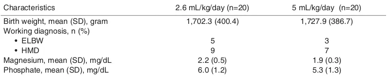
During the study period data were collected IURPVXEMHFWVZKRPHWWKHLQFOXVLRQFULWHULD EXW VL[ VXEMHFWV FRXOG QRW ILQLVKHG WKLV VWXG\ EHFDXVHWZRRIWKHPGLHGDQGIRXUVXEMHFWVZHUH GLVFKDUJHG DJDLQVW PHGLFDO DGYLFH 7KH VXEMHFW characteristics based on birth weight, working diagnosis, magnesium and phosphate level is depicted in Table 1.
Table 1. Subject’s characteristics
Characteristics 2.6 mL/kg/day (n=20) 5 mL/kg/day (n=20)
Birth weight, mean (SD), gram Working diagnosis, n (%)
Ŗ'.$9 Ŗ*/&
Magnesium, mean (SD), mg/dL Phosphate, mean (SD), mg/dL
1,702.3 (400.4)
Note: ELBW = extremely low birth weight, HMD = hyaline membrane disease
Table 2. Ionized calcium level at baseline and 48 hours of age both groups
Factor-B
Ionized calcium level based on time, mean (SD), mg/dL
B1 B2
Factor-A Treatment
A1 4.93 (0.38) 4.46 (0.37)
A2 4.69 (0.41) 4.80 (0.95)
In this study there were two confounding factors, i.e. gestational age and diagnosis of severe asphyxia. The analysis of both confounding factors is depicted inTable 2.
Normal ionized calcium level of preterm
QHZERUQV LV PJG/The mean of ionized
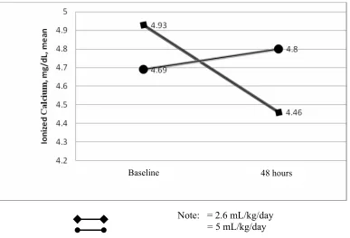
FDOFLXPOHYHODWEDVHOLQHLQJURXSP/NJGD\ZDV 6'PJG/ZKHUHDVDWKRXUVRIDJH ZDV6'PJG/,QJURXSP/NJGD\ the mean of ionized calcium level at baseline was 6'PJG/ZKHUHDVDWKRXUVRIDJH ZDV6'PJG/7KHGLIIHUHQFHRIPHDQ of both doses on ionized calcium level is depicted inFigure 1.
,Q JURXS P/NJGD\ WKH ORZHVW OHYHO RI LRQL]HGFDOFLXPRQKRXUVZDVPJG/ZKHUHDV WKHKLJKHVWRQHZDVPJG/,QJURXSP/NJ day, the lowest level of ionized calcium was 3.59 mg/ G/WKUHHVXEMHFWVZKHUHDVWKHKLJKHVWRQHZDV PJG/WKUHHVXEMHFWV
Repeated measures analysis of variance was used WRDQDO\]HWKHHIIHFWRIDGPLQLVWUDWLRQRIFDOFLXP JOXFRQDWHERWKGRVHVRQKRXUVLRQL]HGFDOFLXPOHYHO which is depicted in Table 2.
As shown in Table 2, there were no statistical GLIIHUHQFHVEHWZHHQHIIHFWVRIDGPLQLVWUDWLRQZLWK FDOFLXP JOXFRQDWH P/NJGD\ DQG P/NJGD\ GRVHV)DFWRU$RQKRXUVLRQL]HGFDOFLXPOHYHO (P=0.33). There were also no significant differences between the change of ionized calcium level based on time (Factor-B) (P=0.20). However, the change between both factors was significantly different (P=0.035).
Discussion
2XW RI VXEMHFWV ZKR PHW WKH LQFOXVLRQ FULWHULD WZRGLHG,WZDVQRWFOHDUWKDWWKHGHDWKRIVXEMHFWV ZDVD´IDLOXUHµGXHWRWKHVLGHHIIHFWRIFDOFLXP gluconate administration or the newborns suffered hypocalcemia or hypercalcemia associated with doses RIFDOFLXPJOXFRQDWHDGPLQLVWUDWLRQ7KHUHLV no known percentage of babies suffered from side HIIHFW RI FDOFLXP JOXFRQDWH K\SRFDOFHPLD RU K\SHUFDOFHPLD%HFDXVHWKHUHZHUHRQO\WZRVXEMHFWV died in this study, we assumed that the cause of death was associated with the underlying diseases, Figure 1. The means of ionized calcium level at baseline and 48 hours of age both groups
Baseline 48 hours
i.e severe asphyxia and HMD. The fact is supported by Klaus and Fanaroff5 that hypocalcemia without
other diseases has good prognosis. Hypercalcemia is usually fatal when ionized calcium level reached more
WKDQPJG/7KUHHVXEMHFWVLQJURXSP/NJGD\
VXIIHUHGIURPK\SHUFDOFHPLDRQKRXUVRIDJHEXW WKHOHYHOPJG/
There were no significant differences between the HIIHFWRIDGPLQLVWUDWLRQZLWKFDOFLXPJOXFRQDWH P/NJGD\DQGP/NJGD\GRVHV)DFWRU$RQ KRXUVLRQL]HGFDOFLXPOHYHO3 DQGDOVRZLWK the alteration of ionized calcium level based on time (Factor-B) (P=0.20). Feed back mechanism by ionized calcium to parathyroid glands of preterm newborns
VWDUWVRQGD\RIOLIH37KHGRVHVRIFDOFLXP
JOXFRQDWH JLYHQ EHIRUH GD\V RI DJH ZRXOG QRW influence the action of parathyroid glands. According WRWKHUHVXOWVERWKGRVHVRIFDOFLXPJOXFRQDWHZDV sufficient for calcium requirement of body to maintain many important biologic functions such as calcium messenger system by which extracellular messengers regulate cell function, activation of cellular enzyme cascades, smooth muscle and myocardial contraction, nerve impulse conduction, and secretory activity of exocrine glands. Our findings agree with Klaus and Fanaroff5WKDWFDOFLXPJOXFRQDWHDGPLQLVWUDWLRQ
RIGRVHP/NJGD\FDQSUHYHQWK\SRFDOFHPLDRI susceptible newborns. Our results also agree with Mainali8WKDWFDOFLXPJOXFRQDWHDGPLQLVWUDWLRQ
of dose 5 mL/kg/day can prevent hypocalcemia of susceptible newborns.
Statistical analysis of interaction between WUHDWPHQWHIIHFWRQKRXUVLRQL]HGFDOFLXPOHYHODQG the alteration of serum ionized calcium level based on time showed significant difference (P=0.035). Physiologically serum calcium level alterations in a few KRXUVDIWHUELUWKFRQWLQXHIRUKRXUVDQGWKHQ stabilize at normal range.3-5 In sick preterm newborns
the serum ionized calcium level must decline below nadir, therefore they will undergo hypocalcemia ZLWKRXWSUHYHQWLYHHIIRUW,QJURXSP/NJGD\WKH pattern of ionized calcium level alteration was suitable with physiologic ionized calcium alteration (Figure 1). This result means that the dose was sufficient for body to maintain many important biologic functions and able to keep ionized calcium level in normal range (not hypocalcemia). This outcome agree with Klaus and Fanarof5 WKDW DGPLQLVWUDWLRQ RI FDOFLXP
JOXFRQDWH GRVH P/NJGD\ LV ZHOO WROHUDWHG E\ newborns.
By contrast, the alteration of ionized calcium level in group 5 mL/kg/day was not physiologic. The alteration formed increasing pattern of ionized calcium level but still in normal range. This pattern means that the dose was sufficient to maintain many important biologic functions but excessive.
In group 5 mL/kg/day, there were three VXEMHFWVH[SHULHQFHK\SRFDOFHPLDDQGWKUHHVXEMHFWV H[SHULHQFHK\SHUFDOFHPLDRQKRXUVRIDJH7KH IDFWRUVFDXVHGK\SRFDOFHPLDRIWKUHHVXEMHFWVZHUH XQFOHDU7KHK\SRFDOFHPLFVXEMHFWVZHUHGLDJQRVHG DV VHYHUH DVSK\[LD WZR VXEMHFWV DQG +0' RQH VXEMHFW 6HYHUH DVSK\[LD PXVW EH FRXQWHG WR EH one of the etiology of the hypocalcemia although no significant difference in statistical analysis of both group. In this study, working diagnosis of severe asphyxia was according to APGAR score only, without laboratory assessment. Theoretically, respiratory distress due to severe asphyxia, HMD, etc will increase calcitonin level, disturb parathyroid hormone (PTH) function, disturb magnesium function to regulate and secrete PTH, and increase phosphate level due to increasing of catabolism process. High concentration of serum calcitonin will rise urinary calcium excretion. Without adequate PTH secretion, this circumstance will decrease ionized calcium concentration. Another possibility ZDVWKRVHVXEMHFWVZLWKK\SRFDOFHPLDH[SHULHQFHG K\SRPDJQHVHPLD RU K\SHUSKRVSKDWHPLD RQ hours of age, therefore hypocalcemia occurred. The limitations of our study associated this hypocalcemia circumstance were no measurement of calcitonin, PDJQHVLXPDQGSKRVSKDWHOHYHOSHUIRUPHGRQ hours of age.
7KRVH VXEMHFWV H[SHULHQFHG K\SRFDOFHPLD DQGK\SHUFDOFHPLDRQKRXUVRIDJHZHUHWUHDWHG according to our protocol. Despite small percentage of hypocalcemia and hypercalcemia in our study, physicians must perform measurement of ionized FDOFLXPOHYHORQKRXUVRIDJH
,Q FRQFOXVLRQ LRQL]HG FDOFLXP OHYHOV LQ hours of age of preterm newborns administered with ERWKGRVHVRIFDOFLXPJOXFRQDWHLVQRWGLIIHUHQW EXW GRVH RI P/NJGD\ \LHOGV SK\VLRORJLF alteration of ionized calcium level compared with 5 mL/kg/day.
References
6LQJKDO$+\SRFDOFHPLDKRPHSDJHRQWKHLQWHUQHW FLWHG-XO\$YDLODEOHIURPhttp://www.eMedicine. com.
2. Namgung R, Tsang RC. Hypocalcemia. In: Rudolph AM, Rudolph CD, Hosteter MK, Lister G, Siegel NJ, editors. 5XGROSK·VSHGLDWULFVVWHG1HZ<RUN0F*UDZ+LOO S
3. Koo WWK, Tsang RC. Calcium and magnesium homeostasis. In: MacDonald MG, Mullet MD, Seshia MM, editors.
Avery’s neonatology: pathophysiology & management of WKHQHZERUQWKHG3KLODGHOSKLD/LSSLQFRWW:LOOLDPV :LONLQVS
5LJR-&XUWLV0''LVRUGHUVRIFDOFLXPSKRVSKRUXVDQG magnesium metabolism. In: Martin RJ, Fanaroff AA, Walsh MC, editors. Fanaroff and Martin’s neonatal-perinatal PHGLFLQHWKHG1HZ<RUN0RVE\S 5. Klaus MH, Fanaroff AA. Care of the high-risk neonate. 5th
HG3KLODGHOSKLD:%6DXQGHUV&RPSDQ\
+XWWQHU .0 +\SRFDOFHPLD K\SHUFDOFHPLD DQG K\SHU magnesemia. In: Cloherty JP, Eichenwald EC, Stark AR,, editors. Manual of neonatal care. 5th ed. Philadelphia: /LSSLQFRWW:LOOLDPV :LONLQVS
&ORKHUW\-33DUULW]$/0DWHUQDOFRQGLWLRQVWKDWHIIHFWWKH fetus. In: Cloherty JP, Eichenwald EC, Stark AR, editors. Manual of neonatal care. 5th ed. Philadelphia: Lippincott :LOOLDPV :LONLQVS
8. Mainali E. Fluid and electrolytes [CD-ROM]. Washington: 86$,'
9. Blackburn ST. Maternal, fetal, and neonatal physiology, a FOLQLFDOSHUVSHFWLYHUGHG6W/RXLV866DXQGHUV 6DQQ/'DYLG/&KD\YLDOOH-$/DVQH<%HWKHQRG0(IIHFW