EXTRACT OF Sargassum echinocarpum ALLEVIATES OXIDATIVE STRESS IN STREPTOZOTOCIN-INDUCED DIABETIC RATS
Muhamad Firdaus
Fakultas Perikanan dan Ilmu Kelautan, Universitas Brawijaya, Malang Jl. Letjen. MT. Haryono No. 165, Malang, Indonesia
E-mail: muhamadfi [email protected]
Abstract
Oxidative stress occured in streptozotocin-induced diabetic rats. The ability of Sargassum echinocarpum to ameliorate the oxidative stress after treatment with streptozotocin was investigated in rats. Adult male rats were intraperitoneally injected with 45 mg/kg of streptozotocin to produce experimental oxidative stress characteristic of diabetes mellitus. Hyperglycemia was observed in blood serum after 10 days
of streptozotocin treatment. There were a signifi cant decrease in the activity of superoxide dismutase
(SOD), catalase (CAT) and glutathione peroxide (GPx) and a signifi cant increase in the levels of
malondialdehyde (MDA) in the serum of diabetic rats. It indicated that there were an increasing lipid peroxidation and oxidative stress in the diabetic rats. Providing S. echinocarpum extract for 90 days on
diabetic rats signifi cantly improved the oxidative stress evidenced by a decreasing of MDA serum level
and an increasing SOD, CAT and GPx activities than streptozotocin-treated rats. These results showed that the extract might improve the clinical manifestation of diabetes mellitus and decrease the oxidative stress in the diabetic rats. This effects appear to be due to its antioxidant properties.
Key words: antioxidants, lipid peroxidation, Sargassum echinocarpum
Introduction
Diabetes mellitus is a common chronic disease. Although both genetic and environmental factors appear to play a role, the cause of diabetes mellitus is still not clear. A large number of studies have demonstrated that oxidative stress and nonenzymatic protein glycation are closely associated with the development of diabetes mellitus (Mehta et al., 2006). Excessive oxidative stress has been implicated in the pathology and complications of diabetes mellitus (Kuyvenhoven & Meinders, 1999). Hyperglycemia generates abnormally high levels of free radicals by a mechanism involving autoxidation of glucose, followed by oxidative degeneration and protein glycation. In addition, nonenzymatic glycosylation of those enzymes that normally detoxify free-radical species may exacerbate oxidative stress in diabetes (Sydow & Münzel, 2003). Thus, clinical complications in diabetes may be due partially to the inability of key antioxidant enzymes to function at normal levels (Jakus, 2000). Conversely, antioxidants are believed to be protective because they may help to protect the human body against damage by reactive oxygen singlet (ROS) (Halliwell, 2009). Antioxidants from natural sources are preferred by consumers due to concerns on the toxic and carcinogenic effects of synthetic antioxidants (Kranl et al., 2005).
Seaweeds have been habitually consumed on Indonesia, especially in coastal society. It has been reported that seaweeds contain a rich and largely untapped source of biologically active substances (Smit, 2004). Marine brown algae contain phloroglucinol phenolics (phlorotannins) (Koivikko et al., 2005, Singh & Bharate, 2006) which are probably good antioxidants, since plant phenolics can behave as ROS scavengers, metal chelators, enzyme modulators and prevent lipid peroxidation (Rice-Evans et al., 1997). The antioxidant activity of this seaweeds has been published (Anggadiredja et al., 1997; Lim et al., 2002; Mori et al., 2003; Wei et al., 2003; Kang et al., 2004; Okada et al., 2004; Iwashima et al., 2005), conversely, nowadays no experiment relating to the anti-oxidative stress activity of S. echinocarpum on diabetic animal model. The aim of this study was to investigate the anti-oxidative stress of S. echinocarpum extract on diabetic rats.
Material and Method
Material
S. echinocarpum was collected on the Coast of Talango Island, East Java. Indonesia, in April 2008. The alga was washed thoroughly with seawater, followed by tap water to remove sand and epiphytes,
by Dr. Augy Syahailatua, Research Centre of Oceanography, Indonesian Institute of Sciences.
Drugs and Chemicals
All the drugs and biochemicals used in this study were purchased from Sigma Chemical Company, Inc., St Louis, MO, USA. The chemicals were of analytical grade.
Experimental Animals
Male Sprague-Dawley rats weighing 150-200 g were procured from Gadjah Mada University, Indonesia. All animal procedures were in accordance with the institutional guidelines for animal research, and approved by the animal research ethics committee of Brawijaya University, Indonesia. They were kept in clean and dry cages with a bedding of paddy husk, fed with standart diet (American Institute of Nutrition (AIN) -93) and water ad libitum.
Extraction of Extract
The dried of S. echinocarpum (2.0 kg) were ground
to a fi ne powder and were macerated three times
with methanol. The suspension was filtered and evaporated under reduced pressure and lyophilized. The green yellow of methanol extract (80 g) was obtained.
Animal Experiment
Animals were divided into 3 groups: normal control, diabetic control, and the diabetic animals were given S. echinocarpum extract (450 mg/kg body weight) by oral gavage. The experimental diabetic rats were obtained by single administration of streptozotocin (45 mg/kg, i.p.), dissolved in freshly prepared 0.1 M
citrate buffer, pH 4.5. Diabetes was confi rmed seven
days latter in streptozotocin induced animals showing blood glucose levels > 200 mg/dl (11.1 mmol/l) as monitored in the blood from tail vein using glucometer. The extract treatment was given for 90 days.
Preparation of Serum Sample
After the 12 weeks treatment period, the whole blood
was obtained by cardiac puncture from the sacrifi ced
rat. The whole blood was allowed to clot for 2 h and further spin at 4000 rpm for 10 min to separate serum from the blood cells. The blood serum acquired was used for lipid peroxide and antioxidant enzymes assays.
Measurement of Lipid Peroxide in Serum
Lipid peroxide assay was determined based on
Ohkawa et al. (1979). A volume of 200 μl Sodium
dodecil sulfonate (SDS) was added with 50 μl
buthylated hydroxy toluene (BHT), 50 μl Ethylene
tetraacetic acid (EDTA), 1500 μl thiobarbituric acid
(TBA) and 1250 μl serum. Solution was vortexed
before added 1500 μl trichloro acetic acid (TCA),
contunued by sentrifuged 500 rpm, for 10 min. Supernatan in reaction tube was heated on water bath
at 80oC, for 20 min. Malondialdehyde determined by
absorbance on spectrophotometer at 532 nm.
Measurement of Superoxide Dismutase Activity in Serum
Superoxide dismutase activity was determined based
on Misra and Fridovich (1972). A volume of 2800 μl
sodium carbonate mixed with 100 μl serum and 100
μl epinephrine. Absorbance was measured at 480 nm
after epinephrine addition.
Measurement of Catalase Activity in Serum
Catalase activity was determined based on Sinha
(1972). A volume of 1000 μl serum was added with 2
ml potassium bichromate and boiled in water bath for 10 min. Absorbance was measured at 570 nm.
Measurement of Glutathione Peroxidase Activity in Serum
Glutathione peroxidase activity was measured by Paglia & Valentine method (1967). A volume of
2000 μl buffer phosphate 0.1 M pH 7.0 mixed with
200 μl serum, 200 μl glutathione 10 mM, and 200 μl
glutathione reductase. After incubated at 37oC, for
10 min, solution added with 200 μl NADPH 1.5 mM.
Finally, 200 μl H2O2 1.5 mM mixed with solution after
incubated at 37oC, for 3 min. Absorbance of solution
was measured at 340 nm.
Results and Discussion
Blood Glucose
The diabetic animals treated with S. echinocarpum
showed signifi cant decrease in the glucose level at
4th , 6th, 8th, 10th and 12th weeks as compared to the
diabetic rats (Figure 1). Even though a signifi cant
antihyperglycemic effect was evident from 4th week
onwards; a furthermore, the decrease in the blood
glucose level was highly signifi cant at 10th week as
compared with the diabetic rats. The S. echinocarpum treatment however, could not restore glucose levels to normal.
There was signifi cant increase in the glucose levels
of diabetic rats throughout the experimental period. The treatment with S. echinocarpum extract showed decrease in the glucose level at different time interval
as compared with the diabetic animals signifi cantly.
possibly done by inhibiting α glucosidase activity of gastrointestinal tract. Polyphenols are well known to bind to proteins in solution and form complexes whose properties depend on the structure of both the polyphenol and the protein. This complexation is responsible for the inhibition of digestive enzymes (Aguie-Beghin et al., 2008).
Effect of Extract on the MDA Level
[image:3.595.62.285.93.242.2]The highest MDA level of serum showed at diabetic rats. However the diabetic group of rats treated with S. echinocarpum extract, showed signifi cant decrease in MDA level of serum (Table 1).
Table 1. Effects of S. echinocarpum extract on MDA
levels in serum of rats.
Groups MDA (nmol/ml)
Normal Diabetic
Diabetic + Sargassum
1.07 ± 0.0474a 17.44 ± 0.4295c 2.87 ± 0.1305b
Values are expressed as mean ± SEM. Different letters indicate signifi cant differences (P < 0.05).
The highest MDA level was obtained on diabetic rats. This suggests that lipid peroxidation is higher in diabetes and supported by earlier reports (Vessby et
al., 2002; Memisogullari et al., 2003). Kuyvenhoven & Meinders (1999) suggested that hyperglycemia can increase lipid peroxidation. The excessive levels of glucose reaching the mitochondria lead to an overdrive of the electron transport chain, resulting in overproduction of superoxide anions, thus become hydrogen peroxide and hydroxyl radical. Interaction between hydroxyl radical and polyunsaturated fatty acid (PUFA) of membrane cell produces malondialdehyde. Lipid peroxidation of cellular structures, a consequence of free radical activity, is thought to play an important role complications of diabetes.
The diabetic rats with S. echinocarpum extract showed decrease in the MDA level as compared with
the diabetic rats signifi cantly. The treatment with S.
echinocarpum extract showed signifi cant prevention in increase in the levels of malondialdehyde. Several studies indicated that phlorotannin of brown algae is able to be protective against lipid peroxidation. The protective nature offered by the brown seaweed extracts may be due to the presence of phlorotannin having free radical scavenging properties (Mori et al., 2003; Wei et al., 2003; Kang et al., 2004).
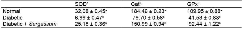
Effects of Extract on the Antioxidant Enzymes Activity
The antioxidant enzymes were decreased in serum of the diabetic rats as compared with the normal. However the diabetic group of rats treated with S.
extract, showed signifi cant increase in the activities
of SOD, catalase and glutathione peroxidise in serum, respectively (Table 2).
The diabetic rats showed decrease in the antioxidant enzymes. The antioxidant enzymes such as SOD,
CAT and GPx are the fi rst line of defence against O2-
and H2O2 mediated injury (Jakus, 2000). The levels
of fi rst line antioxidant enzymes were signifi cantly
depleted in diabetic rats. This deficient function of free radical scavenging enzymes leads to the accumulation of highly reactive free radicals and
Bl
o
o
d
g
lu
co
se
(mg
/d
[image:3.595.57.286.493.540.2]l)
Figure 1. Glucose levels of normal ( ), diabetic
( ), and diabetic rats added Sargassum echinocarpum extract ( ).
Table 2. Effects of Sargassum extract on antioxidant enzymes activity in serum of rats.
SOD1 Cat2 GPx3
Normal Diabetic
Diabetic + Sargassum
32.08 ± 0.45a 6.99 ± 0.47c 25.18 ± 0.36b
184.46 ± 0.23a 79.70 ± 0.58c 150.99 ± 0.94b
109.95 ± 0.88a 41.53 ± 0.83c 92.44 ± 1.22b 1 SOD activity expressed as U/min/ml
2 Cat activity expressed as μmol H
2O2/min/ml 3 GPx activity expressed as mU/min/ml
[image:3.595.93.492.653.702.2]consequent degenerative changes (Kuyvenhoven & Meinders, 1999). In the present study, the activities
of SOD, CAT and GPx were signifi cantly improved
in diabetic rats treated seaweed extract, which may support the defensive nature of the S. echinocarpum extracts against diabetic-induced serum oxidative stress. The effects of phloroglucinol on cell viability might involve dual actions: direct action on oxygen
radical scavenging, as shown by H2O2, OH radical
scavenging and indirect action through induction of antioxidant enzymes. Antioxidant enzymes would be potential target molecules mediating antiapoptotic function of ERK pathway against oxidative stress (Kang et al., 2006).
Conclusion
This study investigated the anti-oxidative stress
effect of S. echinocarpum extract on serum in
diabetic rats and its possible mechanisms. The mechanism of this effect involves inhibition of glucose absorption, alleviation of the malondialdehyde level and enhancement of the antioxidant defence level; therefore, it rendered protection from oxidative stress which is usually associated with diabetes.
Acknowledgment
This study was fi nancially supported by Directorate of
Higher Education via Hibah Bersaing Project.
References
Anggadiredja, J., R. Andyani, Hayati & Muawanah. 1997. Antioxidant activity of Sargassum polycystum (Phaeophyta) and Laurencia obtuse (Rhodophyta) from Seribu Islands. J. Appl. Phycol. 9: 477-479.
Aguie-Beghin, V., P. Sausse, E. Meudec, V. Cheyneir & R. Douillard. 2008. Polyphenol-casein complexes at the air/water interface and in solution: effects of polyphenol structure. J. Agric. Food Chem. 56: 9600-9611.
Halliwell, B. 2009. The wanderings of a free radical. Free Radical Biology & Medicine 46: 531-542.
Iwashima, M., J. Mori, X.Ting, T. Matsunaga, K. Hayasih, D. Shinoda, H. Saito, U. Sankawa & T. Hayashi. 2005. Antioxidant and antiviral activites of plastoquinones from the brown alga Sargassum microcantum, and a new chromene derivative
converted from the plastoquinones. Biol. Pharm. Bull. 28: 374-377.
Jakus, V. 2000. The role of free radicals, oxidative stress and antioxidant systems in diabetic vascular disease. Bratisl Lek Listy. 101: 541-551.
Kang, H.E., H.Y. Chung, J.Y. Kim, B.W. Son, H.A. Jung & J.S. Choi. 2004. Inhibitory phlorotannins from the edible brown alga Ecklonia stolonifera on total reactive species (ROS) generation. Archives of Pharmacal Research 27: 194-198.
Kang, K.A., K.H. Lee, S. Chae, R. Zhang, M.S. Jung, Y.M. Ham, J.S. Baik, N.H. Lee & J.W. Hyun. 2006. Cytoprotective effect of phloroglucinol on oxidative stress induced cell damage via catalase activation. J. Cell. Biochem. 97: 609-620.
Koivikko, R., J. Poponen, T. Honkanen & V. Jormalainen. 2005. Contents of soluble cell-wall-bound and exuded phlorotannins in the brown alga Fucus vesiculosus, with implications on their ecological functions. J. Chem. Ecol. 31: 195-212.
Kranl, K., K. Schlesier, R. Bitsch, H. Hermann, M. Rohe & V. Böhm. 2005. Comparing antioxidative food additives and secondary plant products-use of different assays. Food Chem. 93:171-175.
Kuyvenhoven, J.P. & A.E. Meinders. 1999. Oxidative stress and diabetes mellitus pathogenesis of long term complications. Eur. J. Int. Med. 10(1): 9-19.
Li, Y., Z.J. Qian, B.M. Ryu, S.H. Lee, M.M. Kim & S.K. Kim. 2009. Chemical components and its antioxidant properties in vitro: An edible marine brown alga, Ecklonia cava. Bioorg. Med. Chem. 17: 1963-1973.
Lim, S.N., P.C.K. Cheung, V.E.C. Ooi & P.O. Ang. 2002. Evaluation of antioxidative activity of extracts from a brown seaweed, Sargassun siliquastrum. J. Agric. Food Chem. 50: 3862-3866.
Mehta, J.L, N. Rasouli, A.K. Sinha & B. Molavi. 2006. Oxidative stress in diabetes: A mechanistic overview of its effects on atherogenesis and myocardial dysfunction. Int. J. Biochem. Cell Biol. 38: 794-803.
Misra, H.P. & I. Fridovich. 1972. The role of superoxide anion in the autoxidation of epinephrine and a simple assay for superoxide dismutase. J. Biol.Chem. 247: 3170-3175.
Mori, J., T. Matsunaga, S. Takahashi, C. Hasegawa & H. Saito. 2003. Inhibiting acivity on lipid peroxidation of extracts from marine brown algae. Phytoter. Res. 17: 549-551.
Ohkawa, H., N. Ohishi & K. Yagi. 1979. Assay for lipid peroxides in animal tissues by thiobarbituric acid reaction. Anal. Biochem. 95: 351-358.
Okada, Y., A. Ishimaru, R. Suzuki & T. Okuyama. 2004. A new phloroglucinol derivative from the browm alga Eisenia bicyclis potential for the effective treatment of diabetic complications. J. Nat. Prod. 67: 103-105.
Paglia, D.E. & W.N. Valentine. 1967. Studies on the quantitative and qualitative characterization of erythrocyte glutathione peroxidase. J. Lab. Clin. Med. 70:158-169.
Rice-Evans, C.A., N.J. Miller & G. Paganga. 1997. Antioxidant properties of phenolic compounds. Trends in Plant Science Reviews 2: 152-159.
Singh, I.P. & S.B. Bharate. 2006. Phloroglucinol compounds of natural origin. Nat. Prod. Rep. 23: 558-591.
Sinha, K.A. 1972. Colorimetric assay of catalase. Anal. Biochem. 47: 389-394.
Smit, A.J. 2004. Medicinal and pharmaceutical uses of seaweed natural products: A review. J. Appl. Phycol. 16: 245-262.
Sydow, K. & T. Mu¨nzel. 2003. Diabetes mellitus, oxidative stress and endothelial dysfunction. International Congress Series 1253: 125-138.
Vessby, J., S. Basu, R. Mohsen, C. Berne & B. Vessby. 2002. Oxidative stress and antioxidant status in type 1 diabetes mellitus. J. Int. Med. 251: 69-76.