Indones. J. Chem., 2015, 15 (2), 201 - 209
201
ANTIBACTERIAL COMPOUNDS FROM RED SEAWEEDS (RHODOPHYTA)
Noer Kasanah
*, Triyanto, Drajad Sarwo Seto, Windi Amelia, and Alim Isnansetyo
Department of Fisheries and Marine Sciences, Faculty of Agriculture, Universitas Gadjah Mada,Jl. Flora, Bulaksumur, Yogyakarta 55281, Indonesia
Received January 15, 2015; Accepted May 5, 2015
ABSTRACT
Seaweeds produce great variety of metabolites benefit for human. Red seaweeds (Rhodophyta) are well known as producer of phycocolloids such agar, agarose, carragenan and great variety of secondary metabolites. This review discusses the red algal secondary metabolites with antibacterial activity. The chemical constituents of red algae are steroid, terpenoid, acetogenin and dominated by halogenated compounds mainly brominated compounds. Novel compounds with intriguing skeleton are also reported such as bromophycolides and neurymenolides. In summary, red seaweeds are potential sources for antibacterial agents and can serve as lead in synthesis of new natural medicines.
Keywords:red algae; bioactive compounds; antibacterial
ABSTRAK
Rumput laut menghasilkan berbagai metabolit manfaat bagi manusia. Rumput laut merah (Rhodophyta) dikenal sebagai penghasil phycocolloids seperti agar, agarose, karagenan dan berbagai macam metabolit sekunder. Tulisan ini membahas metabolit sekunder dari alga merah dengan aktivitas antibakteri. Kandungan kimiawi dari alga merah yaitu steroid, terpenoid, acetogenin dan didominasi oleh komponen halogen terutama brominated senyawa. Senyawa baru dengan struktur kimia menarik juga dilaporkan seperti bromophycolides dan neurymenolides. Dari studi ini terbukti bahwa rumput laut merah merupakan sumber potensial untuk agen antibakteri dan dapat berfungsi sebagai senyawa penuntun dalam sintesis obat-obatan alami yang baru.
Kata Kunci:alga merah; senyawa bioaktif; antibakteri
INTRODUCTION
Seaweeds are benthic, macroscopic algae (macroalgae), eukaryotic, multicellular organisms, simplified marine plants that lack of the highly specialized structures (true leaves, stems and roots) and reproductive. Seaweed typically consists of thallus, which is sometime provided with leaf like blade and a root like holdfast. They are able to tolerate salt due to adjustment of the cytoplasm osmolarity and contain photosynthetic pigments (chlorophyll) to produce food and oxygen from carbon dioxide, and the water. Seaweeds are classified based on their pigment. Chlorophytes is green algae that contain pigment chlorophyll as major component. Phaeophyta is brown algae due to the brown pigment called fucoxantin. Rhodophyta is red algae because of the red pigments called phycoerythrine, phycocyanin, phycobilins,
chlorophyll a, β-carotene and unique xanthophyl. Red
Algae are essentially marine and important for marine environment in the formation of tropical reefs by contributing to glue the reef structure and resist to wave
[1]. The chemistry of red algae is more diverse than green or brown algae.
Seaweeds have been used since human life in various ways as food, feed, fertilizer and medicine. As a food source seaweed contains low calories, dietary fibers, carbohydrate, minerals, vitamins and essential amino acids. Therefore, seaweeds provide nutritional requirement for human and have beneficial to human health [2-4]. Seaweeds have been one of the richest and most promising sources of bioactive primary and secondary metabolite [5-6]. The primary metabolites called phycocolloid compounds such as carragenan, agar, and alginate are used as gelling, stabilizing and thickening agents in food, cosmetics, and pharmaceutical industries [7]. Thus, seaweeds have contribution to the economic growth. Other primary metabolites are polysulfated polisaccharides such as laminaran, rhamnan sulfate, galactosyl glycerol and fucoidan exhibit bioactivities such as antioxidant, antiallergic, anti-HIV, anticancer and anticoagulant [8-11]. Varieties of secondary metabolites are synthesized by seaweeds such as carotenoids, polyphenols, terpenoids,
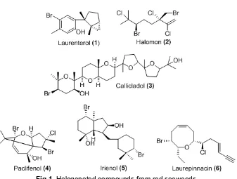
Fig 1.Halogenated compounds from red seaweeds
xanthophylls, chlorophyll, vitamins, alkaloid, saturated and polyunsaturated fatty acid [12-14].
Rhodophyta is rich in diversity of secondary metabolites than brown and green algae. Red algae is the most important source of biologically active metabolites in comparison to other algal. Most of the bioactive substances isolated from marine algae are chemically classified as brominated, aromatics, nitrogen-heterocyclic, nitrosulphuric heterocyclic, sterols, dibutanoids, proteins, peptides and sulphated polysaccharides. The chemistry of Rhodophyta is dominated by compounds from family Rhodomelaceae. Red algae are the main producers of halogenated compounds such as laurenterol (1), halomon (2), callicladol (3) etc as presented in Fig 1 [15-16]. Bromophenols were detected in families Ceramiaceae, Delesseriaceae, Bonnemaisoniaceae, Rhodophyllaceae, Corallinaceae and Rhodomelaceae [17]. The halogenated compounds exhibited diverse biological activities including anti bacterial, antifungal, anti inflammation, ichtiotoxic, cytotoxic and insecticidal activity. In addition, the red algae also produce terpenoid, polyether, and acetogenin along with some amino acid, shikimate, acetate and nucleic acid derivatives [14,18].
Indonesia is a mega biodiversity country and has the highest marine species richness. About 45 percent of world’s seaweed species are in Indonesia. Based on the report of Siboga expedition there are approximately 782 species of seaweeds with number of 196 species are green algae, 134 species are brown algae and 452 species are red algae. Potential of seaweed in Indonesia
has contributed to the increase in revenue coastal communities in Riau, Sumatra, Java, Bali, West Nusa Tenggara, East Nusa Tenggara, Sulawesi and Maluku. Eastern Indonesia is called as a barn of seaweed due to high diversity of seaweed species. Various types of seaweed found in Indonesia but few are known to have economic value for food industry and commodity export such as Gracilaria, Gelidium, Hypnea, Sargasum, and Turbinaria. Production of seaweed Indonesia in 2014 reached 10 million tons. Most of seaweeds in Indonesia are unexplored, unidentified, under development and not fully utilized. According to FAO seaweed is very versatile product and can be diversified to food, cosmetics, pharmaceutical, bioethanol, animal feed, fish feed, integrated aquaculture and wastewater treatment. In Indonesian itself, seaweed production is still limited to the food industry and raw material exports. In efforts to use seaweed as a food industry, cosmetics, pharmaceutical, medicine and agriculture still needs to learn to countries that have experts in seaweed processing. Therefore, future actions still need to study the sustainable utilization of seaweed to improve life and welfare of Indonesian.
Indones. J. Chem., 2015, 15 (2), 201 - 209
203
compounds, terpenoid, steroid and long chain fatty acid (unpublished data).
ANTIBACTERIAL ACTIVITY
The incidence of bacterial resistance to antibiotics and other antibacterial agents increase each year. Several ways are available to protect and against infectious diseases such as using vaccine and antibiotics. Each approach has advantages and disadvantages. Some infectious diseases cannot be prevented by vaccine and antibiotics are still a drug of choice [19]. New antibiotics or antibacterial agents could be discovered from traditional and nontraditional resources. There is an urgently need for novel antimicrobial agents from different sources to combat emergence and reemergence infectious diseases [20-21]. The biodiversity of marine environment provides chemical diversity as source of bioactive compounds which promising to have potential and therapeutic applications [22-26]. Marine algae represent a great source of tremendous diversity of complex natural products and could be a promising source of novel bioactive compounds. Compounds with cytostatic, antioxidant, antiviral, antihelminthic, antifungal and antibacterial activities have been detected in green, brown and red algae [27-31]. It has been reported that seaweeds possess potent and broad spectrum of biological activities against the pathogenic microbes of medical, agricultural, aquaculture and environmental importance [32-34].
Several works have been reported in the searching for broad spectrum activity of seaweed extracts. This review only focuses on the antibacterial activity. Numerous extracts of seaweeds have been examined for antibacterial activity against pathogen in human, agriculture and fish. Extracts of seaweeds Codium decorticatum, Caulerpa scalpelliformis, Gracilaria crassa, Acanthophora spicifera, Sargassum wightii and Turbinaria conoides from Gulf of Mannar exhibited antibacterial activity against Vibrio parahaemolyticus, Salmonella sp, Shewanella sp, Escherichia coli, Klebsiella pneumoniae, Streptococcus pyogenes, Staphylococcus aureus, Enterococcus faecalis, Pseudomonas aeruginosaandProteus mirabilis[35].
Screening of 44 species seaweed extracts from Gran Canary, Canary Island, yielded 28 species showed antibacterial activity with Asparagopsis taxiformis and Cymopola barbatawere the strongest activity [36].
The antibacterial activity of Codium adhaerens Anderson,Sargassum wightiiGreville andAcanthophora spicifera(Vahl.) Boergs collected from intertidal region of the Mandapam coastal water showed weak activity against human pathogenic bacteria S. aureus, V. cholerae, S. dysentriae, S. bodii, S. paratyphi,
P. aeuroginosa and Klebsiella pneumonia [37]. Ethanolic extract ofGracilaria edulis,Calorpha peltada, Hydrocolthres sp were tested against bacteria isolated from fish Escherichia coli, Enterobacter aerogenes, Staphylococcus aureus, Pseudomonas aeruginosa, Streptococcus faecalis and Bacillus cereus. Results showed that ethanolic extract of Gracilaria edulis inhibited the growth of all test organisms except Bacillus cereusandEnterobacter aerogenes. Ethanolic extracts of Calorpha peltada was active against Escherichia coli, Staphylococcus aureus and Streptococcus faecalis, while extract of Hydroclothres sp. only inhibited the growth of Pseudomonas aeruginosa[38].
Dichloromethane, methanol and water extracts of 26 species of cultivated seaweeds were screened for their antibacterial activities against five fish pathogenic bacteria strains Aeromonas salmonicida ssp. salmonicida, Aeromonas hydrophila ssp. hydrophila, Pseudomonas anguilliseptica, Vibrio anguillarum, Yersinia ruckeri. The dichloromethane extracts of Asparagopsis armata, Ceramium rubrum, Drachiella minuta,Falkenbergia rufolanosa,Gracilaria corneaand Halopitys incurvus showed strong antibacterial activities. V. anguillarum and P. anguilliseptica were the two most susceptible bacteria strains. The screening results confirmed that seaweeds as a potential source of antimicrobial compounds or as a health-promoting food for aquaculture [33].
All those studies are preliminary for searching antibacterial agents from algae. They screened extract to look at the activity and none of reports were determined the constituent of bioactive compounds. There numerous research in Indonesia as well that done on screen antibacterial activity of seaweed and limited compounds were reported as antibacterial agents.
ANTIBACTERIAL COMPOUNDS FROM RED ALGAE
Numerous bioactive compounds have been isolated and identified from seaweeds. This review focus on antibacterial compounds isolated from Red Algae (Rhodopytha) such asLaurenciaspp.,Gracillaria spp.,Acanthophoraspp and other rare species.
Laurenciaspp.
Red algae of the genus Laurencia (Rhodomelaceae, Ceramiales) mainly found in tropical, subtropical and temperate coastal waters are known to be prolific sources of a wide variety of halogenated secondary metabolites, such as C15-acetogenins, C15-,
C20-, and C30-terpenoids.A series of compound
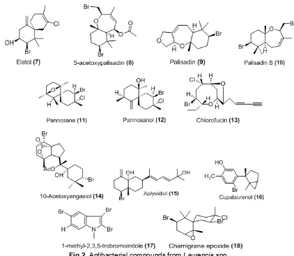
Fig 2.Antibacterial compounds fromLaurenciaspp.
glandulifera collected from the island of Crete and showed activity as antistaphylococcal with MIC 8-256 mg/mL [39].
A halogenated sesquiterpene alcohol, elatol (7) is commonly found in many species of Laurencia, and known for its potent antibacterial activity. The compound was isolated for the first time inL. microcladia, collected in the Southern Brazilian coast. Studies showed that elatol has ecological roles as antiherbivore activity and potential defense against infection by microorganisms [40]. Antimicrobial compounds 5-acetoxypalisadin B (8), palisadin A (9) and palisadin B (10) were isolated from L. saitoi[41].
Antibacterial activity of Malaysian L. pannosa was tested against three species marine bacteria Chromobacterium violaceum, Proteus mirabilis, and Vibrio cholerae. Three halogenated sesquiterpene metabolites were identified as compounds responsible for antibacterial activity pannosanol (11), pannosane (12) and 3(Z) Chlorofucin (13). Pannosanol and pannosane are novel metabolites with camigrane framework. Pannosanol showed MIC 60 µg/disk against
Proteus mirabilis and 100 µg/disk against Chromo-bacterium violaceum and Vibrio cholera. Pannosane and (3Z) Chlorofucin showed activity against Chromo-bacterium violaceum with MIC 60 and 100 µg/disk, respectively [42].
Indones. J. Chem., 2015, 15 (2), 201 - 209
205
Fig 3.Antibacterial agents fromAcanthopora spicifera
Fig 4.Antibacterial steroid, terpenoid and eiconosoid fromGracilariaspp.
Acanthophoraspp.
The genus Acanthophora is one of the most abundance red algae on reef flat with wide distribution in tropic and subtropics regions, but investigation of this species is very rare. The chemical constituent of Achanthopora is rich of non halogenated steroid. Some sterols were isolated and identified from A. spicifera as 6-hydroxycholest-4-ene-3-one, cholest-4-ene-3,6-dione (19), cholest-5-ene-3β-ol, 5α-cholestane-3,6-dione (20),
β-sitosterol, saringosterol, 5α-cholestane-3,6-dione,
cholest-4-ene-3-one (21),
11α-hidroxy-5α-cholestane-3,6-dione, cholest-5-en-3α-ol and
choles-4-ene-3α,6β-diol [44-45]. It has been reported that some sterol showed activity as antibacterial and cytotoxic as well.
Gracilariaspp.
Fig 5.Antibacterial compounds fromAsparagopsis taxiformis
Fig 6.Antibacterial compounds fromC. Serratus
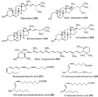
industrial biotechnology due to the commercially product phycocolloid called agar and agarose widely used in food, pharmaceutical and cosmetic industries [46-49]. In addition Gracilaria spp. produces bioactive metabolites such as steroid (22-25), terpenoid (26) and eicosanoid acid (27-30) derivates and with antibacterial activity [50]. The various bioactivities of genus Gracilaria was reviewed comprehensively by deAlmeida et al. [51].
Asparagopsisspp.
The red algae Asparagopsis taxiformis collected from the southwestern coast of India was tested for several activities including antibacterial. Chemical constituent were checked by TLC, HPLC and GC-MS
analysis of active fraction yielded compounds 4,5-dimethyl-1H-pyrrole-2-carboxylic acid, 14-methylpenta-decanoic acid (31), octadec-9-enoic acid 2,3-dihydroxy-propyl ester (32), 9-octadecanoic acid, and octadecanoic acid (33), chlorobenzene (34) [52].
Indones. J. Chem., 2015, 15 (2), 201 - 209
207
Callophycus serratus
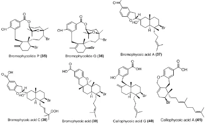
Callophycus serratus is a marine red alga found throughout the tropical and subtropical regions in Southeast Asia, Pacific and Africa. Series of novel diterpene-benzoate macrolide (Bromophycolides), diterpene-benzoic acid (callophylic acids) and diterpene phenol (callophycols) were isolated from the Fiji red algae C. serratus with varied bioactivities including antifungal, antibacterial, anticancer, antimalarial, antitubercular [54-57]. Bromophycolides P and Q (34-35) showed activity as antibacterial against methicillin-resistant Staphylococcus aureus (MRSA) and Vancomycin-Resistant Enterococcus faecium (VREF). The tetrahydropyran ring plays an important role in the antibacterial activity [58]. Bromophycoic acid A, C, E and callophycoic acid G (37-41) were active against MRSA with bromophycoic acid A and callophycoic acid G were the most potent at MIC 1.6 µg/mL. Bromophycoic E, callophycoic acid G and A were active against VREF at MIC 1.6, 3.1 and 0.8 µg/mL, respectively [59].
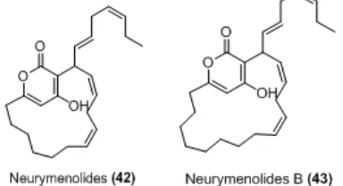
Neurymenia fraxinifolia
The less explored red algae N. fraxinifolis from Taveuni, Fiji was screened for antibacterial activity and yielded two novel compounds neurymenolide A and B (42-43) were identified having macrocyclic α-pyrones
ring and derived from acetogenic pathway. Neurymenolide A exhibited activity against methicillin-resistant Staphylococcus aureus (MRSA) and vancomycin-resistantEnterococcus faecium(VREF) with IC50 of 2.1 μM and 4.5 μM, respectively. The activity of
neurymenolide B was less potent than neurymenolide A in any bioassay panel. This suggest that macrocyclic ring and aromatic hydroxy group are restrict and important for bioactivity relationship [60].
Rhodomellaspp.
Rhodomella confervoides produce mainly bromophenol compounds with various activities such as citotoxic and antibacterial [61-62] onfervoides was investigated for antibacterial activity. Five compounds (44-48) were identified by MS and NMR spectroscopic analyses yielded two new brominated compounds (bromophenol) and three known compounds which are 3-bromo-4-[2,3-dibromo-4,5-dihydroxyphenyl] methyl-5-(hydroxylmethyl) 1,2-benzenediol and 3-bromo-4-[2,3-dibromo-4,5-dihydroxyphenyl] methyl-5- (ethoxymethyl) 1,2-benzenediol, 3-bromo-4-[2,3-dibromo-4,5dihydroxy-phenyl] methyl-5-(methoxymethyl) 1,2-benzenediol,
4,4′- methylenebis [5,6-dibromo-1,2-benzenediol] and
bis(2,3-dibromo-4,5-dihydroxybenzyl) ether. Compound bis (2,3-dibromo-4,5-dihydroxybenzyl) ether was the most active against five strains of bacteria with the MIC less than 70µg/mL [63].
CONCLUSIONS AND FUTURE DIRECTION
The algal chemistry is considered rich of functional group characteristics and broad spectrum of bioactivities. Seaweeds have been used as traditional
Fig 7. Antibacterial Neurymenolides from Neurymenia fraxinifolia
remedies and potential sources of new antibacterial agents. Research and development continue to drive the study of marine algae and their potential metabolites for human health. Development of seaweed is supported by the facts that seaweeds could be collected, renewable, can be cultivate and fast growing. The investigation data on the chemical constituents and bioactivities showed the potential as antibacterial agents or leads for the synthesis novel antibiotics. Further studies are needed to carry out taxonomically classify and standardize methods for cultivating, harvesting and extracting of potential seaweed. Identifying the active compounds is necessary to gather the pharmacochemical profiles and pharmacologic effects. This review demonstrated that halogenated compounds, steroids, terpenoids and fatty acid possess bioactivity as antibacterial and potential to be developed.
Finally, we conclude that red algae are a potential source for antibacterial agents and can serve as lead in synthesis of new natural medicines. Novel compounds with intriguing chemistry and interesting ring system can be used as template for the design and development of new antibacterial compounds. In addition, interdisciplinary collaboration research in natural products, biology, chemistry, chemical ecology and biomedical sciences is important to deliver products that benefit for human life.
ACKNOWLEDGEMENT
Authors would like to thank to Universitas Gadjah Mada for support our research in Indonesian Seaweeds under grant scheme Penelitian Unggulan Perguruan Tinggi Negeri Research Grant contract No. LPPM-UGM/1428/LIT/2013, LPPM -UGM 442/LIT/2014 and New Zealand Aid.
CONFLICTS OF INTEREST
The authors declare no conflict of interest.
REFERENCES
1. Castro, P., and Huber, M.E., 2013, Marine Biology, McGraw-Hill, New York, 102–113.
2. Lordan, S., Roos, P.R., and Stanton, C., 2011,Mar. Drugs, 9(6), 1056–1100.
3. Holdt, S.L., and Kraan, S., 2011, J. Appl. Phycol., 23(3), 543–597.
4. Dawczynski, C., Schäfer, U., Leiterer, M., and Jahreis, G., 2007, J. Agric. Food Chem., 55(25), 10470–10475.
5. El Gamal, A.A., 2010,Saudi Pharm. J., 18(1), 1–25.
6. Bhakuni, D.S., and Rawat, D.S., 2005, Bioactive Marine Natural Products, Anamaya Publisher, New Delhi, 1–10.
7. Laurienzo, P., 2010,Mar. Drugs, 8(9), 2435–2465. 8. Jiao, G., Yu, G., Zhang, J., and Ewart, H.S., 2011,
Mar. Drugs, 9(2), 196–223.
9. Pomin, V.H., and Maurão, P.A., 2008, Glycobiology, 18(12), 1016–1027.
10. Ngo, D.H., and Kim, S.K., 2013, Int. J. Biol. Macromol., 62, 70–75.
11. Lee, J-C., Hou, M-F., Huang, H-W., Chang, F-R., Yeh, C-C., Tang, J-Y., and Chang, H-W., Cancer Cell Int., 13, 1–7.
12. Takaichi, S., 2011,Mar. Drugs, 9(6), 1101–1118. 13. Güven, K.C., Percot, A., and Sezik, E., 2010,Mar.
Drugs, 8(2), 269–284.
14. Maschek, J.A., and Baker, B.J., 2008, “The Chemistry of Algal Secondary Metabolism” inAlgal Chemical Ecology, Amsler, C.D., Springer-Verlag Berlin Heidelberg, Germany, 1–24.
15. Kladi, M., Vagias, C., and Roussis, V., 2003, Phytochem. Rev., 3(3), 337–366.
16. Cabrita, M.T., Vale, C., and Rauter, A.P., 2010, Mar. Drugs, 8(8), 2301–2317.
17. Pedersén, M., Saenger, P., and Fries, L., 1974, Phytochemistry, 13(10), 2273–2279.
18. Ayyad, S.E., Al-Footy, K.O., Alarif, W.M., Sobahi, T.R., Bassaif, S.A., Makki, M.S., Asiri, A.M., Al Halwani, A.Y., Badria, A.F., and Badria, F.A., 2011, Chem. Pharm. Bull., 59(10), 1294–1298.
19. Spížek, J., Novotná, J., Rezanka, T., and Demain, A.L., 2010, J. Ind. Microbiol. Biotechnol., 37(12), 1241–1248.
20. Smith, V.J., Desbois, A.P., and Dyrynda, E.A., 2010,Mar. Drugs, 8(4), 1213–1262.
21. Kasanah, N., and Hamann, M.T., 2004,Curr. Opin. Invest. Drugs, 5(8), 827–837.
22. Kijjoa, A., and Sawangwong, P., 2004,Mar. Drugs, 2(2), 73–82.
23. Jha, R.K., and Zi-rong, X., 2004,Mar. Drugs, 2(3), 123–146.
24. Sashidhara, K.V., White, K.N., and Crews, P.A., 2009,J. Nat. Prod., 72(3), 588–603.
25. Newman, D.J., and Cragg, G.M., 2004, J. Nat. Prod., 67(8), 1216–1238.
26. Capon, R.J., 2010,Aust. J. Chem., 63, 851–857. 27. Kelman, D., Posner, E.K., McDermid, K.J.,
Tabandera, N.K., Wright, P.R., and Wright, A.D., 2012,Mar. Drugs, 10(2), 403–416.
28. Boopathy, N.S., and Kathiresan, K., 2010, J. Oncol., 2010, 1–18.
29. Liu, M., Hansen, P.E., and Lin, X., 2011, Mar. Drugs, 9(7), 1273–1292.
Indones. J. Chem., 2015, 15 (2), 201 - 209
209
31. Beutler, J.A., McKee, T.C., Fuller, R.W., Tischler, M., Cardellina II, J.H., Snader, K.M., McCloud, T.G., and Boyd, M.R., 1993,Antiviral Chem. Chemother., 4(3), 167–172.
32. Jiménez, E., Dorta, F., Medina, C., Ramírez, A., Ramírez, I., and Peña-Cortés, H., 2011,Mar. Drugs, 9(5), 739–756.
33. Bansemir, A., Blume, M., Schröder, S., and Lindequist, U., 2006,Aquaculture, 252(1), 79–84. 34. Manilal, A., Sujith, S., Kiran, G.S., Selvin, J., Shakir,
C., Gandhimathi, R., and Lipton, A.P., 2009, Ann. Microbiol., 59(2), 207–219.
35. Lavanya, R., and Veerappan, N., 2011, Adv. Biol. Res., 5(1), 38-44.
36. del Val, A.G., Platas, G., Basilio, A., Cabello, A., Gorrochategui, J., Suay, I., Vicente, F., Portillo, E., del Río M.G., Reina, G.G., and Peláez, F., 2001,Int. Microbiol., 4(1), 35–40.
37. Seenivasan, R., Rekha. M., Indu, H., and Geetha, S., 2012, J. Appl. Pharm. Sci., 2(10), 159–169. 38. Kolanjinathan, K., Ganesh, P., and Govindarajan,
M., 2009, Eur. Rev. Med. Pharmacol. Sci., 13(3), 173–177.
39. Kladi, M., Vagias, C., Stavri, M., M. Rahman, M.M., Gibbons, S., and Roussisa, V., 2008, Phytochem. Lett., 1(1), 31–36.
40. Dos Santos A.O., Veiga-Santos P., Ueda-Nakamura, T., Filho, B.P., Sudatti, D.B., Bianco, E.M., Pereira, R.C., and Nakamura, C.V., 2010,Mar. Drugs, 8(11), 2733–2743.
41. Ji, N-Y., Li, X-M., Li, K., and Wang, B-G., 2009, Helv. Chim. Acta, 92(9), 1873–1879.
42. Suzuki, M., Daitoh, M., Vairappan, C.S., Abe, T., and Masuda, M., 2001,J. Nat. Prod., 64(5), 597–602. 43. Vairappan, C.S., Ishii, T., Lee, T.K., Suzuki, M., and
Zhaoqi, Z., 2010,Mar. Drugs, 8(6), 1743–1749. 44. Wahidulla, S., D’Souza, L., and Govenker, M., 1998,
Phytochemistry, 48(7), 1203–1206.
45. Lang, K.L., Palermo, J.A., Falkenberg, M., and Schenkel, E.P., 2007, Biochem. Syst. Ecol., 35, 805–808.
46. Prakash, O., Roy, R., and Bhakuni, D.S., 1989, J. Nat. Prod., 52(4), 686–692.
47. Marinho-Sariano, E., 2001, J. Biotechnol., 89(1), 81–84.
48. Francavilla, M., Franchi, M., Monteleone, M., and Caroppo, C., 2013,Mar. Drugs, 11(10), 3754–3776. 49. Andriamanatoanina, H., Chambat, G., and Rinaudo,
M., 2007,Carbohydr. Polym., 68, 77–88.
50. Gerwick, W.H., and Bernart, M.W., 1993, Mar. Biotechnol., 101–149.
51. de Almeida, C.L.F., Falcão, H.d.F., Lima, G.R.d.M., Montenegro, C.d.A., Lira, N.S., de Athayde-Filho, P.F., Rodrigues, L.C., de Souza, M.d.F.V., Barbosa-Filho, J.M., and Batista, L.M., 2011,Int. J. Mol. Sci., 12(7), 4550–4573.
52. Manilal, A., Sugathan Sujith, S., Sabarathnam, B., Kiran, G.S., Selvin, J., Chippu Shakir, C., and Lipton, A.P., 2010, Braz. J. Oceanogr., 58(2), 93–100.
53. Paul, N.A., de Nys, R., and Steinberg, P.D., 2006, Mar. Ecol. Prog. Ser., 306, 87–101.
54. Lin, A.S., Stout, E.P., Prudhomme, J., Le Roch, K., Fairchild, C.R., Franzblau, S.G., Aalbersberg, W., Hay, M.E., and Kubanek, J., 2010, J. Nat. Prod., 73(2), 275–278.
55. Kubanek, J., Prusak, A.C., Snell, T.W., Giese, R.A., Fairchild, C.R., Aalbersberg, W., and Hay, M.E., 2006,J. Nat. Prod., 69(5), 731–735.
56. Kubanek, J., Prusak, A.C., Snell, T.W., Giese, R.A., Hardcastle, K.I., Fairchild, C.R., Aalbersberg, W., Raventos-Suarez, C., Hay, M.E., 2005, Org. Lett., 7(23), 5261-5264.
57. Lane, A.L., Elizabeth P. Stout, E.P., Hay, M.E., Prusak, A.C., Hardcastle, K., Fairchild, C.R., Franzblau, S.G., Le Roch, K., Prudhomme, J., Aalbersberg, W., and Kubanek, J., 2007, J. Org. Chem., 72(19), 7343–7351.
58. Lane, A.L., E. Stout, E.P., Lin, A-S., Prudhomme, J., Le Roch, K., Fairchild, C.R., Franzblau, S.G., Hay, M.E., Aalbersberg, W., and Kubanek, J., 2009,J. Org. Chem., 74(7), 2736–2742.
59. Teasdale, M.E., Shearer, T.L., Engel, S., Alexander, T.S., Fairchild, C.R., Prudhomme, J., Torres, M., Le Roch, K., Aalbersberg, W., Hay, M.E., and Kubanek, J., 2012, J. Org. Chem., 77(18), 8000–8006.