* corresponding author: [email protected]
Association study of LMP-1 expression and
promoter methylation status of tumor
suppressor gene RASSF1A in
nasopharyngeal carcinoma
Ageng Brahmadhi1, Susanna Hilda Hutajulu2,3, Dewi Kartika Paramita3,4, Harijadi5,
Rina Susilowati3*
1Master Student of Biomedical Science, 2Department of Internal Medicine, 3Molecular Biology Laboratory, 4Department of Histology and Cell Biology,
5Department of Anatomy Pathology, Faculty of Medicine, Gadjah Mada University, Yogyakarta,
Indonesia
ABSTRACT
Nasopharyngeal carcinoma (NPC) is a cancer originating from nasopharyngeal epithelial tissue. Genetic susceptibility, exposure to carcinogens, and Epstein-Barr virus (EBV) infection are the main factors in NPC development. Latent membrane protein 1 (LMP-1) is a product of EBV genome, which is able to interact with various intracellular signaling pathways that leads to expression of many proteins, e.g DNA methyltransferase. The increase expression of DNA methyltransferase could induce hypermetylation of tumor suppressor genes (TSG). Ras-association domain family 1A (RASSF1A) is one of TSG that frequently hypermethylated in NPC cases. The aim of this study is to determine the association between LMP-1 expression and promoter methylation status of RASSF1A in NPC patients. The research subjects were 36 NPC patients of the Dr. Sardjito Hospital, Yogyakarta, Indonesia. Latent membrane protein 1 was stained immunohistochemically using monoclonal antibody OT21C. Ras-association domain family 1A methylation status was examined by methylation specific PCR (MSP) of DNA isolated from nasopharyngeal brushing. Chi-square analysis was conducted to examine the association between LMP-1 expression and methylation status of RASSF1A with 95% confidence interval. Latent membrane protein 1 was expressed in 44.4% subjects. The scores of LMP-1 expression were ranged from 0-8 (average of 1.56 ± 2.16). Ras-association domain family 1A methylated in 66.7% of subjects. Statistical analysis showed that there was a relationship between LMP-1 expression and methylation status of RASSF1A (p<0.05). Statistical analysis also showed association between LMP-1 expression score and RASSF1A methylation status (p<0.05). It can be concluded that there was an association between the expression of LMP-1 and RASSF1A methylation status in NPC patients.
Keywords:LMP1 - RASSF1A – NPC – hypermethylation - DNA methyltransferase - methylation specific PCR
INTRODUCTION
Nasopharyngeal carcinoma (NPC) is cancer originated from nasopharyngeal squamous epithelial cells. The incidence of NPC is scattered in distinct populations in several areas world wide, such as China, East Asia, Southeast Asia, Artic, Alaska, North Africa and North America.1This incidence pattern indicates the contributions of genetic and environmental factors in the development of this cancer.1 However 95% NPC cases are related to Epstein-Barr virus (EBV) infection which indicates that EBV is a primary factor in NPC development.2,3
NPC cells immortalization. Latent and expresed in 80-90% of NPC. Latent membrane protein 1 allegedly associated with inhibition of transcription and inactivation of TSG through hyper-methylation.5,6Hypermethylation of several TSGs in NPC cases have been reported, including hypermethylation of RASSF1A (66.7% -84%), death-associated protein kinase (DAP-kinase) (76%), p16 (46%), tumor suppressor in lung cancer 1 (TSLC1), retinoic acid receptor beta (RAR) (80%), methylguanine DNA methyltransferase (MGMT) (20%) and glutathione S-transferases (GSTP) (13%).7-10
Ras-association domain family 1A inactivation has been reported in various types of tumors.11It has a major role in intracellular signal transduction pathways, such as apoptosis, cell cycle arrest and
cell migration.3,11 When RASSF1A is
hypermethylated, the RASSF1A expression is silenced. The lack of RASSF1A expression in a cell will develop unregulated proliferation. The loss of RASSF1A expression also causes the cell to migrate easily, leading to tissue invasion or metastasis.11 Therefore, to increase further understanding about NPC carcinogenesis, this study aims to determine whether there is any association between LMP-1 expression with methylation status of RASSF1A in NPC patients.
MATERIALS AND METHODS
Subjects
The subjects of this study were NPC patients at Dr. Sardjito Hospital, Yogyakarta, Indonesia in 2003-2009. The subjects were diagnosed by otolaryngologist based on result of CT-scan and biopsy examination. Based on Estimated Difference Proportion calculation,12the minimum number of subjects for this study was 20 subjects. Stages of cancer were classified by TMN method based on classification of the American Joint Committee on Cancer (AJCC).13Stage I and II were classified as early stage, while stage III and IV were classified as late stage.14,15Paraffin blocks of nasopharyngeal biopsy were prepared for histopathological and immunohistochemical examination. Nasopharyngeal brushing was performed and DNA extracted with Boom’s method.16This study has been approved
by The Ethics Research Committee of Faculty of Medicine, Gadjah Mada University, Yogyakarta.
LMP-1 immunohistochemistry
Nasopharyngeal carcinoma paraffin blocks were sliced in 5 m thickness, then placed on object glass. The specimens were stained with Streptavidin-Biotin labeled method to determine LMP-1 expression 140Antibody of OT21C kindly provided by Professor Jaap Middeldorp (Vrije Universiteit EBV Group Medisch Centrum/VUMC, Amster-dam). LMP-1 positive control was obtained from tissue that was known expressing LMP-1. Whereas the negative control was samples without OT21C monoclonal antibody. Positive LMP-1 expression was marked by the appearance of brown color in the cytoplasm. LMP-1 expression examination was carried out on five visual field by three examiners, and the scoring expression score was calculated quantitatively based on color intensity and percentage of immunopositive cells per visual field with a scale of 0-12 according to the previous
studies.8,17For analysis, LMP-1 expression scores
were grouped into low expression, middle expression and high expression groups. Cut point was determined by Binning method using SPSS. Latent membrane protein 1 expression score of less than 1 was grouped in low expression group, and LMP1 expression score of 1-3 was grouped in middle expression. While the expression score more than 3 was grouped in the high expression group.
RASSF1A methylation specific PCR
NPC MSP and USP was used as positive control for MSP and USP. While the negative control used PCR mix without template. To analize product of PCR, Fermentas FastRuler ™ Low Range DNA Ladder (Fermentas Cat. No: # SM1103) was used as DNA ladder.
Statistical analysis
Association between methylation status of tumor suppressor gene RASSF1A and LMP-1 expression was analyzed by Chi-square with 95% confidence interval. Chi square analysis was also conducted to determine the association between RASSF1A methylation status with age and stage of the subject.
RESULTS
Subjects characteristics
The subjects in this study were 36 subjects aged 26-72 years with an average age of 47.55 years.
Most of the subjects was male (72.2%). The results showed that 88.9% of research subjects were at an advanced stage (stage III and IV).
LMP-1 expression examination
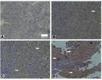
The LMP-1 immunohistochemistry staining showed the variation of LMP-1 expression (FIGURE 1). The LMP-1 expression intensity varied in each subject. The weak, medium and strong intensities were found in the subjects being examined. In some subjects, LMP-1 was expressed uniformly at the entire field examined. While on the other subject, LMP1 was expressed in a scattered or expressed in a group at a particular location. Sixteen subjects (44,4%) expresed LMP-1 (expression score
1). Among them, 14 subjects were in advanced stage. The LMP-1 expression score obtained was ranged between 0-8, with an average value of 1.56 ± 2.16.FIGURE 1. Immunohistochemistry staining of latent membrane protein 1 in NPC biopsies. Positive LMP-1 expression was marked by the appearance of brown color on the cytoplasm. Figure A showed no expression of LMP-1. B, Arrow indicating Expression of LMP-1 with weak color intensity. C, Arrow indicating expression of LMP-1 intermediate level and spread >50% of
RASSF1A methylation status
Methylated specific PCR results showed that hypermethylation of RASSF1A occurred in 24 subjects (66,7%). Figure 2 represented the results of RASSF1A MSP NPC patients. PCR band in MSP was an indicator that hypermethylation occurred in the sample. In case PCR band appeared in both USP and MSP, sample was considered as hypermethylated since there was possibility that the sample also contaminated cells. Figure 2 showed that RASSF1A hypermethylation occured in the subject no 1 and 3, while the subject no 2 was unmethylated.
FIGURE 2. The results of RASSF1A MSP electrophoresis. RASSF1A hypermethylation occured in subject no. 1 and 3.
While subject no. 2 was unmethylated.
Association of LMP-1 expression and RASSF1A methylation status
Table 1 showed the distribution of LMP-1 expression and methylation status of RASSF1A. Latent membrane protein 1 positive subject had more RASSF1A hypermethyation compared to LMP-1 negative subject. Fourteen subjects of 16 LMP-1 positive subjects had RASSF1A hypermethylation. On the other hand, 10 out of 20 LMP-1 negative subjects had RASSF1A hypermetylation.
Variable n MSP RASSF1A χ2
p Unmethylated Methylated
LMP-1 expression
5.625 0.018
Positive 16 2 14
Negative 20 10 10
Phi value = 0.395
TABLE 1. Chi square analysis of LMP-1 expression with RASSF1A methylation status
Chi-square analysis was conducted to determine the association between LMP-1 expression with RASSF1A methylation status. The Chi-square test results showed that there was an association between expression of LMP-1 with RASSF1A methylation status (p= 0.018) and phi value was 0.395. The result indicated that there was a tendency that RASSF1A hypermethylation occured in LMP-1 positive subjects.18 To further analyzed the association between LMP-1 expression score with RASSF1A methylation status, LMP-1 expression score were grouped into low, middle and high expressions. Data were analyzed using cross tabulation to determine the association between the score of LMP-1 expression with methylation status of RASSF1A. Table 2 showed that there was an association between the score of LMP-1 expression with RASSF1A methylation status (p=0.010).
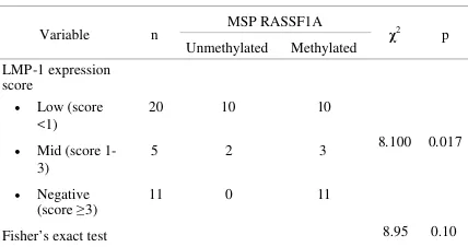
Table 2. Chi square analysis of LMP-1 expression score with RASSF1A methylation status.
Variable n MSP RASSF1A χ2 p
Unmethylated Methylated LMP-1 expression
score
8.100 0.017 Low (score
<1)
20 10 10
Mid (score 1-3)
5 2 3
Negative
(score≥3) 11 0 11
Fisher’s exact test 8.95 0.10
Phi value = 0.474
DISCUSSION
Examination of LMP-1 Expression
been reported which ranged from 50-100%.8,17,19 Latent membrane protein 1 expression score in this study ranged between 0-8 of the maximum score (12) that can be obtained. This score was lower than previous studies. By using the same method, Hariwiyanto17reported that LMP-1 expression score ranged from 2-12 with an average of 7.6 (± SD = 2.6). While other studies reported that LMP-1 score ranged from 3-11.8
The different LMP-1 expression score in this study, possibly caused by the differences in types of antibody used. The antibody used to detect the expression of LMP-1 in this study was a monoclonal antibody that recognized a conformational epitope at residues 290 to 318, overlapped with 11 amino acid repeat in LMP-1.20 Khabir et al.,8 and Hariwiyanto,17used antibodies CS1-4. The CS1-4 was a pool of four types of monoclonal antibodies that recognized the C-terminus of LMP-1. The target was the variable region epitopes on 11 amino acids between residues 205 to 308, and also at 386 the end residue of C-terminus.21,20 Different target antigen epitopes influenced the sensitivity and specificity of antibodies, it was influential on the final assessment of LMP-1 expression.7
Jiwaet al.,7reported that there were different
immunoreactivity between CS1-4 monoclonal antibody with the S12 (OT17). The S12 monoclonal antibody was known to be more sensitive in detecting LMP-1 compared with CS1-4. The S12 also provided better color and contrast than CS1-4. Based on author’s knowledge, research on immunoreactivity comparison between antibody CS1-4 with OT21C has never been done, therefore the level of sensitivity between the two antibodies can not be compared.
RASSF1A methylation status
In this study, RASSF1A hypermethylated in 66.7% subjects. This was in line with previous studies which reported that the frequency of RASSF1A hypermethylation in NPC ranged from 66.7%-84%.2,7,12Statistical analysis showed that there was no relationship between RASSF1A methylation status with age of study subjects (p = 0.637). The RASSF1A methylation status was also
not associated with stage NPC subjects (p = 0.708). The result was in contrast with previous studies which stated that the incidence of TSG promoter methylation increased with the increasing age of subjects.4,11,21-23
Association of LMP-1 expression and RASSF1A methylation status
The result successfully demonstrated the relationship between LMP-1 expression in NPC biopsy with hypermethylation of RASSF1A promoterin DNA that was isolated from nasopharyngeal brushing. The result showed a tendency of hypermethylation of RASSF1AinsubjectwithhighLMP-1expressionscore. This result indicated that LMP-1 expression can be used as marker of RASSF1A hypermethylation incidence. However, intracellular mechanism of LMP-1 in inducing RASSFLMP-1A hypermetylation still need further study.
Latent membrane protein 1 is known to induce tumor suppressor genes hypermetylation through DNA methyltransferase (DNMT1, and DNMT3b DNMT3a) activities.24,25 There was a report of phosporylation of DNA methyltransferase by LMP-1 signaling via c-Jun NH2-terminal kinase (JNK).26 The LMP-1 activates JNK, and phosphorylates transcription factor Jun. The phosphorylated c-Jun binds the activator protein 1 (AP-1) and transactivates the DNMT1 promoter. Elevated DNMT1 expression leads to hypermethylation of TSG.6In addition, LMP-1 may inhibit expression of RASSF1A through the NF-B.20 If the mechanism of LMP-1 in inducing hypermethylation RASSF1A can be recognized, it will be very useful in the development of therapeutic agents. This will open the possibility of application of 3,4-dihydroxybenzalacetone (DBL) for inhibiting NF-B,27or application of azacytidine to inhibit DNA methyltransferase28 activity as one of the NPC therapies.
CONCLUSION
ACKNOWLEDGMENT
We thank Profesor Jaap Middeldorp for the generous gift of monoclonal antibody OT21C. Part of this research was funded by RISBIN IPTEKDOK 2009. We also thank Mrs Agustin, Ms. Sumartiningsih, and Mr. Y. Suhardi for their skillful technical assistance.
REFERENCES
1. Chang ET, Adami HO. The enigmatic epidemiology of nasopharyngeal carcinoma. Cancer Epidemiol Biomarkers Prev 2006;15(10):1765-77.
2. Lo KW, Kwong J, Hui ABY, Chan SYY, To KF, Chan ASC,et al.High frequency of promoter hypermethylation of RASSF1A in nasopharyngeal carcinoma. Clin Cancer Res 2001;61(5):3877–81.
3. Donninger, H; Vos MD, Clark GJ. The RASSF1A Tumor Suppressor. J Cell Sci 2007; 120:3163-72.
4. Chou J, Lin YC, Kim J, You L, Xu Z, He B, et al. Nasopharyngeal carcinoma—Review of the molecular mechanism of tumorigenesis. Head Neck 2008; 30 (7):946–63.
5. Titcomb CP. High incidence of nasopharyngeal carcinoma in Asia. J Insur Med 2001; 33:235–8.
6. Tsai CL, Li HP, Lu YJ, Hsueh C, Liang Y, Chen CL,et al.Activation of DNA methyltransferase 1 by EBV LMP-1 involves c-Jun NH2-terminal kinase signaling. Cancer Res 2006;66(24):11668-76.
7. Jiwa, NM, Oudejans JJ, Dukers DF, Vos W, Horstman A, van der Valk P,et al.Immunohistochemical demonstration of different latent membrane protein-1 epitopes of Epstein-Barr virus in lymphoproliferative diseases. J Clin Pathol 1995;48:438-42.
8. Khabir AH, Karray, Rodriguez S, Rosé M, Daoud J, Frikha M,et al.Boudawara T, Epstein-Barr virus latent membrane protein 1 abundance correlates with patient age but not with metastatic behavior in north African nasopharyngeal carcinomas. J Clin Virol, 2005;2(39): 1-7.
9. Kwong J, Lo KW, To KF, Teo PML, Johnson PJ, Huang DP. Promoter hypermethylation of multiple genes in nasopharyngeal carcinoma. Clin Cancer Res 2002; 8: 131–7.
10. Lo, KW, Cheung ST, Leung SF, Hasselt AV, Tsang YS, Mak KF,et al. Hypermethylation of the pl6 gene in nasopharyngeal carcinoma. Cancer Res 1996;56(6): 2721-5.
11. Agathanggelou A, Cooper WN, Latif, F. Role of the ras-association domain family 1 tumor suppressor gene in human cancers. Clin Cancer Res 2005;65:3497-508. 12. Lwanga SK, Lemenshow S. Sample size determination
in health studies: a Practical Manual. Genewa: WHO, 1991.
13. Greene FL, Compton CC, Fritz AG, Shah JP, Winchester DP.AJCC cancer staging atlas. New York: Springer, 2006. 14. Cheng SH, Tsai CSYC, Yen L, Jian JJ, Chu NM, Chan
KY,et al.Concomitant radiotherapy and chemotherapy for early-stage nasopharyngeal carcinoma. J Clin Oncol, 2000;18:2040-45.
15. Tiong TS, Selva KS. Clinical presentation of nasopharyngeal carcinoma in Sarawak Malaysia. Med J Malaysia 2005;60(5):624-8.
16. Boom R, Sol CJA, Salimans MMM, Jansen CL, Wertheim-Van Dillen PME, J Van Der Noorda. Rapid and simple methods for purification of nucleic acid. J Clin Microbiol 1990;28(3):495-503.
17. Hariwiyanto B. Peran protein EBNA1, EBNA2, LMP1 dan LMP2 virus Epstein-Barr sebagai faktor prognosis pada pengobatan karsinoma nasofaring [Disertasi]. Yogyakarta: Program Doktor Ilmu Kedokteran dan Kesehatan Fakultas Kedokteran Universitas Gadjah Mada, 2009.
18. Morgan GA, Leech NL, GLockner GW, Barrett KC. SPSS for introductory statistics-Use and intrepretation 2nd Ed. New Jersey:Lawrence Erlbaum Associaties
Publishers, 2004.
19. Murono S, Inoue H, Tanabe T, Joab I, Yoshizaki T, Furukawa M,et al.Induction of cyclooxygenase-2 by Epstein–Barr virus latent membrane protein 1 is involved in vascular endothelial growth factor production in nasopharyngeal carcinoma cells. PNAS 2001;98(12): 6905–10.
20. Meij P, Vervoort MB, Aarbiou J, van Dissel P, Brink A, Bloemena E,et al.Restricted low-level human antibody responses against Epstein-Barr virus (EBV)-encoded latent membrane protein 1 in a subgroup of patients with EBV-associated diseases. J Infect Dis 1999;179:1108– 15.
21. Pan ZG, Kashuba VI, Liu XQ, Shao JY, Zhang RH, Jiang JH,et al.High frequency somatic mutations in RASSF1A in nasopharyngeal carcinoma. Cancer Biol Ther 2005;4: 1116-22.
22. Euhus DM, Bu D, Milchgrub S, Xie XJ, Bian A, Leitch AM,et al.DNA Methylation in benign breast epithelium in relation to age and breast cancer risk. Cancer Epidemiol Biomarkers Prev 2008;17(5):1051-59.
23. Boks MP, Derks EM, Weisenberger DJ, Strengman E, Janson E, Sommer IE,et al.The relationship of DNA methylation with age, gender and genotype in twins and healthy controls. PLoS ONE 2009;4(8):1-8.
24. Sidhu S, Deep JS, Sobti RC, Sharma VL, Thakur H. Methylation pattern of MGMT gene in relation to age, smoking, drinking and dietary habits as epigenetic biomarker in prostate cancer patients. Gen Engineering Biotechnol J 2010;8:1-11.
25. Tsai CN, Tsai CL, Tse KP, Chang HY, Chang YS. The Epstein–Barr virus oncogene product, latent membrane protein 1, induces the downregulation of E-cadherin gene expression via activation of DNA methyltransferases. PNAS 2002;99(15):10084–9.
27. Tan SH, Ida H, Goh BC, Hsieh W, Loh M, Ito Y. Analyses of promoter hypermethylation for RUNX3 and other tumor suppressor genes in nasopharyngeal carcinoma. Anticancer Res 2006;26:4287-92.