238 Recent Patents on Materials Science 2013, 6, 238-252
Development of Porous Calcium Phosphate Bioceramics for Bone Implant
Applications: A Review
Abreeq R. Naqshbandi
1, Iis Sopyan
1,*and Gunawan
1,21Department of Manufacturing and Materials Engineering, Faculty of Engineering, International Islamic University
Malaysia, P.O. Box 10, Kuala Lumpur 50728, Malaysia; 2Department of Mechanical Engineering, Faculty of Engineering, Sriwijaya University, Indralaya 30662, Indonesia
Received: February 13, 2013; Accepted: March 25, 2013; Revised: April 5, 2013
Abstract: The present review briefly outlines the most recent patents and journals on various aspects of porous calcium phosphate bioceramics including techniques of preparation, properties and bone implant applications. Bioactive ceramics are a class of materials that have capability to bond directly with the host bone. These materials can be easily assimilated by the body and are considered to be biodegradable. Researches have revealed that artificial bones made from hydroxya-patite or a combination of hydroxyahydroxya-patite (HA) and tricalcium phosphate (TCP) is a perfect substitute for natural bone owing to its excellent biocompatibility and properties close to that of human bone. Bioceramics made of HA are available in dense and porous forms. Several efforts on the fabrication of porous calcium phosphate bioceramics have been carried upon in the field of clinical orthopaedics. The realisation of these efforts can be observed from the fact that numerous pat-ents have been filed on methods of preparing porous calcium phosphate bioceramics for bone implant applications. A number of porous HA ceramics have been developed for applications in both tissue engineering and drug delivery sys-tems. Porous bodies are decomposable in human body and provide a surface for proliferation and growth of cells that are infiltrated from the surrounding tissues so that a new bone grows into the pores and prevents any movement or loosening of the implants. Consequently, these can be used for filling the damaged bone, repair of fractured bone and even can be used as hard tissue replacements. Several processing techniques have been employed for fabrication of porous scaffolds. Among prominent techniques are gel casting, slip casting, camphene-based freeze casting and polymeric-sponge method. Keywords: Bioactive, bioceramics, calcium phosphate, hydroxyapatite (HA), tricalcium phosphate (TCP).
1. INTRODUCTION
Bone is a hard endoskeleton tissue found in almost all vertebrates. Bones protect the vital organs of the body and are collectively known as skeleton. In addition, bones are also responsible for the production of red and white blood cells, mineral storage and facilitate body movement in con-jugation with muscles. Bone has a complex structure and consists of both organic and inorganic components. Table 1
gives detailed information of the chemical composition of the most important human normal calcified tissues [1]. The in-organic composition is formed mostly of calcium phosphate whereas organic part is composed of collagen [2]. The inor-ganic to orinor-ganic ratio of bone is approximately 75-25% by weight and 65-35% by volume.
Numerous researches have been conducted in the field of clinical orthopaedics for detecting the materials that have potential of triggering natural regeneration process of dam-aged or lost bone tissue. These materials have capability of being in contact with bodily fluids and tissues for prolonged periods of time. One of the key factors in a biomaterial’s
*Address correspondence to this author at the Department of Manufacturing and Materials Engineering, Faculty of Engineering, International Islamic University Malaysia, P.O. Box 10, Kuala Lumpur 50728, Malaysia; Tel: +603 6196 4592; Fax:+603 6196 4477; E-mail: [email protected]
usage is its biocompatibility and functionality. There are 4 essential elements that are responsible for bone healing: (1) osteogenic cells (2) osteoinductive signals that are provided by growth factors; (3) an osteoconductive matrix; and (4) adequate blood and nutrient supply [3]. Biomaterials used as bone grafts are identified on the basis of osteogenicity (pres-ence of bone forming cells), osteoconductivity (ability to function as a scaffold) and osteoinductivity (ability to stimu-late bone formation) [4]. Various biological and synthetic bone substitutes have been used so far for clinical applica-tions. Among the biological substitutes, corals have been widely used for bone implant applications since corals have skeletons similar to cortical and cancellous bone whereas among the artificial materials for biomedical purpose, cal-cium phosphate biomaterials have been widely used as bone substitute materials. Hydroxyapatite (HA) (Ca10(PO4)6(OH)2),
is considered to be a perfect mineral replacer due to its simi-larity of chemical composition and crystallographic structure to the biomineral in the natural tissues [5]. It has been used widely for various bones and tooth implants and has good biocompatibility and bioactivity.
HA bonds strongly to the bone and favours osseointegra-tion of bone implant, necessary for minifying the damages to the surrounding tissues. Calcium phosphate-based ceramics such as HA are available in porous as well as in dense form. Porous ceramic implants provide a surface for proliferation
and growth of cells that are infiltrated from the surrounding tissues so that a new bone grows into the pores and prevent any movement or loosening of the implants. In fact porous HA ceramics are proven to mimic the porous structure of the mineral phase of the living bone [6] However, the dimen-sions and pore morphology of these ceramics have a consid-erable effect on bone prostheses [7]. Most of mammalian cells are anchorage dependent, i.e. they need a substrate for growth. Several studies suggest that ceramic scaffolds are suitable for cell proliferation, growth and cell attachment [6]. As a result, numerous studies have been conducted in the field of tissue engineering for development of porous ce-ramic scaffolds for bone replacement applications.
Tricalcium phosphate (TCP) is also a bio-absorbable and biocompatible material and has a crystalline structure and chemical composition similar to that of bone. Moreover its rate of biodegradation is higher than HA [8]. In comparison with other bone substitutes, TCP is characterized by well defined physical and crystalline properties, high level of uni-formity of chemical composition and purity.
Several other materials have also been used as bone-graft substitutes such as bioactive glasses, glass-ceramics, crystal-line phase materials etc. These substances have been used either alone or in combination with acrylic polymers or other group of polymers. These materials have been proven to be osteoconductive as well as biocompatible with host tissues. However, for being an effective bone substitute, these mate-rials should have an appropriate structure and mechanical properties especially the porosity, pore-size and size of the interconnections between each pore [9].
2. APPLICATIONS OF POROUS CALCIUM PHOS-PHATE
HA is a perfect substitute for natural bone owing to its non-toxicity, excellent biocompatibility [10] and properties close to that of human bone. A number of porous HA ceramics have been developed for applications in both tissue engineer-ing [11, 12] and drug delivery system [6, 13-15]. This type of drug delivery system by the use of a bioactive matrix, helps in releasing a therapeutic agent in situ so that an anti-infection
action is produced which is associated to osteoconductivity of materials. The use of bioactive porous ceramics for the deliv-ery of antibiotics has been helpful in improving the recurrence of orthopedic infections [13]. Porous structures allow growth of tissue and bone around a supporting network and allow passage of nutrients/substances across them.
Many other applications include cell loading [6, 16] and chromatography analysis [16]. Some of the recent progresses in tissue engineering that have been achieved include thera-pies for regeneration of new skin as a replacement for burnt skin [17], development of bone-grafts [18], dental ligaments [19], dentin [20], dental enamel [21] and integrated tooth tissues [22,23]. The use of bio-ceramic scaffold depends upon its properties in vivo and in vitro. Bioceramics must
exhibit proper mechanical strength in order to sustain in vivo
stresses and should be mechanically stable with the sur-rounding tissues [24,25]. Various studies reveal that cell cul-ture of mammalian cells in vitro is substrate dependant, i.e.
these cells require solid substrate for growth. One of the techniques used for cell cultivation is micro-carrier culture technique. This technique offers a practical high yield culture of such cells and thus is suitable for large-scale operations. A number of micro carriers have been used so far; however use of ceramic micro-carriers has introduced new possibilities for cell culture owing to their good thermal and mechanical resistances.
In recent years, a number of porous HA ceramics have been developed to serve as graft material and scaffold for bone formation. A newly developed porous HA ceramic is NEOBONE® (Covalent Material, Tokyo, Japan). It has a
porosity of about 75%, with macropores of 100-200m that
are fully interconnected by openings of about 40m in
di-ameter. Yoshikawa and Myoui [26] and Yoshida et al. [27]
have successively made use of these ceramics for the treat-ment of bone tumours and fractures. Another newly devel-oped HA ceramic is Apaceram-AX® (HOYA, Tokyo, Japan).
It has a higher porosity of 85% and contains macropores of 50-300m in diameter [28]. The detailed material
informa-tion of NEOBONE® and Apaceram-AX® is summarized in
Table 2.
3. RECENT PATENTS ON POROUS CALCIUM PHOSPHATE
3.1. Method for Producing a Porous Sintered Body of Calcium Phosphate-based Ceramic
The porous sintered body of calcium phosphate-based ceramic can be used as an artificial dental root, a bone rein-forcing material and a carrier for culture of cells or biologi-cal tissues. The patent US7514024 [29] issued to Matsumoto discloses a method for preparing porous calcium phosphate-based ceramic having a porosity of 80% or more. The method involves a series of steps starting with the
prepara-Table 1. Chemical andStructural Similarities between HA, Enamel, Dentin, and Bone [1].
Composition, wt% Enamel Dentin Bone HA
Calcium (Ca) 36.5 35.1 34.8 39.6
Phosphorous (P) 17.1 16.9 15.2 18.5
Ca/P ratio 1.63 1.61 1.71 1.67
Total inorganic component (%) 97 70 65 100
Total organic component (%) 1.5 20 25 --
tion of slurry comprising of calcium phosphate-based ce-ramic powder having an average particle diameter of 0.5 to 80m, a water-soluble high molecular compound which can
be preferably a cellulose derivative such as methylcellulose, carboxymethylcellulose, etc. and a non-ionic surface active agent preferably a fatty acid alkanolamide. This is followed by vigorous stirring at a temperature of 5 to 20ºC and under a stirring condition of 50W/L or more in order to froth the slurry and a gas is passed (e.g., air, nitrogen, argon etc.) dur-ing this time so as to aid the froth formation. The frothed slurry is then solidified into gel and drying is carried out at a temperature range of 80ºC to less than 100ºC. The dried green body is then degreased at a temperature of 300ºC to 900ºC so as to remove water-soluble high molecular com-pound and the non-ionic surface active agent. The green de-greased body is then sintered at 1000ºC to 1250ºC to get the desired strength. The weight ratio of the total of the calcium phosphate-based ceramic powder, the water-soluble high molecular compound and the non-ionic surface active agent is maintained at 20 to 50 weight % based on 100 weight % of the slurry. The resulting porous sintered body of calcium phosphate-based ceramic has a porosity of 80% and an aver-age pore diameter ranging from 5 to 1500m.
3.2. Porous Calcium Phosphate Networks for Synthetic Bone Material
The use of resorbable and osteoconductive synthetic bone graft material comprising a calcium phosphate mineral ma-trix lattice interpenetrated by a substantially interconnected network of pores has been described in US7758896 [30]. The patent discloses a method for making a synthetic bone graft wherein simultaneous formation of mineral matrix and interconnected network of removable inorganic porogen takes place by controlled spinodal decomposition of a molten mixture of inorganic materials. Spinodal decomposition is a thermodynamic separation of a single-phase liquid into in-terconnected liquid phases with continuous networks. The patent describes a method which involves selection of cal-cium oxide and calcal-cium phosphate components including a removable inorganic porogen that can be a transition metal oxide, in an appropriate ratio so as to provide a desired po-rosity in the product. This is followed by mixing and the mixture of the chemical components so obtained is heated in order to melt the mixture. The molten mixture is then cooled at a controlled rate so that spinodal decomposition of the mixture into a material, consisting of a primary phase of HA and a secondary phase of inorganic porogen, takes place. The inorganic porogen is then removed to leave behind a porous material having a substantially interconnected network of
pores. The patent also presents porous materials having compositions of HA and TCP (as the primary phase) in which the interconnected pores are formed by the spinodal decomposition of a mixture of calcium oxide, calcium pen-toxide and a removable inorganic porogen. The porosity of porous materials of such a composition ranges from 30% to about 60% and pore size from 3 to 700m.
3.3. Porous Ceramic Composite Bone Grafts
Synthetic bone grafts have a significant advantage over natural grafts as their use limits the transmission of disease, occurrence of adverse immunological response and have relatively low costs. The patent US7875342 [31] discusses the use of porous ceramic composite comprising of a sin-tered porous matrix body of calcium phosphate-based com-pound and a biodegradable polymer and a method to prepare the same. The method involves impregnating reticulate-structured organic foam with an aqueous slurry of calcium-phosphate compound which is prepared by combining the ceramic medium with water and dispersing agent. This is followed by drying so that a green body of slurry coated foam structure is formed. It is then pyrolized and sintered at about 900-1300ºC to form a fused ceramic porous implant having interconnected pore-structure. Typical thickness of the coating varies from 10 to about 100m. The patent also
discloses the use of biodegradable coating on the external and internal surfaces of the porous structure so as to improve the physical and mechanical properties of the sintered matrix body. This biodegradable polymer can be photosensitive polymer, polycaprolactone, polyethylene glycol, polyester, polyanhydride etc.
3.4. Porous Composite Containing Calcium Phosphate and Process for Producing the Same
The use of porous body containing calcium phosphate ceramic as an implant has a little likelihood of getting re-jected by the host as it is highly biocompatible. However, as these implants come in contact with soft tissues such as gum, subcutaneous tissue, internal organ etc., cells in the soft tis-sue tend to enter into the porous artificial bone thereby pre-venting the growth of a bone tissue. The patent US8039090 [32] issued to Kawamura et al. discusses method for the
formation of a dense layer on the surface of porous body containing calcium phosphate ceramic which substantially prevents the cells of soft tissues from participating in the bone formation process. The method involves the formation of a porous composite with a porous layer containing a cal-cium phosphate ceramic and a dense layer formed on the part of the porous layer at a position that it comes in contact with
Table 2. Material Properties of NEOBONE® and Apaceram-AX® [28].
Sample Name NEOBONE Apaceram-AX
General name HA HA
Molecular formula Ca10(PO4)6(OH)2 Ca10(PO4)6(OH)2
Sintering temperature 1200 C 1050 C
a soft tissue when implanted in a human body and the pore size and porosity of the dense layer being smaller than the porous layer. The method includes formation of slurry from calcium phosphate ceramic/collagen composite and collagen and pouring the same into a moulding die. This is followed by rapid freezing and drying so that a porous body is formed comprising of a porous layer and a dense layer on the porous layer. The dense layer is then removed except for a portion that comes in contact with a soft tissue when implanted in a human body. The ceramic/collagen composite used in this method is apatite/collagen composite which is produced by adding an aqueous solution of phosphoric acid or its salt and an aqueous calcium salt solution to a collagen solution. The resulting porous composite has a porosity of the porous layer of about 90-98%.
3.5. Porous Calcium Phosphate Bone Material
The use of self-hardening, porous calcium phosphate compositions that represent approximate chemical composi-tions of natural bone has been described in US8147860 [33]. The patent discloses a formable, self-hardening, porous cal-cium phosphate composition that is prepared using a calcal-cium phosphate source, effervescent agent and a biocompatible cohesiveness agent (e.g., binder). This composition is then combined with a biological fluid and results in the produc-tion of a formable paste which hardens and reacts to form a poorly-crystalline apatite (PCA) calcium phosphate. PCA calcium phosphate remodels into bone when introduced at an implant surface. The nature of calcium phosphate powders and the presence of biocompatible cohesiveness agent allow many biological active fluids to pass through without com-promising any of the mechanical properties or formability of the implant. In other words, the implant material retains its cohesiveness upon being introduced at an implant site
in vivo.
3.6. Method for Producing Porous -tricalcium Phos-phate Granules
Biomaterials used for bone implant applications should be able to enhance the regenerative process particularly when used in conjugation with bioactive fluids. The patent US8173149 [34] describes the use of porous ceramic mate-rial having a desired composition, pore size, porosity and granule size for improving the bone regeneration process in a human or animal. The patent discloses the use of porous
-TCP granules for bone implant applications and methods for producing the same. The method involves mixing of TCP powder with a pore-forming agent that can be in the form of bead or resin. Pore-forming agent decomposes into gaseous products at high temperature without leaving behind any solid residue. It can be any thermally decomposable material such as naphthalene, polymers of polyacrylates, copolymers of methyl acrylate and methyl methacrylate or mixtures of polystyrene, cellulose powder, acrylic resins etc. This is fol-lowed by the addition of a granulating solution in order to form a friable mass which is then sieved to form granules. The use of granulating solution is to enhance the formation of granules. The sieved granules are then dried at a tempera-ture of 90-110ºC followed by heating at 700-800ºC so as to remove the pore-forming agent. The formation of porous
-TCP takes place after sintering the granules at 1000-1200ºC. The resulting porous -TCP consists of pores that are
sur-rounded by the skeleton of sintered TCP. The patent also discusses an alternative method for the formation of porous
-TCP which involves blending of TCP powder and
pore-forming agent in order to achieve homogeneity. The ho-mogenized mixture is then pressed into slugs using a press, rotary tablet machine or chilsonators followed by heating and sintering. The pore diameter of -TCP granules of this
invention ranges between 50-125m whereas total porosity
of -TCP is 70%. The patent also provides a method for
in-ducing bone-formation in a mammal by implanting a compo-sition of porous -TCP and binder or a bioactive agent.
3.7. Porous Materials Coated with Calcium Phosphate and Methods of Fabrication Thereof
Calcium phosphate coatings allow bone ingrowth into and around an implant device by supporting the formation of chemical bonds between the device and natural bone. The patent US20120270031 [35] describes a method of coating porous medical implants internally with a layer of Calcium phosphate. The method involves submerging a porous mate-rial in an aqueous solution that contains calcium ions, phos-phate ions, carbonates, NaCl, and HCl, and has a tempera-ture less than 100ºC and an initial pH of 6.0-7.5. The porous material to be coated generally comprises of composite ma-terial and calcium phosphate particles and has a macropore size of 0.5-3.5mm in diameter. The solution is agitated con-tinuously at a speed of 200-400 revolutions per minute so that carbon dioxide gas is removed from the solution and the pH is increased gradually. Agitation enables the internal coating of pores extending within the volume of porous ma-terial. The thickness of the calcium phosphate coating is ad-justed by controlling the contact time or immersion rate. The porous particles coated with calcium phosphate are then separated and mixed with a carrier which can be sodium alginate, gelatine, lecithin, and glycerol etc., followed by the addition of a fluid to form moldable or injectable porous material. The fluid is selected from the group consisting of water, sterilized water, blood, bone marrow aspirate etc. This moldable or injectable porous material has its individual par-ticles coated with a layer of calcium phosphate, preferably HA. The thickness of calcium phosphate layer ranges be-tween 0.5-50m. The patent discusses that this moldable
porous material can be used for various clinical applications including bone repair and regeneration. The patent further discloses that the porous material can be formed into sheet with a preferred thickness of 0.5-2.0mm and pore size range of 200-800m before coating with calcium phosphate.
3.8. Biocompatible Ceramic-polymer Hybrids and Cal-cium Phosphate Porous Body
con-taining Ca(NO3)2, (NH4)2HPO4, (NH2)2CO and HNO3. The
solution is heated in the temperature range of 80-90ºC to produce HAp fibers with axes about 60-100m in length.
The use of carbon-beads is to create space between the fi-bers. Since carbon-beads and the HAp fibers have different densities they tend to separate out. Thus to homogenize the slurry, agar is added so that carbon-beads and fibers can be better dispersed. This slurry containing agar is poured into moulds having a porous bottom and a selected pressure of 30MPa is applied in order to form a pre-compact which is then calcined at 1200-1300ºC for about 3-5 hours. Carbon-beads vaporize leaving behind pores within HAp ceramics. The resulting HAp ceramics have an interconnected pore-structure with a pore diameter of 10m and a porosity of
about 40-70%. The patent also discloses the incorporation of poly-L-lactic acid esters into the pores of porous HAp ce-ramics by enzyme catalysis. HAp, lactic acid and lipase are mixed in a reaction container and then degassed by freeze-thaw and vacuuming. This is followed by flushing with an inert gas while maintaining a proper temperature so as to allow polymerization of the reaction mixture. The resulting HAp-PLLA hybrid is polished into the final product. PLLA is found to improve the mechanical properties of HAp ce-ramics.
3.9. Ion Substituted Calcium Phosphate Coatings
Ion-substituted calcium phosphate plays an important role in bone formation. The patent US20120087954 [37] describes a method for the formation of a surface coating of an ion-substituted calcium phosphate on a substrate. The method involves a substrate that is pre-treated so as to acti-vate its surface. Pre-treatment is carried out with the help of heat treatment, hydrolysis, oxidation, acid or base treatment, UV radiation etc. A portion of this substrate is then im-mersed in the aqueous solution comprising calcium ions, sodium ions, magnesium ions, phosphate ions, Sr2+, F- etc. at a temperature of 20-100ºC with pH of the solution being maintained in the range of 6.0-8.0. The incubation or immer-sion time is around 1-3 days so that a coating of desired thickness is formed on the substrate. The patent also de-scribes the use of two aqueous solutions in order to create additional layers of different chemistry and morphology. The obtained surface coating has about 25-60% cationic substitu-tion of calcium and 10-25% anionic substitusubstitu-tion of phos-phate and hydroxide and a composition comprising 1.5-3% fluoride, 3-8% strontium and 0.5-2% silicon. The morphol-ogy of coating is in the form of sheets, flakes, porous struc-tures, spikes and rods. The invention claims that thickness of the coating can be controlled between 10nm-100m.
3.10. Delayed-setting Calcium Phosphate Pastes
The use of delayed-setting calcium phosphate pastes in the preparation of delivery vehicles for biologically active agents is described in US8216359 [38]. The patent discusses the use of calcium phosphate pastes for delivery vehicles and discloses the method to prepare the same. The paste that is used consists of a calcium phosphate material, an osteogenic protein which acts as a biologically active agent and a non-aqueous liquid which contains less than 5% water. Option-ally, a bioerodible material is also added so as to introduce
porosity in the paste. This paste is then exposed to moisture either before or after implantation and turns into a hardened calcium phosphate material upon hydration. This is followed by applying the delayed-setting paste to the surface of a bone implant so as to promote bone growth thereof. The patent also relates to an implant device comprising of delayed-setting calcium phosphate pastes which can be combined, mixed, and stored without setting or hardening for years to-gether depending upon the application involved.
3.11. Bioactive Bonegraft Substitute
The use of calcium phosphate, resorbable collagen and bioactive glasses for the preparation of bonegrafts is dis-cussed in US8303967 [39]. The method involves forming a homogeneous mixture of calcium phosphate, collagen and bioactive glass in a weight ratio of 75:10:15. Bioactive glass is present either in the form of coating on collagen or as a separate component in the mixture of collagen and calcium phosphate. The pH of the mixture is monitored so that bioac-tive glass does not undergo premature leaching necessary for osteoactivity. The mixture is then made to undergo heating, freeze-drying and many cross-linking techniques to yield a porous bonegraft with a total porosity of 65-95%.
4. PROPERTIES OF POROUS CALCIUM PHOS-PHATE
For developing porous scaffolds, it is important that they meet certain requirements of bone tissue engineering and integrate well with the bone healing process. Some of the important aspects that need to be considered while develop-ing these scaffolds are: appropriate micro- and macroscopic structural morphology including pore size, pore interconnec-tivity, biocompatibility, osteoconducinterconnec-tivity, mechanical strength and biodegradability [40]. The idea is that if an im-planted porous ceramic is progressively replaced by natural bone, its biomechanical properties should more and more resemble with natural bone. Extensive research efforts have been done in order to make use of synthetic HA/calcium phosphate as a bone substitute material in biomedical appli-cations [41, 42].
almost exponentially with increasing porosity [44, 45]. How-ever, by changing the pore geometry, it is possible to influence the strength of porous bioceramics. The compres-sive strength of porous calcium phosphate bioceramics is reported to increase from 2MPa to 20MPa after 3 months of implantation [26].
Pore characteristics are crucial in bone engineering due to its correlation with the degree of bone in-growth. Porosity determines the movement of cells across pores. A good bio-ceramic should have an interconnected porosity ranging be-tween 55-70% and the pore size should range from 150-700m as in natural bone [30]. Pore size distribution for an
ideal scaffold [46] is given in Table 3 as under:
5. PREPARATION METHODS OF POROUS CAL-CIUM PHOSPHATE BIOCERAMICS
5.1. Fabrication of Porous HA Ceramics with Controlled Pore Characteristics by Slip Casting
Porous HA ceramics with controlled pore characteristics were fabricated using slip casting method by Yao et al. [47].
HAp powders with dispersant are milled in a ball jar fol-lowed by the addition of Spherical polymethyl methacrylate (PMMA) particles and polyvinyl alcohol (PVA). In this method (PMMA) is used as a porosifier. The so formed slur-ries, free of foam, are cast into plaster molds. After de-molded and dried at ambient temperature for 72h, the green blocks are heated to burn out the PMMA particles and other volatiles, followed by the treatment at 1200ºC for 2h for den-sification purpose. The pore characteristics of sintered po-rous HA bioceramics can be controlled by changing the size and content of PMMA. For an average particle size of added PMMA of 305, 134 and 62m, the average pore size of
po-rous HA ceramics is 290, 96 and 45m respectively.
5.2. Fabrication of Porous Silicon-incorporated HA Using Natural Coral as a Calcium Source
Natural coral has a porous structure with all its pores interconnected throughout the skeleton as depicted in Fig. (1) [48]. its microstructure resembles that of bone and can be
used as an excellent starting material for synthesis of porous HA.
Moreover, it has been suggested that the incorporation of silicate ions creates defect sites in HA and are favourable to dissolution [49]. In this method porous silicon doped HA has been prepared by hydrothermal treatment and solvo-thermal treatment of the natural coral in silicon acetate saturated
Fig. (1). SEM micrograph of natural coral [48].
acetone solution [48]. The resulting porous silicon doped HA possessed uniformly permeable micropores and uniform pore volume of about 70% with pore size distributions ranging from 200-300m. The compressive strength of the porous
silicon doped HA was found to be 5.5 MPa.
5.3. Highly Porous HA Bioceramics with Interconnected Pore Channels Using Camphene-based Freeze Casting
In this method, HA/camphene slurries are prepared through ball milling. The slurries are prepared with different (as received and calcined) HA contents (10, 15, and 20 vol %) and the temperature during milling is maintained at 60°C followed by casting into the moulds at room temperature. By removing the frozen camphene network via sublimation, a well defined three-dimensional interconnected pore channel system is easily produced [50]. Camphene can be frozen and easily sublimed near room-temperature and hence can be used to produce highly porous HA bioceramics with com-pletely interconnected pore channels. Freeze casting method has been considered a preferred method since it can produce well defined pore structures on a finer scale [51-53]. In addi-tion to it, the use of calcined HA powders produces homoge-neous slurries with different HA contents. Thus this method allows the fabricated samples/bodies to have completely interconnected pore channels with porosity being controlled by the initial HA content. Moreover these porous bodies have higher strength and smaller pore size due to the densifi-cation of the HA walls resulting from sintering of highly packed HA powder networks at 1250°C for 3 h.
Table3. Pore Size Distribution for an Ideal Scaffold in Bone Tissue Engineering Applications [46].
Pore Size (m) Biological Function
< 1 Protein interaction, responsible for bioactivity
1-20 Cell attachment, their orientation of cellular growth (directionally)
100-1000 Cellular growth and bone ingrowth
5.4. Preparation of Porous HA Through Polymeric Sponge Method
Yet another method to fabricate porous ceramics is through replication of the polymeric sponge substrate [54]. Ceramics developed this way have controllable pore size and can be made into various complex shapes useful for different applications [55]. This method is carried out by impregnat-ing the cellulosic sponges with prepared HA slurry. Porous HA prepared by the polymeric sponge method has shown to have interconnected pores but poor mechanical strength for load bearing applications. However, this method results in an adequate pore size distribution as is required for osteocon-duction to take place. The slurries are made from commer-cial HA powders while making the use of cellulosic sponges for impregnation. All porous samples contained micropores of 0.2-1m and macropores of 100-500m in diameter. The
effect of processing parameters viz; sintering rate, stirring time and HA concentration on the physical properties was studied. Higher sintering rates resulted in a higher apparent density and higher compressive strength and thus better me-chanical strength. The average compressive strength of the porous bodies varied between 1.8 and 10.5MPa for a de-crease in porosity from 59.8% to 34.3%; concluding that compressive strength is inversely dependent on porosity. Stirring slurry for a prolonged period of time also resulted in higher compressive strength due to its better homogeneity.
5.5. Spark Plasma Sintering of Macroporous Calcium Phosphate Scaffolds from Nanocrystalline Powders
Macroporous -TCP scaffolds are fabricated via Spark
plasma sintering (SPS) technique using nanocrystalline pow-ders [56]. SPS is a new technique used for the fabrication of various solid nanostructured materials viz; ceramics, cermets and alloys [57]. In this technique nanocrystalline -TCP
powders are first synthesized by a chemical precipitation reaction. The precipitates so obtained are dried at 80°C for 24h followed by calcination of powders at 700 and 800º C for 2h. The obtained nanocrystalline -TCP powders are then
mixed with polyethylene glycol particulates and the mixtures are pressed in a stainless steel die. The prepared green disks are heat-treated at 400°C to remove the organic substances, and then they are presintered. The presintered sample is then placed into a graphite die for spark plasma sintering. The obtained macroporous scaffolds were found to have a poros-ity of 55-70% and macropore size of (300-500m) whereas,
the compressive strength and elastic modulus improved by 50-100%.
5.6. Manufacturing of Highly Porous Calcium Phosphate Bioceramics Via Gel-casting Using Agarose
Gel-casting technique is the method resulting in the pro-duction of scaffolds most closely mimicking the structure of trabecular bone [58]. This method uses agarose as gelling agent [59]. Agarose solution is prepared by adding agarose powder to distilled water followed by heating at 12°C and 1.4 bar in an autoclave. Meanwhile, HA slurries are prepared from HA powders synthesized by precipitation method. Prior to foaming, agarose solution is added to HA slurry while maintaining the temperature at 60°C. The foaming suspen-sion containing agarose is then poured into a mould which is
further cooled by water to transform the liquid state to a gelled state. The whole process is followed by de-moulding, drying and sintering. Characterization of the ceramic foams was carried out using different techniques in order to moni-tor cellular microstructure of the sinters by analyzing in terms of estimation of cell sizes (spherical pores) and win-dow sizes (interconnected pores). XRD study revealed that additives used in the gel-casting process did not influence the phase composition of the investigated materials. The obtained porous (P = 90%) CaP scaffold had macropores (spherical~500m and interconnecting windows~100m in
diameter) and a small amount of micropores (0.2-0.9m) as
seen in Fig. (2). The advantage of this technique is that it is
environmental friendly.
Fig. (2). SEM micrographs of the cross-section of sintered CaP foam [59].
5.7. Biphasic Calcium Phosphate Macroporous Scaffolds Derived from Oyster Shells
Oyster shells are considered to be biocompatible and bioactive materials for bone grafting applications [60]. This method uses HA powders obtained from the hydrothermal treatment of oyster shells for the fabrication of porous scaf-folds [61] via polymeric replication method as reported in previous works [62, 63]. The overall process flowchart of the method is shown in Fig. (3).
The resulting scaffolds, comprised of a biphasic structure of HA/-TCP, and open macropores with pore size in the
range of 200-500m and interconnected micropores from
100nm to 500nm. In addition, O-HA scaffolds showed excel-lent permeability and a high porosity upto 91.4% + 1.2%. Cell-culture results confirmed their non-cytotoxic behavior and a better biocompatibility.
5.8. Fabrication of Mesoporous Carbonated HA Micro-spheres by Hydrothermal Method
biological fluids, resorbability and formation of bone-like apatite after soaking in simulated body fluids (SBF) [67-69]. A number of natural sources have been used as starting ma-terials for the preparation of porous HA by hydrothermal method or by treatment with phosphate buffer solution [70-72]. Some of them include corals [73], algae [74], cuttlefish [75], and nacre [76, 77]. In this work calcium carbonate mi-crospheres (CCMs) (prepared from nacre) are used as start-ing materials for the fabrication of mesoporous carbonated HA microspheres (MCHMs) via hydrothermal method [78]. The chemical elements of nacre mainly include O (48.026 wt.%), Na (0.281 wt.%), Al (0.215 wt.%), Si (0.058 wt.%), S (0.022 wt.%), Ca (40.957 wt.%), Sr (0.083 wt.%), and other elements such as C, H, and N (10.4 wt.%). The preparation of CCMs from nacre is mentioned elsewhere [79]. A sche-matic diagram of the overall method is shown in Fig. (4).
The resulting MCHMs have a diameter of ~5μm and are composed of many nanoparticles within the whole micro-spheres. These nanoparticles aggregate to form mesopores with a pore size of 4.5-14.0nm among them.
5.9. Production of Porous Calcium Phosphate (CaP) Ce-ramics with Highly Elongated Pores Using Carbon-coated Polymeric Templates
This method produces porous calcium phosphate ceram-ics with highly elongated pores. The elongated pore structure
ensures good compressive strength of these ceramics. CaP/ camphene slurry is cast into stretched polymeric sponges, precoated with a very thick carbon coating layer used as a template. This is followed by heat treatment at 800°C for 3h in order to remove the carbon-coated template and then at 1250°C to sinter CaP walls. The resulting sample has a highly elongated pore structure with a pore size of 512 ±
96m and a porosity of about 38 vol% where as compressive
strength values are as high as 22 MPa in the direction paral-lel to the pore elongation [80].
5.10. Porous Alumina-HA Composites Through Protein Foaming–consolidation Method
Porous HA ceramics have been commonly used for bone implant applications due to their excellent bioactivity and compatibility. However, these ceramics have a disadvantage of being mechanically weak thereby have limited use in load-bearing applications [6]. Various attempts have been made to improve the strength of porous HA ceramics one of them being incorporating alumina into HA powders. Jun
et al. [81] successfully fabricated porous alumina-calcium
phosphate composites using polyurethane sponges. The re-sulting porous bodies were found to have a porosity of 75-90% and a compressive strength of 6MPa.
We have reported the fabrication of porous alumina ce-ramics using egg yolk as a pore forming agent via protein
Fig. (3). Schematic of the basic process: (a) Hydrothermal conversion of oyster shells to ha nano-powders and (b) Polymer replication tech-nique, which is used to fabricate the macroporous scaffolds [61].
Oyster Shell Waste
Crashed Cleaned
Autoclave
220oC, 6h
Ca2+ PO42
OH
-HA nano-powders
(a)
HA nano-powders
+
Stuff well
PVA Solution Polyurethane sponge
Squeezed and dried
Polyurethane sponge with HA slurry
Macroporous scaffold Sintered
foaming-consolidation method [82]. The obtained porous bodies showed good pore connectivity with a pore diameter of approximately 100-500m. However, in the present
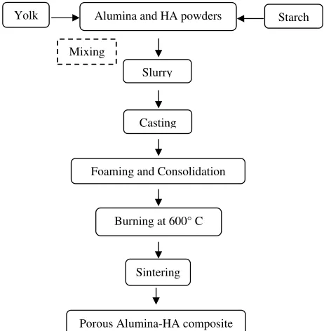
method, protein foaming-consolidation method has been used to fabricate porous alumina-HA composites [83]. The flowchart of the method followed is given in Fig. (5).
Fig. (5). Flowchart of fabrication of porous alumina-HA compos-ites through protein foaming-consolidation method.
Fig. (6). SEM micrograph of porous alumina-HA ceramics sintered at (a) 1350 and (b) 1550°C [83]. Reproduced by the permission of authors.
Alumina and HA powders, obtained via sol-gel technique [84] are mixed with yolk and starch in a proportionate ratio in order to make a slurry. Yolk, used as the protein source for foaming, is taken from chicken egg. It consists of 25 wt% protein and 24 wt% lipids. Starch flour acts as a binder whereas Darvan 821A (40 wt% aqueous solution of ammo-nium polyacrylate; R.T. Vanderbilt, USA) as the dispersing agent. The resulting slurries are cast into cylindrical open stainless steel molds followed by foaming and consolidation at 180°C for 1 h. The green bodies obtained after consolida-tion are burnt at a temperature of 600°C for removal of yolks and then sintered between 1200-1550°C for 2h.
This method produces porous composites with 26-77 vol% shrinkage and 46%-52% porosity. The micrograph of porous alumina-HA ceramics sintered at 1350 and 1550°C respectively, shows that with an increase in sintering tem-perature the grains get strongly bonded together as a result of progressive fusion of particles as depicted in Fig. (6).
Fig. (4). Schematic diagram of Fabrication of mesoporous carbonated HA microspheres by hydrothermal method.
Deionized water CCMs
(prepared from nacre)[89]
Disodium hydrogen phosphate (Na2HPO4·12H2O) (5.0 g)
Polytetrafluoroethylene (Teflon)-lined stainless steel
Hydrothermal Reactions Mixing
140° C, 12 h
Drying
MCHMs
80° C, 48 h
Alumina and HA powders
Slurry
Foaming and Consolidation
Sintering
Porous Alumina-HA composite
Yolk Starch
Mixing
Casting
Fig. (7). Micro CT cross-sectional images of HA thickness with color coded porous bodies having HA-to-alumina mass ratio of (a) 0.30 and (b) 1.0 w/w [83]. Reproduced by the permission of authors.
The effect of HA loading and yolk addition on the prop-erties of porous samples was also studied. Fig. (7a-7b)
shows Micro CT images of HA thickness with HA-to-alumina mass ratio of 0.3 and 1.0 w/w respectively. The thickness of HA increased with an increase in HA loading in the slurry. Sample of 0.3 w/w ratio shows thicker HA layer in the middle part of porous body than the upper part, whereas in 1.0 w/w ratio sample, HA particles precipitate only in the surface part.
It was also observed that an increase in HA content in the slurry led to a decrease in pore size. The 3D images of colour coded porous bodies with corresponding pore size distribu-tions is shown in Fig. (8). The pore size of 0.3 ratio sample
was found to be in the range of 10-460m and of 1.0 ratio
sample in the range of 10-350m as seen in Fig. (8b) and
Fig. (8d) respectively.
On contrary an increase in the yolk amount in slurry leads to an increase in pore size due to higher foaming ca-pacity of slurry. This result is attributed to the fact that am-phiphilic character of protein decreases the surface tension of slurry and leads to better foaming property [85].
Biocompatibility tests revealed good compatibility of the cells to the porous microcarriers as the cells attached and grew at the surface of microcarriers at 8-120 cultured hours which is depicted in Fig. (9). The cell growth on porous
alumina microcarrier has been observed to be 0.015 h-1 which increased with an increase in HA-to-alumina mass ratio. Figure 9a shows the microstructure of porous alumina
after being sintered at 1550°C without cell seeding. Figure
9b shows an initial stage of attachment and proliferation of
the cells on porous alumina surfaces only in 12 cultured hours. After 120 hours, Vero cells have started to respond in different manners to every sample. The Vero cell density for pure alumina samples was lower Fig. (9c) than that of the 1.0
w/w ratio sample Fig. (9d).
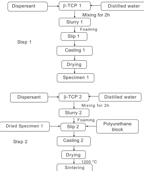
5.11. Fabrication and Properties of Porous -TCP Ce-ramics Prepared Using a Double slip-Casting Method Using Slips with Different Viscosities
This method produces porous -TCP scaffolds with
en-hanced mechanical properties and has a better control over the microstructures of the scaffolds [86]. Scaffolds are pro-duced by combining two methods viz; double slip-casting and polymer sponge method. In step 1, Slurry 1 has been obtained by adding a solution of 1.5 wt% deflocculant to
-TCP1 powder with a solid content of 58.5 wt% followed by dilution with water. After that 0.5 wt% of foaming agent has been added to it to make a slip-1. The specimen is obtained
Fig. (8). 3D images of color coded pores with corresponding pore size distribution of bodies with HA-to-alumina mass ratio of (a, b) 0.30 and (c, d) 1.0 w/w [83]. Reproduced by the permission of authors.
!
"
#
!
"
1 mm
1 mm
$
by dipping a polyurethane block into slip-1, after which it’s dried under vacuum for 5 min to yield dried specimen-1. Slip-2 has been obtained by adding -TCP 2 powder with a
solid content 62.5 wt% to a solution of 1.5 wt% deflocculant followed by dilution with water to make slurry-2. After that dried specimen-1 has been dipped into slip-2, and then the sample is dried under vacuum for 5 min followed by heating at a rate of 0.5°C / min to 250°C and then incubated at 250ºC for 30 min to decompose the deflocculant and the polyure-thane template. The samples are again heated at a rate of 3°C /min to 1200°C, incubated for 3h at 1200°C, and cooled down to room temperature at a rate of 3°C/min to yield the final samples. The series of steps involved in this method are shown in Fig. (10) as under:
The resulting scaffolds have a porosity of 61.4% and an open, uniform, interconnected porous structure with a bi-modal pore size of 100-300m and a very high flexural
strength of 56.2MPa.
6. CURRENT & FUTURE DEVELOPMENTS OF BIO-CERAMIC BONE IMPLANTS
A well designed bioceramic scaffold in terms of structure and properties plays an important role in directing and en-hancing cell growth and cell proliferation to surrounding tissues [14, 87-89]. Porous HA ceramics have been exten-sively used as bone graft substitutes [90, 91]. Moreover their interconnected structure favors bone ingrowth and osseoin-tegration [58]. The strength of these ceramics/scaffolds de-pends upon various factors e.g., pore size, pore shape, and pore orientation [92]. Variation in preparation technique al-lows production of porous HA with different porosity, pore connectivity, surface and mechanical properties. Several
Fig. (10). Series of steps involved in fabrication of porous -TCP
ceramics [86].
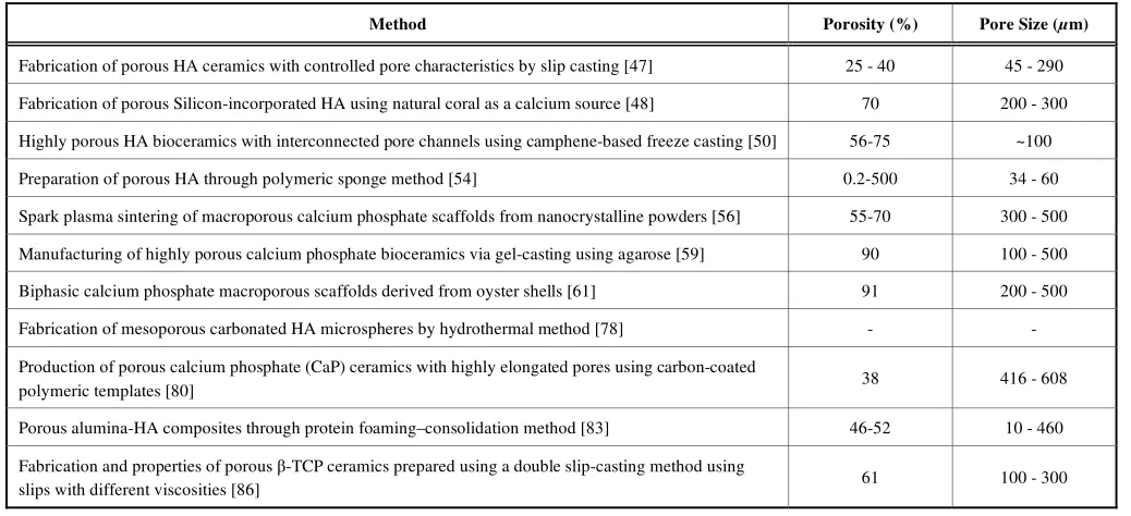
methods for the preparation of porous HA have been high-lighted in this article through a thorough review of the most recent patents and journals. Some of the methods are tabu-lated in Table 4 below:
Fig. (9). The morphology of surface of (a) Porous alumina without cell seeding (b) Initial attachment and proliferation of Vero cells on sur-faces of the alumina-HA porous bodies (c) Vero cell density of Pure alumina sample (d) Vero cell density of 1.0 w/w ratio sample [83]. Re-produced by the permission of authors.
%"&" ! b' %"!((#)!
*+ ,
(
-(&
"!
%-
&$
%"&" ! b' %"!((#)!
(
-(&
"!
%-
!
* + ,
(- !, ($
%#&$
!&
However, due to the poor mechanical properties of the porous HA bioceramics, their use for load bearing applica-tions is severely restricted [93, 94]. Several attempts have been made to improve the mechanical strength of porous HA bioceramics e.g., doping with metals or blending with high performance reinforcing materials or organic polymers, in-cluding introducing secondary phase [95, 96] and optimizing sintering techniques [97] etc. Kim et al. [98] used poly (
-caprolactone) (PCL) and HA composite coatings on the sur-face of the porous HA scaffolds to improve the mechanical properties of the scaffold. Yook et al. [99]used polystyrene
(PS) polymer as a binder to HA/camphene slurries. The PS binder increased the green strength of the sample thereby preventing the sample cracking. The compressive strength was significantly increased from 1.1+0.2 to 2.3+0.5 MPa, while the pore size was decreased from 277+47 to 170+29μm. Zhang et al. [100]developed a novel method for
the fabrication of porous bodies. The method was developed by combining the conventional freeze casting and gel cast-ing. Hydantoin epoxy resin was used in the freeze casting which promoted the gelation process and led to a higher compressive strength of the green body. However, after sin-tering freeze-gelcast samples exhibited a twice higher com-pressive strength and 15.6% higher porosity. Lee et al. [101]
used bone ash for the fabrication of biphasic calcium phos-phate bioceramics. Bone ash, used as a raw material for bone china, was treated with NaOH solution and then calcined to obtain calcium phosphate powders. The bone ash derived-biphasic calcium phosphate ceramics showed high biostabil-ity in liquid environment when -compared with commercial calcium phosphate ceramics. Yang et al. [102] developed a
strong layered HA/TCP-zirconia scaffold composite by em-ploying newly developed slip-deposition and coating-substrate co-sintering technique. The developed scaffolds exhibited a bending strength of 321 MPa. Also from the
in vitro cell culture study, it was indicated that the coatings
had no cytotoxicity.
For developing a bone graft, its in vivo resorption rate
becomes a matter of concern since bone graft should be rap-idly resorbable and replaced by new bone. In a recent patent application, Yang et al. [103] related to a composite of
-calcium sulfate hemihydrates and amorphous -calcium phos-phate having resorption time of 3-6 months. The composite consists of calcium sulfate which has a bone regeneration and angiogenic effect. The preparation method involves dis-solving the solutions containing calcium ion and sulphate ion separately in calcium chloride solution. The process is fol-lowed by heating and mixing of two solutions to produce
-calcium sulphate hemihydrates (-CSH). Solid -CSH is
separated from the solution after filtration. Amorphous cal-cium phosphate (ACP) is formed by adding a phosphate compound to the solution of calcium chloride. -CSH and
ACP are further mixed to produce the desired composite (
-CSH/ACP).
Recently, attention has been paid to the development of resorbable tissue scaffold that can have load bearing strength and at the same time sufficient porosity so that growth of bone tissue is promoted effectively. In a patent of Liu et al.
[104], one such prosthetic device is described. The patent discloses the use of bioactive glass fibers comprising of cal-cium carbonate, phosphorus pentoxide, silica and sodium carbonate, bonded into a porous matrix having a pore size distribution in order to facilitate the growth of bone tissue for the treatment of bone defects. Bioactive glass fibers of such a composition have capability to form silica-rich layer and a calcium phosphate film on the surface that readily bonds glass material to bone. The rigid three-dimensional matrix of bioactive tissue scaffold is formed by heating the mixture of bioactive glass fiber, binder, pore forming agent and liquid.
Preparation of calcium phosphate particles in the nano to micrometer range with controlled morphology without the use of surfactant or template has been proposed by Engqvist and Xia [105]. The invention presents a method for the
Table 4. Porosity and Pore Size of Porous Samples Produced by Different Methods.
Method Porosity (%) Pore Size (m)
Fabrication of porous HA ceramics with controlled pore characteristics by slip casting [47] 25 - 40 45 - 290
Fabrication of porous Silicon-incorporated HA using natural coral as a calcium source [48] 70 200 - 300
Highly porous HA bioceramics with interconnected pore channels using camphene-based freeze casting [50] 56-75 ~100
Preparation of porous HA through polymeric sponge method [54] 0.2-500 34 - 60
Spark plasma sintering of macroporous calcium phosphate scaffolds from nanocrystalline powders [56] 55-70 300 - 500
Manufacturing of highly porous calcium phosphate bioceramics via gel-casting using agarose [59] 90 100 - 500
Biphasic calcium phosphate macroporous scaffolds derived from oyster shells [61] 91 200 - 500
Fabrication of mesoporous carbonated HA microspheres by hydrothermal method [78] - -
Production of porous calcium phosphate (CaP) ceramics with highly elongated pores using carbon-coated
polymeric templates [80] 38 416 - 608
Porous alumina-HA composites through protein foaming–consolidation method [83] 46-52 10 - 460
Fabrication and properties of porous -TCP ceramics prepared using a double slip-casting method using
manufacture of ion-substituted calcium phosphate particles with controlled morphology and structure via a surfactant-free biomineralization process. Calcium phosphate particles are synthesised with mineralisation and precipitation meth-ods comprising the basic steps of preparing a salt solution and precipitating calcium phosphate particles from the salt solution. The solution comprises calcium, and phosphate ions and one or more of magnesium, sodium, potassium, chloride, carbonate or sulphate ions. The morphology of cal-cium phosphate particles is controlled by adjusting the centration of substituted ions. Use of such particles with con-trolled morphology is helpful in stimulating regeneration of damaged bone by inserting ion-substituted calcium phos-phate into the body.
In a recent patent, Kjellin and Andersson [106] provided highly crystalline synthetic nano-sized HA with a specific area in the range of 150m2/g to 300m2/g. It is the highest
specific area ever presented. The patent discloses that nano-sized HA crystals resemble HA particles that are present in the living body and are suitable in the biomimicking of body tissue for the making of body implants. Thus HA of this invention is suitable for being deposited on the surface of an implant to make it bioactive in order to stimulate bone growth process.
A patent based on the fabrication of an implantable de-vice comprising of bioceramic particles has been published recently by Gale et al. [107]. The patent relates to a method
for fabricating a stent comprising of biodegradable polymer and bioceramic particles. Bioceramic particles are made from biodegradable bioceramics such as tetracalcium phos-phate, amorphous calcium phosphos-phate, -TCP, -TCP and
various types of bioglass materials. The use of bioceramics is aimed at inhibiting infection that may result during implanta-tion and to increase the fracture toughness of the stent. Bio-ceramic particles disperse the strain over a large volume of the stent and increase its flexibility and resistance to crack-ing durcrack-ing deployment.
In addition to the efforts made in developing bioceramic implants as a replacement for fractured or diseased bones, several techniques have been introduced to form bioceramic coating on the metal implants as well. In a patent appeared on February 2013, Liu et al. [108] indicated a method to
develop a gradient bioceramic coating on the surface of tita-nium alloy which can be implanted in order to restore defects in human sclerous tissues. The method involves mixing of powdery composite ceramics of calcium phosphate and cal-cium carbonate with rare earth oxide and powdery titanium on the surface of a titanium alloy. This is followed by clad-ding treating the surface with carbon dioxide laser process-ing system so that synthesis and coatprocess-ing of HA on the sur-face of titanium alloy are completed in one step.
In a patent published in January 2013 by Betz et al.
[109], the use of coherent aggregate of elongate bone parti-cles was made for the preparation of osteoimplant. The ob-ject of the invention was to provide a low density implant which possesses an open pore structure to allow the passage of blood and other bodily fluids and yet retain its original shape. The implant was prepared by mixing elongate demin-eralised bone particles with an aqueous wetting agent fol-lowed by moulding into a desired shape. The excess
mois-ture was removed through heating at elevated temperamois-ture in absence of pressure. The osteoimplant formed from such a process does not require time-consuming rehydration prior implantation and can attain any shape and configuration. In yet another recent patent issued to Burkinshaw [110], a composite bone graft kit comprising of an allograft bone component and a synthetic bone substitute (such as -TCP,
HA and poly-lactic acid) is disclosed. The composite is ar-ranged in a core/outer layer structure and consists of a mesh casing wherein allograft bone component and synthetic bone substitute are in contact with each other. The patent also de-scribes the use of a bone graft syringe with composite bone graft disposed within the syringe. This bone graft syringe is further claimed to be connected with a delivery syringe for delivery of an injectable component such as cell concentrate, platelet rich plasma or a bone marrow aspirate.
A great progress has been accomplished in the field of bio-ceramics so far; however major advances are needed to be done in order to develop ceramics with improved proper-ties depending upon the specificity of disease and demand. A large increase in active elderly people has alarmingly raised the need for load-bearing bone graft substitutes. Owing to this demand, new strategies for enhancing the mechanical properties of porous scaffolds have to be proposed and de-veloped.
CONFLICT OF INTEREST
The authors confirm that this article content has no con-flict of interest.
ACKNOWLEDGEMENTS
The authors would like to thank Ministry of Higher Edu-cation (MOHE) for the partial support provided through the FRGS programme (Project No. FRGS11-025-0173).
REFERENCES
[1] Dorozhkin SV. Calcium orthophosphates. J Mater Sci 2007; 42: 1061-95.
[2] Currey JD. Bones: Structure and mechanics. Princeton, Princeton University Press 2002.
[3] Einhorn TA. Enhancement of fracture-healing. J Bone Joint Surg Am 1995; 77: 940-56.
[4] Ozer K, Chung KC. The use of bone grafts and substitutes in the treatment of distal radius fractures. Hand Clinics 2012; 28: 217-23. [5] Engel E, Castaño O, Salvagni E, Ginebra MP, Planell JA.
Biomaterials for Tissue Engineering of Hard Tissues. Strategies in Regenerative Medicine: Integrating Biology with Materials Design ed: Springer Science+Business Media LLC.; 2009.
[6] Sopyan I, Mel M, Ramesh S, Khalid KA. Porous hydroxyapatite for artificial bone applications. Sci Technol Adv Mater 2007; 8: 116-23.
[7] Aza PND, Luklinska ZB, Santos C, Guitian F, Aza SD. Mechanism of bone-like formation on a bioactive implant in vivo. Biomaterials 2003; 24: 1437-45.
[8] Nuss KMR, Rechenberg BV. Biocompatibility issues with modern implants in bone - a review for clinical orthopedics. Open Orthop J 2008; 2: 66-78.
[9] Frieb W, Warner J. Biomedical Applications. In: Schüth F, Sing KSW, Weitkamp J, Eds. Handbook of Porous Solids. Weinheim: Wiley-VCH; 2002; 5: 2923-62.
[11] Yarlagadda PKDV, Chandrasekharan M, Shyan JYM. Recent advances and current developments in tissue scaffolding. Biomed Mater Eng 2003; 15: 159-77.
[12] Liu Y, Schoenaers J, Groot KD, Wijn JD, Schepers E. Bone healing in porous implants: a histological and histometrical comparative study on sheep. J Mater Sci Mater Med 2000; 11: 711-7.
[13] Krajewski A, Ravaglioli A, Roncari E, Pinasco P, Montanari L. Porous ceramic bodies for drug delivery. J Mater Sci Mater Med 2000; 12: 763-71.
[14] Komlev VS, Barinov SM, Girardin E, Oscarsson S, Rosengren Å, Rustichelli F, et al. Porous spherical hydroxyapatite and fluorhydroxyapatite granules: Processing and characterization. Sci Technol Adv Mater 2003; 4: 503-8.
[15] Meurice E, Leriche A, Hornez J-C, Bouchart F, Rguiti E, Boilet L, et al. Functionalisation of porous hydroxyapatite for bone substitutes. J Eur Ceram Soc 2012; 32: 2673-8.
[16] Starling, L.B., Stephan, J.E. Calcium phosphate microcarriers and microspheres. US6210715 (2001).
[17] Hohlfeld J, Roessingh Ad, Hirt-Burri N, Chaubert P, Gerber S, Scaletta C, et al. Tissue engineered fetal skin constructs for paediatric burns. Lancet 2005; 366: 840-2.
[18] Warnke PH, Springer ING, Wiltfang J, Acil Y, Eufinger H, Wehmöller M, et al. Growth and transplantation of a custom vascularised bone graft in a man. Lancet 2004; 364: 766-70. [19] Park CH, Rios HF, Jin Q, Bland ME, Flanagan CL, Hollister SJ,
et al. Biomimetic hybrid scaffolds for engineering human tooth-ligament interfaces. Biomaterials 2010; 31: 5945-52.
[20] Sakai VT, Zhang Z, Dong Z, Neiva KG, Machado MA, Shi S, et al. SHED differentiate into functional odontoblasts and endothelium. J Dent Res 2010; 89: 791-6.
[21] Chen H, Tang Z, Liu J, Sun K, Chang SR, Peters MC, et al. Acellular synthesis of a human enamel-like microstructure. Adv Mater 2006; 18: 1846-51.
[22] Duailibi MT, Duailibi SE, Young CS, Bartlett JD, Vacanti JP, Yelick PC. Bioengineered teeth from cultured rat tooth bud cells. J Dent Res 2004; 83: 523-8.
[23] Ikeda E, Morita R, Nakao K, Ishida K, Nakamura T, Takano-Yamamoto T, et al. Fully functional bioengineered tooth replacement as an organ replacement therapy. Proc Natl Acad Sci USA 2009; 106: 13475-80.
[24] Kemppainen JM, Hollister SJ. Tailoring the mechanical properties of 3D-designed poly(glycerol sebacate) scaffolds for cartilage applications. J Biomed Mater Res A 2010; 94: 9-18.
[25] Saito E, Kang H, Taboas JM, Diggs A, Flanagan CL, Hollister SJ. Experimental and computational characterization of designed and fabricated 50:50 PLGA porous scaffolds for human trabecular bone applications. J Mater Sci Mater Med 2010; 21: 2371-83.
[26] Yoshikawa H, Myoui A. Bone tissue engineering with porous hydroxyapatite ceramics. J Artif Organs 2005; 8: 131-6.
[27] Yoshida Y, Osaka S, Tokuhashi Y. Clinical experience of novel interconnected porous hydroxyapatite ceramics for the revision of tumor prosthesis: A case report. World J Surg Oncol 2009; 7: 76. [28] Yamasaki N, Hirao M, Nanno K, Sugiyasu K, Tamai N, Hashimoto
N, et al. A comparative assessment of synthetic ceramic bone substitutes with different composition and microstructure in rabbit femoral condyle model. J Biomed Mater Res B Appl Biomater 2009; 91B: 788-98.
[29] Matsumoto, T. Method for producing a porous sintered body of calcium phosphate-based ceramic. US7514024 (2009).
[30] Hyers, R., Sansoucie, M. Porous calcium phosphate networks for synthetic bone material. US7758896 (2010)
[31] Smith, T.J.N., Jason, H., Sydney, M.P., Reginald, S. Porous ceramic composite bone grafts. US7875342 (2011).
[32] Kawamura, K., Nakajima, T., Shoji, D., Tanaka, J., Kikuchi, M., Ikoma, T. Porous composite containing calcium phosphate and process for producing the same. US8039090 (2011).
[33] Rosenberg, A.D., Pélichy, L.D.G., Bondre, S., Strunk, M. Porous calcium phosphate bone material. US8147860 (2012).
[34] Dalal, P.S., Diamaano, G.R., Kulkarni, S.C. Method for producing porous -tricalcium phosphate granules. US8173149 (2012). [35] Guan, L., Davies, J.E. Porous materials coated with calcium
phosphate and methods of fabrication thereof. US20120270031 (2012).
[36] Aizawa, M., Rikukawa, M., Shigemitsu, Y., Nagashima, H. Biocompatible ceramic-polymer hybrids and calcium phosphate porous body. US20120136088 (2012).
[37] Xia, W., Lindahl, C., Engqvist, H., Thomsen, P., Lausmaa, J. Ion substituted calcium phosphate coatings. US20120087954 (2012). [38] Lee, D.D., Lee, Y.M., Rosenberg, A.D., Pélichy, L.D.G.D., Sutaria,
M., Tofighi, A.N. Delayed-setting calcium phosphate pastes. US8216359 (2012).
[39] Clineff, T.D., Koblish, A., Bagga, C.S., Erbe, E.M., Nagvajara, G.M., Darmoc, M.M. Bioactive bonegraft substitute. US8303967 (2012).
[40] Tripathi G, Basu B. Processing and biological evaluation of porous HA/poly(methyl methacrylate) hybrid composite. Int J Adv Eng Sci Appl Math 2011; 2: 161-7.
[41] Hutmacher DW, Schantz JT, Lam CXF, Tan KC, Lim TC. State of the art and future directions of scaffold-based bone engineering from a biomaterials perspective. J Tissue Eng Regen Med 2007;1:245-60.
[42] Habraken WJEM, Wolke JGC, Jansen JA. Ceramic composites as matrices and scaffolds for drug delivery in tissue engineering. Adv Drug Delivery Rev 2007; 59: 234-48.
[43] Dorozhkin SV. Calcium orthophosphates as bioceramics: State of the art. J Funct Biomater 2010; 1: 22-107.
[44] Pecqueux F, Tancret F, Bouler J-M. Young's modulus of macroporous bioceramics: Measurement and numerical simulation. Biocer Develop Appl 2010; 1: 1-3.
[45] Tancret F, Boulerb JM, Chamousseta J, Minoisa LM. Modelling the mechanical properties of microporous and macroporous biphasic calcium phosphate bioceramics. J Eur Ceram Soc 2006; 26: 3647-56.
[46] Sánchez-Salcedo S, Arcos D, Vallet-Regí M. Upgrading calcium phosphate scaffolds for tissue engineering applications. Key Eng Mater 2008; 2: 19-42.
[47] Yao X, Tan S, Jiang D. Fabrication of hydroxyapatite ceramics with controlled pore characteristics by slip casting. J Mater Sci Mater Med 2005; 16: 161-5.
[48] Kim YH, Song H, Riu DH, Kim SR, Kim HJ, Moon JH. Preparation of porous Si-incorporated hydroxyapatite. Curr Appl Phys 2005; 5: 538-41.
[49] Porter AE, Patel N, Skepper JN, Best SM, Bonfield W. Comparison of in vivo dissolution processes in hydroxyapatite and silicon-substituted hydroxyapatite bioceramics. Biomaterials 2003; 24: 4609-20.
[50] Lee EJ, Koh YH, Yoon BH, Kim HE, Kim HW. Highly porous hydroxyapatite bioceramics with interconnected pore channels using camphene-based freeze casting. Mater Lett 2007; 61: 2270-3. [51] Deville S. Freeze-casting of porous biomaterials: Structure,
properties and opportunities. Materials 2010; 3: 1913-27.
[52] Araki K, Halloran JW. Porous ceramic bodies with interconnected pore channels by a novel freeze casting technique. J Am Ceram Soc 2005; 88: 1108-14.
[53] Song JH, Koh Y-H, Kim H-E, Li LH, Bahn HJ. Fabrication of a porous bioactive glass-ceramic using room-temperature freeze casting. J Am Ceram Soc 2006; 89: 2649-53.
[54] Sopyan I, Kaur J. Preparation and characterization of porous hydroxyapatite through polymeric sponge method. Cer Int 2009;35:3161-8.
[55] Tian J, Tian J. Preparation of porous hydroxyapatite. J Mater Sci 2001; 36: 3061-6.
[56] Zhang F, Lin K, Chang J, Lu J, Ning C. Spark plasma sintering of macroporous calcium phosphate scaffolds from nanocrystalline powders. J Eur Ceram Soc 2008; 28: 539-45.
[57] Kumar R, Prakash KH, Cheang P, Khor KA. Microstructure and mechanical properties of spark plasma sintered zirconia-hydroxyapatite nano-composite powders. Acta Mater 2005; 53: 2327-35.
[58] Jones JR, Hench LL. Regeneration of trabecular bone using porous ceramics. Curr Opin Solid State Mater Sci 2003; 7: 301-7. [59] Potoczek M, Zima A, Paszkiewicz Z, Slosarczyk A. Manufacturing
of highly porous calcium phosphate bioceramics via gel-casting using agarose. Ceram Int 2009; 35: 2249-54.
[60] Duplat D, Chabadel A, Gallet M, Berland S, Bédouet L, Rousseau M, et al. The in vitro osteoclastic degradation of nacre. Biomaterials 2007; 28: 2155-62.
[61] Yang Y, Yao Q, Pu X, Hou Z, Zhang Q. Biphasic calcium phosphate macroporous scaffolds derived from oyster shells for bone tissue engineering. Chem Eng J 2011; 173: 837-45.
![Table 1. Chemical and Structural Similarities between HA, Enamel, Dentin, and Bone [1]](https://thumb-ap.123doks.com/thumbv2/123dok/2953552.1704608/2.612.64.579.82.208/table-chemical-structural-similarities-ha-enamel-dentin-bone.webp)
![Table 2. Material Properties of NEOBONE® and Apaceram-AX® [28].](https://thumb-ap.123doks.com/thumbv2/123dok/2953552.1704608/3.612.63.575.82.172/table-material-properties-neobone-apaceram-ax.webp)
![Table 3. Pore Size Distribution for an Ideal Scaffold in Bone Tissue Engineering Applications [46].](https://thumb-ap.123doks.com/thumbv2/123dok/2953552.1704608/6.612.334.571.107.361/table-pore-distribution-ideal-scaffold-tissue-engineering-applications.webp)
![Fig. (2). SEM micrographs of the cross-section of sintered CaP foam [59].](https://thumb-ap.123doks.com/thumbv2/123dok/2953552.1704608/7.612.345.559.214.354/fig-sem-micrographs-cross-section-sintered-cap-foam.webp)
![Fig. (3). Schematic of the basic process: (a) Hydrothermal conversion of oyster shells to ha nano-powders and (b) Polymer replication tech-nique, which is used to fabricate the macroporous scaffolds [61]](https://thumb-ap.123doks.com/thumbv2/123dok/2953552.1704608/8.612.112.514.56.393/schematic-hydrothermal-conversion-polymer-replication-fabricate-macroporous-scaffolds.webp)
![Fig. (7). Micro CT cross-sectional images of HA thickness with color coded porous bodies having HA-to-alumina mass ratio of (a) 0.30 and (b) 1.0 w/w [83]](https://thumb-ap.123doks.com/thumbv2/123dok/2953552.1704608/10.612.111.519.431.708/micro-sectional-images-thickness-porous-bodies-having-alumina.webp)