www.elsevier.com / locate / bres
Research report
Effect of experimental pain from trigeminal muscle and skin on motor
cortex excitability in humans
a,b b c a
Antonietta Romaniello
, Giorgio Cruccu , Anne S. McMillan , Lars Arendt-Nielsen ,
a,d ,
*
Peter Svensson
a
Center for Sensory-Motor Interaction, Orofacial Pain Laboratory, Aalborg University, Fredrik Bajers Vej 7, D-3 9220, Aalborg S, Denmark
b
Department of Neurological Sciences, University ‘La Sapienza’, Rome, Italy
c
Oral Rehabilitation, Faculty of Dentistry, University of Hong Kong, Hong Kong, China
d
Department of Prosthetic Dentistry and Stomatognathic Physiology, Royal Dental College, University of Aarhus, Aarhus, Denmark
Accepted 15 August 2000
Abstract
The pathophysiology of many orofacial pain syndromes is still unclear. We investigated the effect of tonic muscle and skin pain on the excitability of the trigeminal motor pathways using transcranial magnetic stimulation (TMS). Motor evoked potentials (MEPs) were recorded in the masseter surface electromyogram (EMG). Magnetic pulses were delivered with a large coil at intensities 1.1 and 1.5 times the motor threshold, and for each intensity, MEPs were recorded at three different clenching levels: 15, 30 and 45% of maximum voluntary contraction (MVC). Baseline, pain and post-baseline recordings were compared in two sessions. Firstly, muscle pain was induced by infusion of hypertonic saline (5.8%) into the left masseter. Secondly, skin pain was induced by topical application of capsaicin (5%) on the left cheek. Muscle and skin pain did not induce significant effects on the amplitude or latency of the MEPs (ANOVAs:
P.0.50). In both sessions, the amplitude of the MEPs increased with the increase of the clenching level and stimulus intensity (P,0.0001; P,0.005) whereas the latency was not significantly changed (P.0.05; P50.11). Muscle pain was associated with an increase in the pre-stimulus EMG activity on the non-painful side compared with baseline (P,0.01), which could be due to compensatory changes in the activation of the painful muscle. The need for voluntary contraction to evoke MEPs in the masseter muscles and compensatory mechanisms both at the brainstem and cortical level might explain the lack of detectable modulation of MEPs. Nonetheless, the present findings did not support the so-called ‘vicious cycle’ between pain — central hyperexcitability — muscle hyperactivity.
2000 Elsevier Science B.V. All rights reserved.
Theme: Sensory systems
Topic: Pain modulation: anatomy and physiology
Keywords: Experimental pain; Transcranial magnetic stimulation; Masseter muscle; Trigeminal physiology
1. Introduction [11,15]. Relatively few studies have dealt with the possible modulation of the trigeminal motor pathways related to Interactions between activity in nociceptive afferent pain [10,32]. Investigations in experimental models and in fibers and motor excitability have been described at spinal patients, however, have shown different effects of pain on and trigeminal level [17,20,23,35]. In humans, there is central nervous system excitability. In particular, there is indirect evidence that painful stimuli may modulate motor evidence of inhibitory effects in experimental pain con-activity at the cortical level, and vice-versa [7,36,41,42]. ditions and no central modulation in patients [10,23,32,44]. Moreover, some clinical conditions involving masticatory In order to bridge the gap between different experimental muscle dysfunction and orofacial pain have been attributed conditions and to test the long-held concept of the so-to primary hyperactivity of the central nervous system called ‘vicious cycle’ between pain — central hyperexcit-ability — muscle hyperactivity, we investigated the effects of tonic muscle and skin pain on the trigeminal motor *Corresponding author. Fax:145-98-154-008.
E-mail address: [email protected] (P. Svensson). pathways excitability in humans with the use of
nial magnetic stimulation (TMS). This technique is now EMG activity remained within the window for more than widely used to assess motor pathways both in healthy 400 ms, the program automatically triggered the magnetic subjects and in patients [3,6,31,40]. In the trigeminal stimulator (MagLite-r25, Dantec, Denmark) [37]. The system TMS provides an indirect measure of the central subjects were asked to maintain the clenching level for excitability of the masticatory system including motor about 2–3 s after each stimulus. The inter-stimulus interval cortex and corticobulbar connections [8,25,27]. was 10–15 s. The duration of EMG activity recorded was The aim of the present study was to investigate the 300 ms and included pre-stimulus (100 ms) and post-modulation of trigeminal motor pathways during tonic stimulus (200 ms) periods.
muscle and skin pain. Moreover, since the recording of Transcranial magnetic stimulation was performed with trigeminal MEPs requires voluntary activation of the the MagLite-r25 and a circular coil (140 mm diameter; masticatory muscles the importance of clenching levels peak magnetic field: 1.9 T). The coil was placed over the was determined in each experimental condition. Finally, midline, 3–4 cm anterior to the vertex with the current different levels of motor cortical activation were examined flowing clockwise. After finding the optimal stimulation using two different stimulus intensities (1.1 and 1.5 times site on the scalp, it was marked with a dark pen and the the motor threshold). coil was fixed in a stable position, so that the same position was kept during the whole experiment. The subject was asked to keep his head still and the position of the coil was checked after each trial. The motor threshold (Th) was 2. Materials and methods
measured while the subject was clenching their teeth at about 30% of maximum. The threshold was determined by 2.1. Subjects
descending and ascending methods and was defined as the minimum stimulus intensity that produced five discrete A total of 17 healthy subjects (12 men and 5 women)
MEPs in both muscles, with peak-to-peak amplitude of at aged 21–42 years (mean age6S.E.M.: 26.261.7)
partici-least 0.10 mV, discernible visually on the monitor from ten pated. The subjects had no history of temporomandibular
consecutive stimuli. The mean Th, measured at 30% MVC, disorders or orofacial pain and they did not take any
was 48.1%61.4 of the maximum output of the magnetic medications. All the subjects fulfilled the inclusion criteria
stimulator. Low-intensity stimuli (1.13Th) had a mean for magnetic stimulation of neural tissue [28]. Informed
intensity of 54.661.6%; the high intensity (1.53Th) had a consent was obtained prior to the study in accordance with
mean intensity of 70.261.8%. the guidelines of the Helsinki Declaration. The Local
Ethics Committee had approved the study.
2.3. Experimental pain 2.2. Recording of MEPs
2.4. Protocol 3. Results
The experiment was performed in two separate sessions. 3.1. Experimental pain In the first session the effect of muscle pain was examined
and in the second session skin pain was studied. Each The mean amount of hypertonic saline infused into the experiment was performed in ten subjects; three subjects left masseter muscle was 2.260.3 ml. The infusion caused participated in both sessions. The recording procedure was a local sensation of ‘intense’ (nine subjects out of ten) pain the same on both occasions: MEPs were recorded at three from the masseter muscle, with a spread toward the upper clenching levels (15, 30 and 45% MVC) and for each or lower molar teeth and the temporomandibular joint clenching level two stimulus intensities were used: 1.1 (3 / 10). The mean pain intensity on the VAS was 5.460.3 (low intensity5L) and 1.5 (high intensity5H) times the cm. Topical application of capsaicin produced a local motor threshold. With this paradigm, MEPs were measured painful sensation described as ‘burning’ (7 / 10). Sponta-prior to the application of experimental pain (baseline), neous pain was located in the capsaicin-treated area only. during pain and 20 min after pain had disappeared (post- The mean pain intensity on the VAS was 2.960.2 cm. baseline). MEPs were recorded when the pain intensity
was constant. In three subjects, attempts were made to 3.2. Motor evoked responses obtain MEPs in the three conditions with the jaw-closing
muscles at rest (0% MVC). The sequence of clenching During clenching, MEPs were obtained bilaterally in the levels and stimulus intensities was randomized. A total of masseter, in all subjects. No responses were detected with 16 EMG sweeps were recorded in each trial and averaged the jaw muscles at rest. The MEPs appeared as biphasic off-line. The onset-latency and peak-to-peak amplitude and reproducible responses within individuals (Fig. 1). were measured on the non-rectified, averaged MEPs. The Furthermore, the MEPs were symmetrical and the overall root-mean-square (RMS) amplitude of the 100 ms preced- four-way ANOVA showed no significant differences in ing the magnetic stimulus (RMS pre) was measured from latency or amplitude between the two sides. The central rectified averaged signals. position of the coil prevented stimulation of the trigeminal root and no early responses were observed on either side [8].
2.5. Statistical analysis
3.3. Effect of tonic pain Mean values6S.E.M. are given in the text, tables and
figures. The onset latencies, the amplitudes of the MEPs Neither muscle pain nor skin pain affected the MEPs and the pre-stimulus EMG activity of both left and right (Figs. 2 and 3). For all clenching levels and stimulus masseter were compared using analysis of variance intensities, latency and amplitude did not change with (ANOVA) with four repeated factors: muscles (two levels: muscle pain (F (2,9)50.19; P.0.50) (F (2,9)50.19; P. painful and non-painful side); clenching level (three levels: 0.50) or skin pain (F (2,9)50.08; P.0.50) (F (2,9)50.30; 15, 30 and 45% of MVC); stimulus intensity (two levels: P.0.50). Pre-stimulus EMG activity remained constant low and high intensity); conditions (three levels: baseline, with skin pain (F (2,9)50.05; P50.93). During and after pain, post-baseline). Post-hoc Tukey tests were performed muscle pain a significant asymmetry of the pre-stimulus to adjust for multiple pair-wise comparisons. Significance EMG activity was observed due to a relative increase in was set at P,0.05 for all the analyses. the pre-stimulus EMG activity in the right masseter
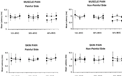
Fig. 2. Effects of different conditions (jBaseline,mPain anddPost-Baseline) on the MEP latency at different clenching levels (15, 30 and 45%) and stimulus intensity (——— Low and — — — High). Experimental tonic pain had no statistically significant effects on the onset-latency (muscle pain: P.0.50; skin pain: P.0.50). The increase of the clenching level and the stimulus intensity tended to shorten the onset latency (P.0.05 and P50.11, respectively).
painful side) (F (2,9)518.3; P,0.01), at 30 and 45% latency shortened by less than 1 ms; this change, however, MVC (Fig. 4). Since the aim of the study was to record the did not reach statistical significance (F (2,9)53.4; P5 masseter MEPs in a standardized condition, the left 0.11). The stimulus intensity did not influence the pre-masseter (painful side) was used to provide feedback for stimulus EMG activity (F (2,9)51.4; P.0.50) (Fig. 4). the optimal control of the clenching level. For this reason
no modulation of the pre-stimulus EMG activity in the left
masseter could be detected. 4. Discussion
3.4. Effect of clenching level The present study showed that tonic muscle and skin pain did not induce detectable change in the motor evoked Even at low levels of voluntary contraction (15% MVC) potentials (MEPs) in the masseter muscles. Muscle pain, and low stimulus intensity reproducible MEPs were ob- however, was associated with an increase in the pre-tained in all subjects with a short latency (mean 6.260.6 stimulus EMG activity in the non-painful masseter. Sys-ms) and a small amplitude (mean 0.560.1 mV). When the tematic changes in pre-stimulus EMG activity of the clenching level was increased the amplitude strongly masseter muscles and stimulus intensity influenced sig-increased from 0.5 mV60.1 at 15% MVC to 1.4 mV60.2 nificantly the amplitude of the MEPs.
at 45% MVC (F (2,9)526.4; P,0.0001) (Fig. 3), and
latency slightly, but not significantly, shortened (F (2,9)5 4.1. Effect of tonic pain on cortical excitability 3.6; P.0.05) (Fig. 2).
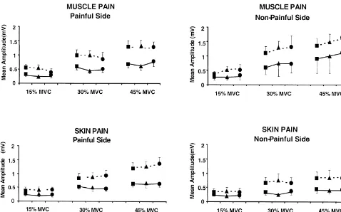
Fig. 3. Effects of different conditions (jBaseline,mPain anddPost-Baseline) on the MEP amplitude during different clenching levels (15, 30 and 45%) and stimulus intensity (——— Low and — — — High). Experimental tonic pain had no statistically significant effects on the amplitude (muscle pain: P.0.50; skin pain: P.0.50). The increase in clenching levels and stimulus intensity induced a significant increase of the MEPs amplitude (P,0.0001 and P,0.005, respectively).
intensity and quality of two modalities of experimental nevertheless have activated structures downstream to the pain no detectable changes in the amplitude and onset- motor cortex, i.e., the corticobulbar axons directly in the latency of the MEPs were found. white matter like electrical anodal stimulation would have Few studies have dealt with pain-induced changes of done; hence an inhibitory effect could have occurred MEPs. Painful laser stimuli applied to the skin of the hand upstream to the site of excitation along the motor path-induced bilateral inhibitory effects on motor cortex ex- ways. In this case the voluntary activation of muscles is citability in humans [42]. However, the trigeminal central inhibited but MEPs would not be affected. TMS is known motor pathways had a normal excitability in a group of to induce D-waves at high intensity (70–80% output) and patients with painful temporomandibular disorders [10]. A their amplitude is smaller than that of D-waves elicited by direct comparison with these studies is difficult because of electrical stimulation [1,5]. The D-wave induced by mag-differences in duration and modality of the nociceptive netic stimulation is also smaller than the second or third input and because extrapolation of results from the spinal components of the I-waves [18]. There is also evidence system to the trigeminal system is complicated by func- that TMS can produce D-waves but that cranial-nerve tional differences [2,14,24,33,35,36,44]. In the present motoneurons require the arrival of I-waves in order to fire study there was no evidence of pain-induced changes of [4,9]. Thus, we can not exclude that the magnetic stimuli motor cortex excitability although we can not entirely in the present study induced some D-waves even at low exclude this possibility. The necessity of voluntary con- intensity (55%), but the contribution of the I-waves to the traction of the jaw-closing muscles to elicit MEPs could, in recorded MEPs was most likely larger than the contribu-part, have masked an inhibitory effect induced by the tion of the D-waves. Hence we believe that the low-nociceptive input. TMS is supposed to induce I-waves intensity TMS would have been able to detect a change in preferentially (due to indirect or transsynaptic activation of cortical excitability if it had occurred.
corticospinal or corticobulbar neurons); the D-wave (due to
direct activation of the corticospinal or corticobulbar 4.2. Effect of tonic pain on lower-motoneuron axons) can occur only at higher stimulus intensities excitability
[5,18,19]. The lowest intensity of the magnetic stimulus
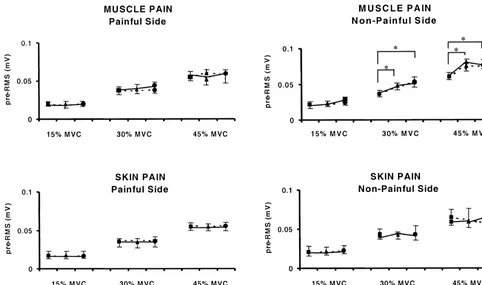
Fig. 4. Effects of different conditions (jBaseline,mPain anddPost-Baseline) on the pre-stimulus EMG activity (pre-RMS) at different clenching levels (15, 30 and 45%) and stimulus intensity (— — — Low and ——— High). Experimental muscle pain induced a significant increase in the pre-RMS on the non-painful side (P,0.01). The bars with the asterix indicate the significant increase in the pre-RMS during Pain and Post-Baseline conditions compared to the Baseline condition, at 30 and 45% clenching level (Tukey test: P,0.05; P,0.005). No effects were observed on the pre-RMS at different stimulus intensity (P.0.50).
was associated with an asymmetry in the pre-stimulus into the cat masseter muscle caused prolonged effects. EMG activity. The relative increase of the pre-stimulus Movements and muscle contraction may increase the firing EMG activity was only observed on the non-painful side rate of muscle nociceptors primed by the chemical (non-feedback muscle) and more pronounced at higher stimulus [44]. To explain post-infusion changes, we sug-clenching levels, probably because muscle pain increases gest that, although the subjects no longer reported pain, the with stronger contractions. Since the painful muscle also muscle may nevertheless have been tender in the post-pain served as the feedback muscle and necessarily had a recording. Regardless of the mechanism, these long-term, constant EMG level, an inhibitory effect on the homonym- pain-related changes in masseter EMG activity suggest that ous motoneurons could have been compensated by an the nociceptive input to motoneurons was so strong that it increased voluntary drive. An increased activity in the could produce excitability changes masking any changes in descending pathways would also have an impact on the corticobulbar excitability.
contralateral motoneuron pool [6] and cause an increase in Tonic cutaneous pain did not induce any effect on the the pre-stimulus EMG activity on the non-painful side pre-stimulus EMG activity. This is consistent with previ-(non-feedback muscle). Other studies have documented ous studies reporting that topical application of capsaicin is that experimental pain in the masseter muscle is associated unable to induce changes in the EMG activity or the with a significant decrease of the maximum voluntary electrically-evoked exteroceptive suppression responses occlusal force and EMG activity during agonist function mediated at brainstem level [21,30].
[35,37,43]. It has been suggested that the inhibitory effect
of muscle pain on motor function can be interpreted as an 4.3. Effects of clenching level and stimulus intensity ‘adaptation’ to pain, in order to limit overall movements
motor cortex, Electroenceph. Clin. Neurophysiol. 77 (1990) 390– latency of the MEPs in the present study. This
phenom-401. enon has been extensively studied in the limbs and may
[2] L. Arendt-Nielsen, T. Graven-Nielsen, H. Svarrer, P. Svensson, The result from enhanced excitability at cortical level (more influence of low back pain on muscle activity and coordination powerful descending volleys) or spinal level (lower thres- during gait: a clinical and experimental study, Pain 64 (1996)
231–240. hold of motoneurons) [12,16,22,39].
[3] R. Benecke, B.U. Meyer, P. Schonle, B. Conrad, Transcranial In the trigeminal system voluntary pre-activation is
magnetic stimulation of the human brain: responses in muscles essential to evoke masseter MEPs. The most likely reason
supplied by cranial nerves, Exp. Brain Res. 71 (1988) 623–632. is that masseteric motoneurons have a high activation [4] A. Berardelli, M. Inghilleri, R. Formisano, N. Accornero, M. threshold or that a relatively small proportion of the Manfredi, Stimulation of the motor tracts in motor neuron disease, J.
Neurol. Neurosurg. Psychiatry 50 (1987) 732–737. trigeminal motoneuronal pool receives monosynaptic,
fast-[5] D. Burke, R. Hicks, S.C. Gandevia, J. Stephen, I. Woodforth, M. conducting cortical projections [8]. However, once some
Crawford, Direct comparison of corticospinal volleys in human background EMG is provided the masseter MEPs are
subjects to transcranial magnetic and electrical stimulation, J. progressively facilitated by further increases in voluntary Physiol. (Lond.) 470 (1993) 383–393.
contraction similarly to limb MEPs and likely with similar [6] L.J. Carr, L.M. Harrison, J.A. Stephens, Evidence for bilateral innervation of certain homologous motoneurone pools in man, J. mechanisms at the segmental and cortical level.
Physiol. (Lond.) 475 (1994) 217–227. The increase in stimulus intensity from 1.1 to 1.5 times
[7] K.L. Casey, S. Minoshima, T.J. Morrow, R.S. Koeppe, A com-the motor threshold also induced a strong increase in com-the parison of human cerebral activation patterns during cutaneous amplitude of the MEPs in the present study. This could be warmth, heat pain, and deep cold pain, J. Neurophysiol. 76 (1996) due to a greater number of excited pyramidal cells and 571–581.
[8] G. Cruccu, A. Berardelli, M. Inghilleri, M. Manfredi, Functional descending fibers or because the increased firing rate
organization of the motor system in man, Brain 112 (1989) 1333– exerted a greater spatial-temporal summation at the
lower-1350. motoneuron synapse. The latency of the MEPs was
[9] G. Cruccu, M. Inghilleri, A. Berardelli, G. Pauletti, M. Manfredi, shortened by less than 1 ms and this small latency gain Cortico-facial and cortico-trigeminal projections. A comparison by confirms findings of previous studies [8,25]. Much of the magnetic brain stimulation in man, Electroenceph. Clin. Neuro-latency gain, which can be observed in MEPs in the hand physiol. 41 (1990) 140–144.
[10] G. Cruccu, G. Frisardi, G. Pauletti, A. Romaniello, M. Manfredi, muscles, may be due to the absence of D-waves with
Excitability of the central masticatory pathways in patients with low-intensity magnetic stimuli [5,16,18]. In contrast, a
painful temporomandibular disorders, Pain 73 (1997) 447–454. relatively long central delay of MEPs in cranial-nerve [11] A. De Laat, H.W. van der Glas, J.F.L. Weytjens, D. van Steenberghe, muscles is probably caused by the need for the arrival of The masseteric post-stimulus electromyographic complex in people the first I-waves, both with low- and high-intensity stimuli with dysfunction of the mandibular joint, Archs. Oral Biol. 30
(1985) 177–180. [4,9].
[12] V. Di Lazzaro, D. Restuccia, A. Oliviero, P. Profice, L. Ferrara, A. In conclusion, the present study shows that experimental
Insola, P. Mazzone, P. Tonali, J.C. Rothwell, Effects of voluntary tonic pain in the trigeminal region (whether from muscle or
contraction on descending volleys evoked by transcranial stimula-skin) is unable to induce detectable modulation of the tion in conscious humans, J. Physiol. (Lond.) 508 (1998) 625–633. MEPs. This finding does not entirely exclude pain-related [13] A.M. Drewes, S. Helweg-Larsen, P. Petersen, J. Brennum, A. changes in cortical excitability, because the magnetic Andreassen, L.H. Poulsen, T.S. Jensen, McGill Pain Questionnaire translated into Danish: experimental and clinical findings, J. Clin. stimulus may also generate some D-waves, thus bypassing
Pain 9 (1993) 80–87. the pyramidal cell and because compensatory mechanisms
[14] T. Graven-Nielsen, P. Svensson, L. Arendt-Nielsen, Effects of at the cortical or brainstem level in addition to the strong
experimental muscle pain on muscle activity and co-ordination voluntary activity may raise the cortical excitability to a during static and dynamic motor function, Electroenceph. Clin. level that hinders the detection of small changes. Overall, Neurophysiol. 105 (1997) 156–164.
the present findings argue against the so-called ‘vicious [15] C.J. Griffin, R.R. Munro, Electromyography of the masseter and anterior temporalis muscles in patients with temporomandibular cycle’ between pain — central hyperexcitability — muscle
dysfunction, Archs. Oral Biol. 16 (1971) 929–949. hyperactivity.
[16] A. Hufnagel, M. Jaeger, C.E. Elger, Transcranial magnetic stimula-tion: specific and non-specific facilitation of magnetic motor evoked potentials, J. Neurol. 237 (1990) 416–419.
Acknowledgements [17] M. Inghilleri, G. Cruccu, M. Argenta, L. Polidori, M. Manfredi, Silent period in upper limb muscles after noxiuos cutaneous stimulation in man, Electroenceph. Clin. Neurophysiol. 105 (1997) The present study was supported by the Danish National
109–115.
Research Foundation. ASM was supported, in part by a [18] K. Kaneko, S. Kaway, Y. Fuchigami, G. Shiraishi, T. Ito, Effects of T.C. White Travel Grant from the Royal College of stimulus intensity and voluntary contraction on corticospinal po-Physicians and Surgeons of Glasgow, UK. tentials following transcranial magnetic stimulation, J. Neurol. Sci.
139 (1996) 131–136.
[19] K. Kaneko, Y. Fuchigami, H. Orita, A. Ofuji, S. Kaway, Effect of coil position and stimulus intensity in transcranial magnetic
stimula-References tion on human brain, J. Neurol. Sci. 147 (1997) 155–159.
¨ ¨
[21] P. Kemppainen, A. Waltimo, T. Waltimo, M. Kononen, A. Per- [33] B.J. Sessle, Acute and chronic craniofacial pain: brainstem mecha-tovaara, Differential effects of noxious conditioning stimulation of nisms of nociceptive transmission and neuroplasticity, and their the cheek by capsaicin on human sensory and inhibitory masseter clinical correlates, Crit. Rev. Oral Biol. Med. 11 (2000) 57–91. reflex responses evoked by tooth pulp stimulation, J. Dent. Res. 76 [34] C.S. Stohler, X. Zhang, J.P. Lund, The effect of experimental jaw (1997) 1561–1568. muscle pain on postural muscle activity, Pain 66 (1996) 215–221. [22] U. Kischka, R. Fajfr, T. Fellenberg, C.W. Hess, Facilitation of motor [35] P. Svensson, L. Arendt-Nielsen, L. Houe, Sensory-motor interactions
evoked potentials from magnetic brain stimulation in man: a of human experimental unilateral muscle pain: a quantitative analy-comparative study of different target muscles, J. Clin. Neurophysiol. sis, Pain 64 (1996) 241–249.
10 (1993) 505–512. [36] P. Svensson, S. Minoshima, A. Beydoun, T.J. Morrow, K.L. Casey, [23] H. Kranz, C. Adorjani, G. Baumgartner, The effect of nociceptive Cerebral processing of acute skin and muscle pain in humans, J.
cutaneous stimuli on human motoneurons, Brain 96 (1973) 571– Neurophysiol. 78 (1997) 450–460.
590. [37] P. Svensson, A. De Laat, T. Graven-Nielsen, L. Arendt-Nielsen,
[24] J.P. Lund, R. Donga, C.G. Widmer, C.S. Stohler, The pain-adapta- Experimental jaw-muscle pain does not change heteronymous H-tion model: a discussion of the relaH-tionship between chronic reflexes in the human temporalis muscle, Exp. Brain Res. 121 musculoskeletal pain and motor activity, Can. J. Physiol. Pharmacol. (1998) 311–318.
69 (1991) 683–694. [38] P. Svensson, T.S. Miles, T. Graven-Nielsen, L. Arendt-Nielsen, [25] G.M. Macaluso, G. Pavesi, M. Bonanini, D. Mancia, P.U. Gennari, Modulation of stretch-evoked reflexes in single motor units in Motor-evoked potentials in masseter muscle by electrical and human masseter muscle by experimental pain, Exp. Brain Res. 132 magnetic stimulation in intact alert man, Archs. Oral Biol. 35 (1990) (2000) 65–71.
623–628. [39] P.D. Thompson, B.L. Day, J.C. Rothwell, D. Dressler, A. Maertens [26] S.B. McMahon, E. Sykova, P.D. Wall, C.J. Woolf, S.J. Gibson, de Noordhout, C.D. Marsden, Further observations on the facilita-Neurogenic extravasation and substance P levels are low in muscle tion of muscle responses to cortical stimulation by voluntary as compared to skin the rat hindlimb, Neurosci. Lett. 52 (1984) contraction, Electroenceph. Clin. Neurophysiol. 81 (1991) 397–402.
235–240. [40] C. Trompetto, C. Caponnetto, A. Buccolieri, G. Marchese, G.
[27] A.S. McMillan, C. Watson, D. Walshaw, J.P. Taylor, Improved Abruzzese, Responses of masseter muscles to transcranial magnetic reproducibility of magnetic stimulation-evoked motor potentials in stimulation in patients with amyotrophic lateral sclerosis, Elec-the human masseter by a new method for locating stimulation sites troenceph. Clin. Neurophysiol. 109 (1998) 309–314.
on the scalp, Archs. Oral Biol. 43 (1998) 665–668. [41] T. Tsubokawa, Y. Katayama, T. Yamamoto, T. Hirayama, S. [28] P. Mortifee, H. Stewart, M. Schulzer, A. Eisen, Reliability of Koyama, Chronic motor cortex stimulation for the treatment of
transcranial magnetic stimulation for mapping the human motor central pain, Acta Neurochir. Suppl. 52 (1991) 137–139. cortex, Electroenceph. Clin. Neurophysiol. 93 (1994) 131–137. [42] M. Valeriani, D. Restuccia, V. Di Lazzaro, A. Oliviero, P. Profice, D. [29] J.Y. Ro, N.F. Capra, Evidence for subnucleus interpolaris in Le Pera, E. Saturno, P. Tonali, Inhibition of the human primary craniofacial muscle pain mechanisms demonstrated by intramuscular motor area by painful heat stimulation of the skin, Clin. Neuro-injections with hypertonic saline, Brain Res. 842 (1999) 166–183. physiol. 110 (1999) 1475–1480.
[30] A. Romaniello, P. Svensson, G. Cruccu, L. Arendt-Nielsen, Modula- [43] K. Wang, P. Svensson, L. Arendt-Nielsen, Modulation of exterocep-tion of exteroceptive suppression periods in human jaw-closing tive suppression periods in human jaw-closing muscles by local and muscles induced by summation of nociceptive and non-nociceptive remote experimental muscle pain, Pain 82 (1999) 253–262. inputs, Exp. Brain Res. 132 (2000) 306–313. [44] K.G. Westberg, P. Clavelou, G. Schwartz, J.P. Lund, Effects of [31] J.C. Rothwell, P.D. Thompson, B.L. Day, J.P.R. Dick, T. Kachi, chemical stimulation of masseter nociceptors on trigeminal J.M.A. Cowan, C.D. Marsden, Motor cortical stimulation in intact motoneurons and interneuron activities during fictive mastication in man. I. General characteristics of EMG responses in different the rabbit, Pain 73 (1997) 295–308.
muscles, Brain 110 (1987) 1173–1190. [45] N. Witting, P. Svensson, H. Gottrup, L. Arendt-Nielsen, T.S. Jensen, [32] G. Schwartz, J.P. Lund, Modification of rhythmical jaw movements Intramuscular and intradermal injection of capsaicin: a comparison