iv
ABSTRACT BOOK ABSTRACT BOOKv
Patronage:Chairman of WFNS Spine Committee President of Indonesian Neurosurgical Society
Chairman of Asian Epilepsy Surgery Society
• Welcome Message ...xxiii
• Scientific Schedule of WFNS SPINE COMMITTEE-INS-FUJITA BANTANE ...1
ABSTrACT PlENAry lECTUrE 1 • Spine Tuberculosis ...14
• Percutaneous Endoscopic Lumbar Discectomy, Possibility and Limitation ...15
• Foreman Magnum Decompression for Type I Arnold Chiari Malformation ....16
• Laminoplasty Techniques for Cervical Myelopathy and Radiculopathy ...17
PlENAry lECTUrE 2 • Problems of Surgery in Geriatric Spine ...18
• Spine Anatomy Differences at A Global Level; Do Our Patients have the same Spines, Spine Disease and Can We Generalize Spine Treatment ...19
• Anterior and Posterior Approach Subaxial Cervical Spine ...20
• Posterior Decompression and Fusion for Spondylotic Myelopathy ...21
lUNCH SyMPOSIUM • Robotic Visualization System ...22
• Introduction Of IORT (Intrabeam) for Neurosurgery ...23
SATEllITE SyMPOSIUM SS 1 - SPINE 1: CErVICAl DEGENErATIVE • Updates in Treatment of Cervical Spondylosis and Spinal Stenosis ...25
• New Technique of Cervical Laminoplasty for Cervical Myelopathy ...26
• Complications of Anterior Cervical Discectomy and Fusion ...27
• Long-Term Follow-Up of Operations for Cervical Disc Herniation ...28
• Surgical Treatment of Cervical OPLL ...29
• Cervical Spinal Cord lnjury ...30
• Tethered Cord Injury: True or False ...31
vi
ABSTRACT BOOK ABSTRACT BOOKvii
SS 3 - SPINE 2: DEFOrMITy
• Lateral Approach for Stabilization and Correction of Lumbar Deformity 33
• Correction and Fixation Surgery for Adult Spine Deformity with
Osteoporosis ...34
• Surgery for Adult Degenerative Scoliosis ...35
• Correction for Spine Deformity ...36
• Pelvic Parameter in Adult Degenerative Deformity ...37
• Spinal Osteotomies for Spinal Deformities ...38
SS 4 - BrAIN 1: VASCUlAr 1 • Embolization of Brain Arterial Venous Malformation ...39
• Transpetrosal Approach for Giant Aneurysms in Posterior Fossa ~ Microanatomy and Actual Operative Procedures ...40
• Treatment of Unusual Internal Carotid Artery Aneurysms:Clipping and Hybrid Method ...41
• New Management and Strategy of Cerebral Aneurysm by Feature in Japan ...42
• Surgical Cliping versus Endovascular Coiling in Cerebral Aneurysm ...43
• Flow Diversion Stent for Large and Giant Internal Carotid Artery Aneurysm: Initial Experience ...44
• Management of Poor Grade Aneurysmal SAH ...45
SS 5 - SPINE 3: MINIMAl INVASIVE • Microscopic Lumbal Decompression ...46
• Minimal Invasive TLIF: Clinico-Radiological Assesment Safety and Reability ...47
• Disc FX Technique for Sacroilliac Joint Syndrome ...48
• Patology and Pathophysiology of Lumbar Herniated Nucleus Pulposus on Minimally Invasive Surgery Approach ...49
• Current Status, Challenges and Future of the Percutaneous Endoscopic Spine Surgery ...50
• Accurate Placement of Percutaneous Pedicle Screws without the Use of Neuronavigation / O-arm Technology and Reduction-fixation of Lumbar Spondylolisthesis by Percutaneous Pedicle Screws and a Minimal Access Approach ...52
SS 6 - SPINE 4: TUMOr • Management of Spinal Intramedullary Tumors ...53
• Metastasis Spine Prognostic Factors ...54
• Cervical Intramedullary Tumors: Surgical and Neurophysiological Monitoring Aspects ...55
• Flip Osteoplastic Laminotomy Flap for Excision of Long Segment Spinal Tumours in Chilldren ...56
• Surgery of Intramedullary Tumors ...57
• Surgery of Spinal Intramedullary Tumors: Optimization of Surgical Safety and Precision ...58
• Minimal Access Corridors in Intra Dural Extra Medullary Tumours and Technical Challenges ...59
SS 7 – BrAIN 2: TUMOr • Surgical Urgency Grouping of Pituitary Tumor Patients...60
• Strategy Management of Malignant Anterior Skull Base Tumors: Personal Experience ...61
• Treatment Strategy for Elderly Meningioma ...62
• Secondary Brain Tumor ...63
• Central Nervous System Hemangioblastomas: Clinical and Surgical Management...64
• Save Radical Resection for High Grade Glioma, Where are we now? ...65
SS 8 – SPINE 5: CErVICAl • Cervical Arthroplasty. Expanding Indications to Slit Discs and Segmental Kyphosis ...66
• Transpedicular Approach in Subaxial Cervical Spine: A Challenge in Cervical Fixation ...67
• Posterior Approach for Odontoid Fracture Type II Fixation ...68
• How to Choose between Anterior and Posterior Approach for OPLL? An Evidence Based Approach ...69
• Fusion vs TDR in Cervical Spine - A Decade and More Than 500 Cases Later - What We Learnt ...71
• C1 C2 Posterior Fixation...72
• Cervical Dislocation Fracture: Anterior-Posterior Stabilization Technique 73 SS 9 – BrAIN 3: TrAUMA • Osteoplastic Procedures for Front Temporal Craniotomy ...74
viii
ABSTRACT BOOK ABSTRACT BOOKix
• Primary Neurosurgical Life Support (PNLS): Effective Simulation
Training for Neurosurgical Management ...76
• Early Decompressive or Late Decompressive Craniotomy for Intracranial Bleeding with Severe GCS (A Proposed for Hospital with NeurotraumaSurgery Facility) ...77
• TBA ...78
• Prognostic Value of Convergent Type of Hemorrhage Visualized by Susceptibility Weighted Image in Diffuse Brain Injury ...79
• Management of Neurosurgery Cases in Lombok Island Earthquake 2018 80 SS 10 –BrAIN 4: TECHNIQUE • Modern Surgical Management of Patients with Symptomatic Low Grade Glioma in Eloquent Areas ...81
• Pitfall Anterior Transpetrosal (Kawase Approach) for Combine Midle and Posterior Fossa Lession ...82
• Strategy of Minimal Invasive Surgery in Spontaneous ICH ...83
• A Technical Method of Extradural Anterior Clinoidectomy. ~Microanatomy and Actual Operative Procedures~ ...84
• One-and-A-Half Cavity Concept for Single Nostril Endoscopic Endonasal Transsphenoidal Hypophysectomy; a Technical Report ...85
• Microvascular Decompression with Keyhole Craniotomy ...86
PlENAry lECTUrE 3 • Spinal Cord Tumor ...87
• Learning Curve MIS Surgery ...88
• Development of Modern Experimental Spinal Cord Trauma and the Importance of Biomechanics ...89
• Image-Guide Neurospine Surgery: Challenges and Solutions ...90
PlENAry lECTUrE 4 • Adjacent Cortico Cancellous Bone Grafts in Anterior Cervical Fusion Newer Concept ...91
• Anterior C1 C2 Fixation for Mobile AAD or Fracture Odontoid ...92
• Achieving a Better Mechanical Stability in Osteoporotic Spine ...93
• Minimally Invasive Management of Metastatic Spine Tumors ...94
SS 11 – SPINE 6: TECHNIQUE • Role of Spinal Navigation (O-arm) in Lumbar Fusion Procedures ...95
• Surgical Strategy for Spinal Infection and Osteoporosis, How I do It? ...96
• MIS Spinal Fixation using O-arm ...97
• Surgical Management for Thoracic Spinal Tuberculosis ...98
• Minimally Invasive Surgery of Spine Tumors ...99
• Transarticular Facet Screw Fixation of the Subaxial Cervical Spine: Advantages and Limitations ... 100
• Clinical outcome of Trans-sacral Epiduroscopic Laser Decompression (SELD) ...101
SS 12 – MISCEllANEOUS • Patien Safety & Ethics ...102
• Lesson Learned from Indonesian Stock Exchange Spine Casualties: a Neurosurgeons Perspective ...103
• Primary Central Nervous System Lymphoma (PCNLS): 7 Years’ Experience in Single Institution ...104
• Beyond the Pillars of Hercules: the Navigation of the Cerebral Aqueduct and the Fourth Ventricle to Manage Intraventricular Blood Clots and Arachnoid Cysts ...105
• Multisegmental Diffuse Intradural Extramedullary Spinal Tumor ...106
• Mixed Pain Concept in Chronic Low Back Pain ...107
• Epidural Analgesia for Post Spine Surgery Pain Management ...108
SS 13 – SPINE 7: TECHNIQUE • Low Back Pain and Sciatica, Surgical versus Nonsurgical Treatment! ....114
• Influence of Indocyanine Green Angiography on Microsurgical Treatment of Spinal Perimedullary Arteriovenous Fistulas ...115
• Metastatic Spinal Cord Compression Tumor In Dept Neurosurgery Faculty Of Medicine Universitas Indonesia – General Hospital Dr. Cipto Mangunkusuma Jakarta ...116
• 100 Case Microdisectomy What I Learn? ...117
• Cervical Spine Anterior Approach, DisCectomy, and Corpectomy ...118
• Infections in Spinal Instrumentation: A Proposal for Management Algorithm using Closed-Suction Irrigation System and Vacuum Assisted Closure (VAC) ...119
• Usefulness of Percutaneous Endoscopic Lumbar Discectomy ...120
x
ABSTRACT BOOK ABSTRACT BOOKxi
• Intradiscal Decompression for Contained Disc Herniation Lumbar Area ...122
• Multiple Inherited Schwannomas, Meningiomas, and Ependymomas (MISME) A Report on Rare Case of Neurofibromatosis Type 2 Tumors ...123
• Evaluation and Emergency Treatment Of The Newborn With Spina Bifida ...124
• Degenerative Cervical Myelopathy: Practical Guide and Update on Current Clinical Evidence in Indonesia ...125
• Surgical Treatment for Osteoporotic Vertebral Fracture in Geriatric Patients ...127
• Craniovertebral Fixation - a New Technique of Occipital Cervical Fixation ...128
SS 15 – FUNCTIONAl • How to make MVD Safe & Efficacious - Personal Experience Gained Through 5120 Cases ...129
• Maximizing Decrease in Drug Dosage and Increase in ON time following Bilateral STN DBS Using Constant Current for Advanced Parkinsons Disease ...130
• Radiofrequency Ablation for Chronic Knee Pain, Single Institute Experiences ...131
• Do’s and Don’ts in Micro Vascular Decompression Surgery ...132
• Stereotactic Surgery in Parkinson, Tremor and Dystonia ...133
• Secondary Trigeminal Neuralgia: Clinical Feature & Surgical Result ...134
• Selective Amygdalo Hippocampectomy with Mini Craniotomy ...135
SS 16 – BrAIN 5: VASCUlAr 2 • Minimally Invasive Strategies for Cerebral Aneurysm Surgery ...136
• Frontline of Endovascular Therapy for Cerebral Aneurysm ...138
• Strategy for Coiling of Wide-Necked Aneurysms and Fusiform Aneurysms ...139
• Surgery for Cerebral AVM...140
• Save Acute Stroke Patient by Endovascular Therapy ...141
• Acute Ischemic Stroke Management in Cipto Mangunkusumo National General Hospital ...142
• Management of CCF In Fac. of Medicine Padjajaran Univeristy / Hasan Sadikin General Hospital ...143
SS 17 – BrAIN 6: VASCUlAr 3 • Result of Early High Flow bypass & Trapping for Ruptured Blood Blister Like ICA Aneurysms ...144
• Table-Side Evaluation of C-Arm CT Perfusion Images Before and Just After Mechanical Thrombectomy Treatment for Acute Ischemic Stroke Patients ...145
• Dual Strategy Approach for Minimally Invasive Aneurysm Surgery ...146
• Lessons Learnt from 200 AVM Surgery: Battles against Cerebral AVMs 147 • How to Manage Intracerebral Hematoma: Concept and Novel Method ..148
• Mobile Computer Application for Classifying Stroke by Ambulance Service ...149
SS 18 – SPINE 9 • Transforaminal Epiduroscopic Besivertebral Nerve Laser Ablation (Tebla) for Chronic Back Pain Combined with Modic Change...150
• CV Junction Maningioma Present with Pregnancy: Case Report and Literature Review Plans and Result ...151
• Whole Spine Concept Imaging for Preoperative Evaluation of Spinal Degenerative Disease ...152
• Endoscopic Removal of Spinal Intradural Tumour via Interlaminar Approach ...153
• Fail Back Surgery Syndrome ...154
• One Stage Transpedicular Unilateral Corpectomy Stabilized by Cervical Titanium Mesh and Transpedicle Screw Fixation for Tubercolosis/Trans Thoracic and Translumbar fot Th 10-11-12 and L1-L2 Disc Prolapse after Filed Laminectomy Surgery ...155
• Penetrating Gunshot Wound of Cervical Spine: Debates, Recommendations, Strategies with Illustrative Case in Civilian ...156
SS 19 – SPINE 10: MINIMAl INVASIVE • Challenges and Complication in Minimal Invasive Spine Surgery ...157
• Short and Mid-Term Follow-up in PDS ...158
• Pitfalls in OLF Surgery ...159
• Low Cost Solution with Percutaneus Endoscopic Lumbal Discectomy for Simple Lumbar Disc Disorder ...160
• Modality for Lumbar Discogenic Pain Syndrome ...161
xii
ABSTRACT BOOK ABSTRACT BOOKxiii
SPECIAl lECTUrE
• Craniocervical Junction Instability: When to Add Occiput to Fusion? ...183
• Role of Epilepsy Surgery in developing Basic Research in Neuroscience ... 184
• Ethical and Legal Aspects in Spine Surgery ...185
SCIENTIFIC SCHEDUlE OF 12TH AESC ... 191
ABSTrACT INDONESIAN EPIlEPSy SCHOOl • Drug Refractory Epilepsy, How do We Diagnose DRE ...194
• Managing Antiepileptic Drug, Starting, Changing, and Stopping AED’s ...195
• Neuroimaging in Epilepsy: Best Imaging Sequence for Best Detection of Epileptogenic Lesion ...196
• EEG and Semiology in Focal or Partial Seizures ...197
• Starting Comprehensive Epilepsy in Surabaya: Challenge, Opportunity and Strategy ...195
• Candidates for Epilepsy Surgery ...196
ASIAN EPIlEPSy SUrGEry CONGrESS SESSION • Establishing Advance Epilepsy Surgery Program in Developing Countries ...197
• Autonomic Changes in Patients with Intractable Epilepsy ...198
• Presurgical Planning of Intracranial Electrode Insertion in Patients with Cortical Migration Disorders ...199
• Identification of Genes Associated with Cortical Malformation using a Transposon-Mediated Somatic Mutagenesis Screen in Mice ... 200
• Utility of Statistical Parametric Mapping Analysis for Detection of Epileptic Foci In [18F] FDC And [11C] Flumazenil Pet Studies ...201
• Multi-Institutional Study of Epilepsy and Glia in Japan ...202
• Stereo-EEG for Periventricular Nodular Heterotopia with Drug-Resistant Epilepsies ...203
• Fully-implantable Wireless ECoG Device ...204
• Cavernoma Related Epilepsy: Controversy on Management ...205
• Epilepsy Surgery for Tuberous Sclerosis Complex ...206
• Vagal Nerve Stimulations (VNS) ...207
• Epilepsy Surgery for Tuberous Sclerosis Complex ...208
SS 20 – PEDIATrIC • Utilization of Endoscopy in Neurosurgery Cases in Cipto Mangunkusumo Hospital, Jakarta, Indonesia ...163
• Curative Resection for Lesional Refractory Epilepsy in Children Outcomes and Local Experience in Hospital Kuala Lumpur ...164
• Neurosurgical Aspect in Syndromic Craniosynostosis ...165
• Changes of Subventricular Zone Neural Stem Cells in Hydrocephalus: An Experimental Animal Model ...166
• A Review in Pediatric Hydrocephalus Ten years Experience with Ventriculoperitoneal Shunt ...167
• Pediatric Spinal Dysraphysm ...168
SS 21 – BrAIN & PErIPHErAl NErVES: TrAUMA • Severe Extracranial Injuries Effect on Outcomes of Traumatic Brain Injuries ...169
• The Hypothermia Therapy in Severe Traumatic Brain Injury: Impartial Perspective ...170
• The Role of Axonal Supercharging in Chronic Peripheral Nerve Injury...171
• Management of Brachial Plexus Injury ...172
• The Influence of Decompressive Craniectomy with Mesh on Peridural Tissue of Wistar Mice with Traumatic Brain Injury ...173
• Penetrating Brain Injury Due to Gunshot Wounds by Low-Velocity Bullets as Air Rifle (Air Guns): A 7 Years Experience of the Neurosurgery Service ...174
SS 22 – BrAIN 7: TECHNIQUE • Endoscopy for Sellae Region Lession ...176
• Tansnasal Endoscopic Surgery for Pituitary Adenoma ...177
• Preoperative Embolization as a Brain Tumor’s Resection Strategy in a Young Woman with No Neurological Deficits: a Case Report ...178
• Awake Craniotomy ...179
• Management of Anterior Skull Base Tumor ...180
• Management of Parasagittal Meningioma...181
xiv
ABSTRACT BOOK ABSTRACT BOOKxv
• Automated Brain Anatomy Labeling and Localization for Stereo -
Electroencephalography (SEEG Anatomy Labeling) ...209
• TBA ...210
• Endoscopic Epilepsy Surgery: Indication and Technique ...211
• Microscopic Corpus Callosotomy: Long Term Outcome ...212
• Evaluation of Cognitive Function in Temporal Lobe Epilepsy...213
• Stigma and Epilepsy Surgery in PWE in Ethiopia ...214
• SEEG Investigation and Surgery Treatment for Insular Epilepsy ...215
• TBA ...216
• TBA ...217
SCHEDUlE OF OrAl PrESENTATION ... 218
ABSTrACT OrAl • OP 001 - Description of 7th Cervical Vertebrae Lamina using 2D CT-Scan Morphometric and 3D Virtual Simulation in Reference to Translaminar Screw Placement Requisites ...244
• OP 002 - Prevalence of Complications Following Cervical Unilateral Open-Door Laminoplasty in Cervical Spondylosis Patients: Systematic Review and Meta-Analysis ...245
• OP 003 - Spinal Epidural Abcess Causing Foot Drop in Pre-Existing Bertolotti’s Syndrome ...246
• OP 004 - Thoracic Medial Branch Blocks in Managing Chronic Facet Joint Pain for Multiple Osteoporotic Compression Fracture: Case Report...247
• OP 005 - Refractory Dorsalgia Caused By Sacro-Iliac Joint Dysfunction in Elderly Managed Successfully By Pulse Radiofrequency Ablation ...248
• OP 007 - A Rare Case of Ochronosis Presenting with Cervical Compressive Myelopathy ...249
• OP 009 - Endoscopic Removal of Spinal Tumor via Interlaminar Approach ...250
• Anton M.J. Sirait...250
• OP 010 - Paraspinal Abscess of Spinal Tuberculosis: Which Is the Best Surgical Approach? ...251
• OP 011 - Spinal Cord Stenosis Due to Cervical Metastasis From Papillary Thyroid Carcinoma: A Case Report ...252
• OP 012 - Cervical Skull Traction Followed by Decompressive Laminectomy, Internal Fixation and Fusion using Titanium Mesh in Grade III Traumatic Spondylolisthesis of C 5-6, Bilateral Facet Dislocation C 5-6 : Case Report ...253
• OP 013 - Biomechanical Properties of Injectable Silicon for Nucleus Pulposus Replacment: Preliminary in Vitro Study ...254
• OP 014 - Comparison Of Surgical Versus Conservative Treatment Of Sciatica Due To Lumbar Disc Herniation ...255
• OP 015 - The Effect Of Psychosocial Factors In The Success Of Conservative Management For Low Backache ...257
• OP 017 - Correlation between Clinical Symptoms and Radiological Findings on Moderate and Severe Head Injury Associated With Atlanto-Occipital Dislocation ...259
• OP 018 - Cranial Trauma Associated Scalp Cerebrovascular Lesions: Our Clinical Experience. ...260
• OP 019 - Evaluating The Impact Of Helmet Use And Government Role On Preventing Head Injury In Indonesian Remote-Border Region ...261
• OP 020 - Management and Evaluation of Orbitocranial Penetrating Brain Injury from a Fishing Gun: A Rare Case Report ...262
• OP 021 - Case Report Compound Open Depressed Displaced Frontal Bone Fracture And Cerebral Prolapse Over Supraorbital Rim ...263
• OP 022 - The Relation Of Glasgow Coma Scale Toward PT and APTT Value among Head Injury Patients in Emergency Department Ulin Hospital ...264
• OP 023 - Surgical Complications and Long-Term Outcome of Bifrontal Decompressive Cranioectomy used for Management of Cases with Refractory Cerebral Edema Following Traumatic Brain Injury ...265
• OP 024 - Subdural Haematoma as A Complication of Spontaneous Intacranial Hypotension: A Rare Case ...266
• OP 025 - Skull Fracture and Massive Epidural Hematoma Secondary to the Mayfield Three-Pin Skull Clamp in Paediatric Patient: A Case Report and Review of The Literature ...267
xvi
ABSTRACT BOOK ABSTRACT BOOKxvii
• OP 027 - Supine Position RetroSigmoid Approach: Case Report ...269
• OP 028 - Potential of Endogenous Cell-Based Therapy for Traumatic Brain Injury ...270
• OP 029 - Temporo-Parietal Subdural Empyema in an Adult
Mimicking Chronic Subdural Hematoma: A Case Report ...271
• OP 030 - Complications Following Cranioplasty: Incidence and
Predictors At RSUP Dr. Sardjito Yogyakarta ...272
• OP 031 - Surgical Interventions Management for Traumatic Brain
Injuries and Spontaneus ICH in the Elderly Patients in Sardjito Hospital 273
• OP 032 - S100B Serum Level as a Mortality Predictor for Traumatic Brain Injury: A Meta-Analysis...274
• OP 033 - Giant Facial Nerve Schwannoma Involving Middle Cranial Fossa ...275
• OP 034 - Pre-Operative Measurement of Diplopia uses
Strabismic-Deviation Values in Sphenoorbital Meningioma Patients ...276
• OP 035 - Awake Craniotomy for Supratentorial Tumor Resection ...277
• OP 036 - Profile of Glioma Patients in Dr. Cipto Mangunkusumo
National Hospital Jakarta-Indonesia: A Descriptive Study ...278
• OP 037 - Immediate Recovery of Severe Vertigo in Patient with Bilateral Cerebellopontine Angle Arachnoid Cyst Following
Microsurgical Treatment ...279
• OP 038 - Progesterone and Estrogen Receptors Positive Status in
Sphenoorbital Meningioma in 16-Year-Old Male: A Case Report ...280
• OP 039 - Emergency Presentation, Management and Primary
Outcome in Patients with Glioblastoma Multiforme ...281
• OP 040 - Male Meningiomas Characteristic in Dr. Kariadi General
Hospital, Semarang: A Descriptive Study ...282
• OP 041 - Clinical Outcome After Awake Craniotomy for Glial Tumor Resection in the Supplementary Motor Area ...283
• OP 042 - Case Report: Sellar Teratoma in Young Children with
Progressive Visual Loss ...284
• OP 043 - Minimally Invasive Approach for Anterior Cranial Fossa
Meningioma, Learning Curve as a Young Neurosurgeon: Case Reports .285
• OP 044 - Glioblastoma, Osteoplasty versus Decompression? - Serial Case ...286
• OP 045 - Challenges Faced in Operating Intracranial Epidermoid
Cysts: A Case Series ...287
• OP 046 - A Case Series of Suspected Solitary Bone Plasmacytoma:
Limited Modalities for Comprehensive Management ...288
• OP 047 - 3D Printing as a Tool Personalized Medicine in
Hyperostosis Sphenoorbita Meningioma ...289
• OP 048
• Distress in Glioblastoma Multiforme Patients And Caregiver: A Qualitative Study of the Status of Medical Knowledge For
Psychosocial Distress Condition. ...290
• OP 048
• OP 049 - A Review Of Brain Implant Device: Current Developments And Applications ...292
• Siti Aminah Hospital, Bumiayu, Indonesia ...292
• OP 050 - Neuronal Migration Disorders In Epilepsy: A Case Report ...293
• OP 052 - Surface Electromyography as an Objective Tool for Evaluating Tremorin Parkinson Disease: Pre and Post Vim
Thalamotomy ...294
• OP 053 - Therapeutic Benefit of Palmitoylethanolamide in the
Management of Neuropathic Pain ...295
• OP 054 - Surgery in Sturge–Weber Syndrome with Uncontroled
Epilepsy: A Case Report ...296
• OP 055 - Trigeminal Neuralgia Management: Some Challenges in
Microvascular Decompression Surgery and Literature Review ...297
• OP 056 - The Role of Neuronavigation in Surgical Management of Cerebral Cavernoma Malformation Related Epilepsy: Case Series
from National Brain Center Hospital, Jakarta ...298
• OP 057 - A Case Report of Teflon Wrapping for Unclippable
Intracranial Aneurysm in Choroid Artery with Giant Thrombus ...299
• OP 058 - Cerebral Cavernoma Malformation Related Epilepsy Cases in National Brain Center Hospital, Jakarta: A Descriptive Study ... 300
• OP 059 - Narrow Cistern as an Anatomical Challenge in
Microvascular Decompression Surgery for Trigeminal Neuralgia:
Case Report ...301
• OP 060 - Correlation Between Ferritin and Glasgow Outcome at Discharge Scale in Spontaneous Intracerebral Hemorrhage Patients Who Underwent Surgical Treatment ...302
• OP 061 - Moyamoya Disease: A Case Report Treated with
xviii
ABSTRACT BOOK ABSTRACT BOOKxix
• OP 062 - Microsurgery for Grade II-III Spetzler-Martin Arteriovenous Malformation with Hemorrhagic Presentation and Cyst Formation in a Pediatric Patient: A Case Report ...304
• OP 062
• OP 063 - Clinical Improvement of Patients Undergoing Endovascular Embolization in Traumatic Carotid Cavernous Fistula: Case Series ...306
• OP 064 - Mini Osteoplastic Craniotomy for Spontaneous Intracerebral Haematoma as Alternative to Minimally Invasive
Technique ...307
• OP 065 - Aggressive Type Dural Arteriovenous Fistula of Transverse-Sigmoid Sinus Junction: Surgical Disconnection as an Option ...308
• OP 066 - Middle Cerebral Artery Infarction Due to Traumatic Internal Carotid Dissection: A Rare Case ...309
• OP 067 - Pharmacoresistant Temporal Lobe Epilepsy Controlled By Bilateral Anterior Thalamic Nuclei Thalamotomy ...310
• OP 068 - Evolution of the Bony Orbit and its Legacy for Predation:
The Supraorbital-Torus’ Appearance and Disappearance Riddle ...311
• OP 069 - Thermoregulation, Parietal Lobe, and Febrile Seizures in an Evolutionary Quest ...312
• OP 070 - Neurosurgery Education for Medical Student in Indonesia ...314
• OP 071 - How to Face the Struggles and Overcome Them, While
Establishing Neurosurgery at a Rural Medical College ...315
• OP 072 - The Effect of Curcumin Extract Toward Mature Brain Derived Neurotrphic Factor (M-Bdnf) Expression After Traumatic
Brain Injury ...316
• OP 073 - Correlation Between Human Epithelial Growth Factor 2 (Her 2) Expression with Histopathological Level on Intracranial
Meningioma Patients at Haji Adam Malik Hospital Medan ...317
• OP 074 - Ventriculo-Sagittal Sinus Shunt for Hydrocephalus: A Case Report...318
• OP 075 - Clinical Profiles of Closed Spina Bifida Patients Undergoing
Surgery in Cipto Mangunkusumo General Hospital from January
2014 – June 2018 ...319
• OP 076 - Our Experience in Surgical Treatment of Arnold Chiari
Malformation Type 1 ...320
• OP 077 - A Case Series of Hydrocephalus as Clinical Indicator of Central Nervous System Relapse in Acute Lymphoblastic Leukemia
in RSUP Dr. Sardjito ...321
• OP 078 - CVJ Anomaly: An Overlooked Cause of Stroke in Young ...322
• OP 079 - Giant Interparietal Enchepaloceles: How We Managed Them ..323
• OP 080 - Short-Term Follow-Up of Additional Gravitational Valve in the Management of Symptomatic Overdrainage in Children with
Fixed Differential Pressure Valve Shunts ...324
• OP 081 - Modified Revised Trauma-Marshall Score: A Propose Tool
Predicts Outcome in Moderate and Severe Traumatic Brain Injury ...325
• OP 082 - Demography, Histopathology and Surgical Outcome of Spinal Tumors in Department Of Neurosurgery Faculty of Medicine Universitas Indonesia – RSUP Nasional Dr. Cipto Mangunkusumo ...326
• OP 083 - Hemichorea Post Stroke Controlled with Unilateral
Pallidotomy. ...327
• OP 084 - Incidence of Intracranial Meningioma in Patients with
Family History of Solid Organ Malignancy ...328
• OP 085 - Late Onset Seizure nd Left Hemiparesis after Unusual
Craniocerebral Penetrating Injury by a Rusty Sickle (CASE REPORT) ...330
• OP 086 - Carotid Cavernous Fistula ...331
• OP 087 - High Filamin-C Expression Predicts Enhanced Invasiveness and Poor Outcome in Glioblastoma Multiforme ...335
• OP 088 - Rapid Improvement in Motoric Strength After Cranioplasty in Patient with Sinking Skin Flap Syndrome: A Case Report ...336
• OP 089 - Surgical Management of Tuberculosis of the Spine: A Retrospective Analysis of 127 Cases in a Tertiary Care Hospital of Bangladesh. ...337
• OP 090 - Post Traumatic Memory Function Disturbance Associated with Depressed Skull Fracture ...338
• OP 091 - Non Surgery Treatment on Massive Corpus Callosum
Hematoma without Disconnection Syndrome: A Case Report ...339
• OP 092 - Iatrogenic Spinal Subdural Haematoma as a Complication of Lumbar Puncture : A Case Report ...340
• OP 093 - Neurosurgical Lesioning for Cancer Pain ...342
• OP 094 - Multiple Meningiomas Treatment in Dr. Cipto
Mangunkusumo Hospital: A Case Report ...343
• OP 095 - Cerebral Collateral Circulation in Total Occlusion of the
Right Internal Carotid Artery ...344
• OP 096 - Chiari Type I Malformation Profile in Cipto Mangunkusumo
xx
ABSTRACT BOOK ABSTRACT BOOKxxi
Dear Friends,It is our great pleasure to invite you to The 5th WFNS Spine Committee Biennial Conference of WFNS which will be held at Bali, Indonesia between October 25th - 27th, 2018.
WFNS scientific committees try to contribute to the education and progress of sub disciplines of neurosurgery. Spine surgery is getting a high interest and Spine Committee Symposia every two years are the largest activity of the committee. I am happy to invite you to Bali, Indonesia to endorse activities in this part of the world. This meeting will be in conjunction with the Annual Meeting of Indonesian Neurological Society, Asian Epilepsy Surgery Congress. On October 25, a one-day cadaver dissection course will be held in Surabaya. The meeting aims to reach a large number of audience, thus contribute to the spine education in this area more effectively. There will be “intense”, and full of excellent lectures from prominent experts, results of implementation of new procedures, case discussions, debate sessions, video demonstrations, and workshops from industry.
The location of our congress is Bali island, one of the most beautiful and exotic place of the world. We really hope that it will endow us with many precious and long-lasting memories to cherish.
We look forward to seeing you in Bali in October 2018. Co-chairman of the WFNS Spine Committee.
Mehmet Zileli Michael G.Fehlings Daniel J.Hoh
WELCOME MESSAGE
• OP 097 - Sacral Chordoma: Operative Management, Radiotherapy and Outcome in Cipto Mangunkusumo Hospital (Case series) ...346
• OP 098 - Management of Delayed CSF Leakage After Frontal
Based Tumor Removal : A Case Report ...347
• OP 099 - Case Series: ...348
• Gamma Knife Preoperative Preparation for Arteriovenous
Malformations (AVMs) ...348
• OP100 - An Unusual Case of Through-And-Through Stab Penetrating Head Injury to Temporal Lobe without Neurologic
Deficit: A Case Report ...349
• OP 101 - Endoscopic Fenestration with Unexpected Intraventricular Slough Deposit followed with iVEL & EVD Implantation in Infant with Infected Multiloculated Congenital
Hydrocephalus: A Case Report ...350
• OP102 - Spontaneous Recovery of Prefrontal Medial Syndrome Following Giant Olfactory Groove Meningioma Resection: A Case
Report...351
• OP103 - Surgery of Left Temporal Region Arachnoid Cyst with
Neuroendoscopy (Case report) ...353
• OP 104 - Incidence and Clinicopathological Features of
Meningioma in RSUP Dr. Sardjito During 2017 ...354
• OP105 - Subfrontal Craniotomy Approach for Management of
Craniopharyngioma : Case Report ...355
• OP 106 - A Giant Pituitary Adenoma: Surgical Excision via
Subfrontal Approach. ...356
118
ABSTRACTBOOK ABSTRACTBOOK119
SS 13– SPINE 7: TECHNIQUE
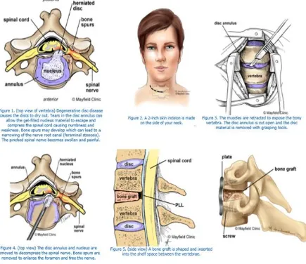
CERVICAL SPINE ANTERIOR APPROACH, DISCECTOMY, AND CORPECTOMY
Ridha Dharmajaya
Department of Neurosurgery, Faculty of Medicine, University of Sumatera Utara/Haji
Adam Malik General Hospital, Medan, Indonesia Corresponding Author: ridhadharmajaya@yahoo.com
Anterior cervical diskectomy and fusion (ACDF) has been used for almost six decades. First introduced in the 1950s, ACDF is now widely used to treat cervical spondylotic radiculopathy and myelopathy with long-term clinical success. ACDF enables removal of compressive lesions of the spinal cord, such as osteophytes, intervertebral disks, and ossfied posterior longitudinal ligaments (OPLLs). It has also been used to treat a range of other cervical diseases (mainly between C2 and T1 vertebrae) related to cervical instability (degenerative, traumatic, oncological, infectious, inflammatory, iatrogenic).
The best predictor of good patient outcomes after surgery is proper preoperative patient selection based on clinical symptoms, physical examination, and imaging studies. ACDF surgery has been shown to have clinical success rates of 97% and 94% for one- and two-level fusions, respectively, at a mean clinical follow-up ≥ 12 months.
Overall, complication rates for ACDF operations vary from approximately 5% to 19%. Surgical complications may be categorized as occurring in the preoperative, intraoperative, or postoperative period. Avoiding irreversible complications is the only logical solution to their management. A majority of the complications that occur during an ACDF are avoidable with appropriate patient selection, careful preoperative planning, meticulous surgical technique, and close follow-up and monitoring of the clinical and radiographic conditions of the treated patient.
Finally, treatment plans must be individualized based on each patient’s underlying pathology and associated medical condition.
Keywords: Cervical spine; Anterior approach; Anterior Cervical Discectomy and Fusion (ACDF); cervical pain; anterior approach; Discectomy; Corpectomy
SS 13 – SPINE 7: TECHNIQUE
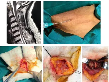
INFECTIONS IN SPINAL INSTRUMENTATION: A PROPOSAL FOR MANAGEMENT ALGORITHM USING CLOSED-SUCTION IRRIGATION SYSTEM AND VACUUM ASSISTED CLOSURE (VAC)
Erliano Sufarnap, Muhammad Faris, Amiril Mukminin, M. Dwikoryanto, Eko Agus Subagio
The management of surgical site infection on the instrumented spine is problematic. The instrumentation is required to stabilize or to maintain spinal deformity correction, and removal could be dangerous.
A case of infections in spinal instrumentation using close-suction irrigation system (CSIS) and vacuum assisted closure (VAC) revealed good result is reported by the authors .The authors also reviewed scientific literatures for the efficacy using close-suction irrigation system (CSIS) and vacuum assisted closure (VAC) on post operative spinal infection..
Fifteen scientific literatures consist of close-suction irrigation system and vacuum assisted closure (VAC) showed good outcome. The spinal instrumentation retained in 75 – 95 % after complete healing from infection. The studies revealed that early postoperative spinal infections were recover completely. These studies reported decreasing number of debridement and irrigation that mandatory in instrumented spinal infection treatment to 30 – 40 %. Duration of hospitalisation also decreased as well.
Cervical Spine Anterior Approach, Discectomy and Corpectomy
Ridha Dharmajaya
aa