Endocrine Pharmacology
Alteration in male reproductive system in experimental cholestasis: Roles for opioids
and nitric oxide overproduction
Samira Kiani
1, Behzad Valizadeh, Bahram Hormazdi, Hoda Samadi, Tahereh Naja
fi
,
Morteza Samini, Ahmad R. Dehpour
⁎
Department of Pharmacology, School of Medicine, Tehran University of Medical Sciences, P.O. Box 13145-784, Tehran, Iran
a b s t r a c t
a r t i c l e
i n f o
Article history:
Received 14 July 2008
Received in revised form 19 April 2009 Accepted 29 April 2009
Available online 13 May 2009
Keywords:
Endogenous opioid Nitric oxide Cholestasis Bile duct ligation Sex hormone LH FSH Testosterone Sperm analysis Male reproductive system (Rat)
Cirrhosis is associated with impairment of the male reproductive system, hypogonadism and feminization. It is important to rule out whether the impairment in the reproductive system exists earlier in the course of cholestatic liver disease to target effective therapies at the best time point. In this study we investigated the role of endogenous opioid and nitric oxide system in alterations of the reproductive system in male rats. We performed sham or bile duct ligation surgery on male Sprague–Dawley rats and treated the animals for seven days with saline, naltrexone, an opioid receptor blocker (20 mg/kg) and N (G)-nitro-L-arginine methyl ester (L-NAME), a nitric oxide synthase inhibitor (10 mg/kg). We then evaluated the plasma level of testosterone, luteinizing hormone (LH) and follicle stimulating hormone (FSH), sperm count and motility as well as biomarkers of cholestasis and nitric oxide productions. The results showed that following cholestasis, total testosterone level decrease and LH level increase in plasma of cholestatic rats and treatment with L-NAME and naltrexone could improve the plasma level of testosterone. Naltrexone could decrease the elevated level of LH in cholestatic animals. In addition, the weight of seminal vesicles and prostate significantly decreased in cholestasis as compared to the control group and treatment with L-NAME and naltrexone could improve the weights of the two organs in cholestasis. Our results demonstrate for the first time that the male reproductive system is impaired early in cholestasis and that endogenous opioid and nitric oxide system contribute to these impairments in the early course of the disease.
© 2009 Elsevier B.V. All rights reserved.
1. Introduction
The male reproductive system can be impaired by acute and chronic diseases of the other organs. It has been recognized that cirrhotic liver disease, irrespective of the underlying etiology, is associated with hypogonadism and feminization parallel with impairments in the serum level of sex hormones (Chen et al., 1995;
Van Thiel et al., 1985). Liverfibrosis and cirrhosis are associated with significant morbidity and mortality worldwide and there is no potent curative treatment for the disease or for the associated complications. Therefore, finding strategies to prevent the progress of associated pathologies early in the course of the disease is quite rational.
Endogenous opioid peptides, mainly methionine enkephalins, accumulate in plasma of cholestatic patients and animals (Thornton and Losowsky, 1998; Yurdaydin et al., 1998). Through interaction with their widespread receptors (mu, kappa and delta) they contribute to
the pathophysiology and manifestations of cholestatic liver disease (Swain et al., 1992; Gaskari et al., 2002). Several studies have reported that endogenous opioid peptides are important contributors to the mechanisms that influence the development and function of the testes (Fabbri et al., 1989). They also modulate sex hormone plasma levels, either via hypothalamic-pituitary axis or directly by their effects on testes (Engelhardt, 1989).
Cholestasis is also accompanied by nitric oxide (NO) overproduc-tion (Nahavandi et al., 2000; Mani et al., 2001). The high incidence of endotoxemia in liver disease and obstructive jaundice, triggers the induction of vascular NO synthesis, thereby leading to increased systemic NO overproduction and systemic hypotension (Vallance and Moncada, 1991). As a well recognized free radical, NO also modulates some of the biological events of the disease (Ebrahimi et al., 2006). Expression of nitric oxide synthase has been shown within Leydig cells (Tatsumi et al., 1997) and the produced nitric oxide can impair testicular steroidogenesis (Kostićet al., 1998).
The aim of the present study was to identify the alterations in the plasma level of sex hormones early in the course of cholestatic liver disease and detect some of the underlying mechanisms that may cause this alteration. With this aim, we have treated bile duct ligated (BDL) male rats with either naltrexone, an opioid receptor blockade,
–
⁎ Corresponding author. Tel.: +98 21 6112802; fax: +98 21 6402569.
E-mail address:[email protected](A.R. Dehpour). 1
Current affiliation: Department of Pulmonary and Critical Care, School of Medicine and Dentistry, University of Rochester, USA.
0014-2999/$–see front matter © 2009 Elsevier B.V. All rights reserved. doi:10.1016/j.ejphar.2009.04.049
Contents lists available atScienceDirect
European Journal of Pharmacology
or N (G)-nitro-L-arginine methyl ester (L-NAME), a nitric oxide synthase inhibitor for seven days.
2. Materials and methods
2.1. Reagents
All materials were purchased from Sigma unless otherwise stated in the text.
2.2. Animals
Adult male Sprague–Dawley rats (215–225 g, Pasteur Institute of Iran, Tehran, Iran) were used throughout this study. The animals were housed in a temperature-controlled room (23 ± 1 °C), on a 12-h regular light/dark cycle with a free access to standard laboratory rat chow and water. They were handled in accordance with the criteria outlined in the“Guide for the Care and Use of Laboratory Animals” (NIH US publication no. 85-23 revised 1985). All experiments were performed in line with the ethical considerations, recommended by the Pasteur Institute of Iran.
The rats were randomly divided into six groups, each consisted of age and weight matched rats (n= 6 in each group). Three groups were sham operated (control groups) and the other three groups under-went bile duct ligation (Gaskari et al., 2002; Cameron and Oakley, 1931). Briefly, midline laparotomy was performed under general anesthesia achieved by intraperitoneal (i.p.) injection of ketamine HCL (Gedoon Richter, Hungary), 50 mg/kg, and xylazine HCL (Bayer, Germany), 10 mg/kg (Ghaffari et al., 2004). The common bile duct was then exposed. In BDL rats, the bile duct became triply ligated with silk, 1 cm distal the duodenum to avoid the ligation of parabiliary pancreatic ductuli, then excised between two proximal ligations; whereas in sham-operated rats, the bile duct became manipulated and no ligation or resection was performed. Finally the abdominal wall was closed in two layers.
2.3. Drug administration and sample collection
One group of sham-operated and bile duct ligated rats was treated with daily subcutaneous administration of isotonic sterile saline solution (normal saline 1 ml/kg/day, s.c.). The second group of sham-operated and BDL rats received daily subcutaneous injection of naltrexone (20 mg/kg/day, s.c.) (Gaskari et al., 2002; Ghaffari et al., 2004) (Iran Daru, Tehran, Iran) for seven consecutive days and the third group received daily subcutaneous injections of L-NAME (10 mg/kg/day, s.c.). Seven days after cholestasis, the animals were sacrificed under sodium pentobarbital (50 mg/kg; i.p.) anesthesia, tissue samples were collected (see below) and blood samples were obtained via cardiac puncture.
2.4. Determination of plasma total bilirubin and liver enzyme activities
Total bilirubin concentration in plasma and the plasma alkaline phosphatase (ALP), alanine aminotransferase (ALT) and
gamma-glutamyl transpeptidase (γ-GT) activities in samples were determined
with commercially available kits (Ziestchemi, Tehran, Iran).
2.5. Plasma nitrite and nitrate concentration
The measurements were done according to the method by
Miranda et al. (2001)in 96 well plates and absorbance at 540 nm was measured using a standard plate reader. Fresh standard solutions of nitrate were included in each experiment.
2.6. Determination of plasma sex hormones
Plasma level of luteinizing hormone (LH), follicle stimulating hormone (FSH) and testosterone was evaluated with commercially available RIA kits by one of the reference laboratories in Iran. The plasma level of testosterone was determined by Spectria RIA kits (Orion Diagnostica, Finland) (Apter and Peter Eriksson, 2006). The analytical detection limit was 0.1 nmol/l and the specificity was around 4.5%.The plasma levels of LH and FSH were determined by RIA kits provided by Kavoshyar Company in Iran. All hormone measure-ments were done by a reference laboratory in Tehran, Iran. Standard curve was obtained according to the kit/manufacturer's instruction. All hormone estimations were performed in the same assay. Intra-assay variation wasb10%, and inter-assay wasb14%.
2.7. Determination of sperm count, motility and morphology
After seven consecutive days, rats were sacrificed and the abdo-men was opened by a longitudinal excision. First, 1 cm of the distal part of the left vas deferens was isolated and placed inside 5 cm3of a buffer solution (Sigma Hank's solution plus BSA 0.2%, glucose 0.09% and NaHCO3 0.35 g/l) in 37 °C for later determination of sperm motility (Wier and Rumberger, 1995; Seed et al., 1990).
Then, the ventral part of prostate, seminal vesicles, testes and epididymides were isolated and weighed with a precise scale. The right caudal epididymis was isolated, weighed, sliced completely, placed inside the buffer solution and incubated in 37 °C for later analysis of total sperm count. Total sperm count was calculated after dilution of right caudal epididymis samples with 0.5% formaldehyde and counting of
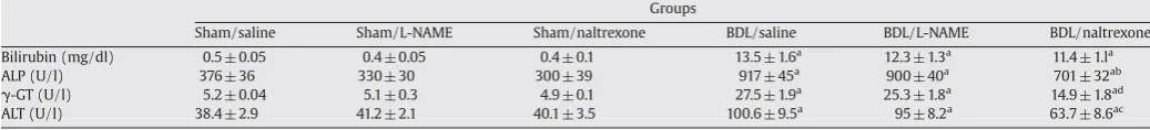
Table 1
Plasma levels of bilirubin, alkaline phosphatase activity (ALP), alanine transaminase activity (ALT) and gamma-glutamyl transpeptidase (γ-GT) in experimental groups.
Groups
Sham/saline Sham/L-NAME Sham/naltrexone BDL/saline BDL/L-NAME BDL/naltrexone Bilirubin (mg/dl) 0.5 ± 0.05 0.4 ± 0.05 0.4 ± 0.1 13.5 ± 1.6a 12.3 ± 1.3a 11.4 ± 1.la
ALP (U/l) 376 ± 36 330 ± 30 300 ± 39 917 ± 45a 900 ± 40a 701 ± 32ab
γ-GT (U/l) 5.2 ± 0.04 5.1 ± 0.3 4.9 ± 0.1 27.5 ± 1.9a 25.3 ± 1.8a 14.9 ± 1.8ad
ALT (U/l) 38.4 ± 2.9 41.2 ± 2.1 40.1 ± 3.5 100.6 ± 9.5a
95 ± 8.2a
63.7 ± 8.6ac
The table shows the plasma levels of bilirubin, alkaline phosphatase activity (ALP), alanine transaminase activity (ALT) and gamma-glutamyl transpeptidase (γ-GT) in sham and bile duct ligated (BDL) rats that were treated with naltrexone (20 mg/dl), L-NAME (10 mg/dl) or saline. a:pb0.001 compared with control groups; b:pb0.01 compared with the BDL/
saline group; c:pb0.05 compared with the BDL/saline group; d:pb0.001 compared with the BDL/saline group.
Fig. 1.Plasma nitrite + nitrate levels in sham-operated and BDL rats given naltrexone, L-NAME or saline are shown above. Plasma level of nitrite +nitrate increased by bile duct ligation and both L-NAME and naltrexone could decrease this elevation.⁎:pb0.001 vs all sham and⁎⁎:pb0.001 vs BDL/saline. Data are shown as mean ± S.E.M.
sperms under light microscopy according to the method described previously (Seed et al., 1990).
Sperm motility was also determined under light microscopy with the magnitude of 10, from a small portion of the vas deferens transferred from the buffer solution to a slide. The immobile sperms were counted. Then the slide wasfixed and the whole number of sperms in the slide was counted.
Sperm morphology was determined with the same samples used for sperm motility study. Slides were prepared from vasa deferentia, air dried and studied by light microscopy at the magnification of 40×. Three hundred sperms were observed. According to previous studies (Seed et al., 1990), the percentage of abnormality in rat sperm is low; therefore, we considered two major and widely accepted groups of normal and abnormal sperms and evaluated sperm morphology based on the ratio of abnormal sperm to normal sperm.
2.8. Statistical analysis
All data are expressed as mean ± S.E.M. Statistical evaluation of data was performed using analysis of variances (ANOVA), followed by the Tukey post hoc test. p-values less than 0.05 were considered statistically significant.
3. Results
3.1. General characteristics
Seven days after bile duct ligation or sham surgery, the survival rate of all three groups of sham-operated rats was 100%. L-NAME and naltrexone treated control rats had a decrease in the amount of their
food intake as compared to the saline treated one (pb0.05). No other
parameter significantly changed. However, 83% of the BDL rats in all groups of treatment survived to seven days (five rats in each group). These rats had jaundice and the color of their urine was intense yellow compared to the weak-yellow urine of the sham groups. No abdominal distension or ascites were noticed in BDL animals. Their food intake
and body weight were significantly less than the sham groups
(pb0.01).
Moreover, there was no obvious difference in the general appearance of naltrexone or L-NAME treated rats compared to the saline treated BDL rats. They still were jaundiced and had dark urine. Also, prolonged drug treatment did not influence the decreased weight and daily food intake in BDL animals.
3.2. Plasma bilirubin level and liver enzyme activities
After one week, plasma bilirubin and liver enzymes (ALT, ALP, and
γ-GT) were significantly elevated in BDL rats compared with sham
controls consistent with biliary obstruction (Table 1). Daily adminis-tration of naltrexone significantly attenuated the increase in plasma ALT, ALP, andγ-GT activities. However, this effect was not observed in
the BDL group treated with L-NAME which did not show significant alteration of liver enzymes as compared to the saline treated ones. Plasma bilirubin, a serum marker of cholestasis, was virtually identical in BDL/saline; BDL/naltrexone and BDL/L-NAME groups (Table 1). Treatment with L-NAME did not significantly alter the plasma level of liver enzymes and bilirubin in BDL animals as compared to the BDL/ saline group.
3.3. Plasma nitrite + nitrate concentration
Plasma nitrite + nitrate level significantly increased after bile duct ligation (Fig. 1). Prolonged treatment with L-NAME could significantly decrease this elevation as compared to the saline treated group (53.50 ± 8.4 vs 29.9 ± 4.8, BDL/saline, BDL/L-NAME, respec-tively, pb0.05). Prolonged treatment with naltrexone could also
decrease the nitrite + nitrate level in BDL rats as compared to the saline treated BDL group. The level of nitrite + nitrate in drug treated BDL animals was not significantly different from the appropriate control groups.
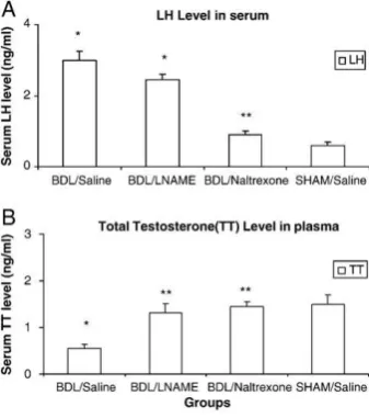
3.4. Sex hormone alterations
As shown inFig. 2, seven days after bile duct ligation the plasma level of total testosterone decreased in BDL rats as compared to the sham-operated ones (Fig. 2) (1.5 ± 0.2 vs 0.55 ± 0.09, in sham/saline and BDL/saline respectively,pb0.05). Prolonged treatment with both
naltrexone and L-NAME could significantly inhibit this decrease in BDL treated rats (pb0.05).
Bile duct ligation was also accompanied by a significant increase in plasma LH level (3 ± 0.25 vs 0.6 ± 0.09 in BDL/saline and sham/saline respectively, pb0.05). Prolonged treatment with naltrexone could
significantly decrease the LH level in BDL rats, but this effect was not observed in L-NAME treated BDL rats.
Fig. 2.The level of luteinizing hormone (LH) and total testosterone (TT) changed significantly in BDL animals as compared to the sham treated ones. Please note that the values of sham/L-NAME and sham/naltrexone were not different from sham/saline groups so to avoid the repetition of data these have been omitted in thefigures and are only mentioned in the text.⁎and ⁎⁎arepb0.05 vs sham/saline and BDL/saline respectively. Data are shown as mean ± S.E.M.
Table 2
Ratio of the weights of prostate, seminal vesicles, epididymis and testes to body weight in experimental groups.
Group
Sham/saline Sham/L-NAME Sham/NTX BDL/saline BDL/L-NAME BDL/NTX
Prostate 9.3 ± 0.6 8.1 ± 0.5 8.0 ± 0.8 6.0 ± 0.3a
9.0 ± 0.8b
9.0 ± 0.9b
Seminal vesicles 7.9 ± 0.4 6.9 ± 0.3 6.8 ± 0.3 5.1 ± 0.4a
7.5 ± 0.7b
7.7 ± 0.5b
Epididymis 28.7 ± 0.6 28.1 ± 0.7 28.8 ± 0.8 28.9 ± 0.5 27.2 ± 1.0 27.8 ± 0.8
Testes 101.7 ± 2.5 101.5 ± 2.9 103.5 ± 2.3 103.3 ± 4.0 102.3 ± 5.9 106.5 ± 5.2
Bile duct ligation was accompanied by a significant decrease in the weight of prostate and seminal vesicles in animals. Treatment with L-NAME and naltrexone could slow down the atrophy in these two organs. No other significant changes were observed in the other organs following cholestasis and treatments. Data are shown as mean ± S.E.M. a:pb0.05 as compared to sham/saline, b:pb0.05 as compared to the BDL/saline group.
The plasma level of FSH did not change significantly following bile duct ligation and the treatment with L-NAME and naltrexone could not change this value either (data not shown). Neither of the treatments could affect the plasma level of hormones in sham treated animals compared to saline treatment.
3.5. Sperm count, motility and morphology
Bile duct ligation did not have any significant effect on sperm count, motility and morphology in BDL and sham-operated rats and naltrexone and L-NAME treatments did not significantly change these parameters (insignificant data, not shown). The weight of seminal vesicles and prostate significantly decreased in BDL animals as compared to control ones, treatment with L-NAME and naltrexone, could increase the weight of both organs in BDL animals, but did not have any significant effects on sham-operated animals (Table 2).
4. Discussion
It has been proposed that both overproduction of opioids (Swain et al., 1992) and retention due to impaired biliary excretion (Thornton and Losowsky, 1998), may contribute to the accumulation of opioid peptides in liver diseases. The accumulation of circulating opioids is expected in different models of cholestasis in which there is an impaired hepatic excretion of bile as the liver is the main route for the elimination of endogenous opioid peptides via their excretion into the bile (Thornton and Losowsky, 1998). Therefore, the elevated plasma opioid peptides have been shown in animals and patients with cholestasis (Swain et al., 1992, 1993; Spivey et al., 1994).
ALT is a cytosolic enzyme of the hepatocytes; so its increased concentration in plasma reflects hepatocyte injury (Plaa and Hewitt, 1982).γ-GT and ALP are enzymes embedded in the hepatocyte plasma
membranes. Damage to the cell membranes and thus, injury to the liver can raise their level in plasma (Plaa and Hewitt, 1982; Bulle et al., 1990). Liver injury during cholestasis also comes with systemic oxidative stress (Ljubuncic et al., 2000; Ottesen et al., 2001). It has been shown that opioids increase free radical formation, enhance NO overproduction and cause systemic oxidative in cholestasis (James et al., 1982; Zhang et al., 2004). Our previous works and the current data have shown that naltrexone treatment leads to a decrease in systemic NO overproduction (Kiani et al., 2007). It also reduces liver injury due to oxidative stress by improving the anti-oxidant capacity of the liver (Ebrahimkhani et al., 2006). This improvement of liver injury is best manifested by the decline in plasma liver enzymes after naltrexone treatment in cholestasis.
Endogenous opioids are involved in different manifestations of liver disease such as pruritus (Bergasa et al., 1995), ascites (Thornton et al., 1998), and hyperdynamic circulation (Gaskari et al., 2002). To our knowledge, data regarding the participation of endogenous opioids in the disturbance of sex hormones during cholestasis are lacking in the literature. In this study, we identified a new role for endogenous opioid system in the process of sex hormone alteration, which occurs in the cholestatic liver.
There are different studies which suggest that opioids, either administered exogenously or produced endogenously, can modulate the hypothalamic-pituitary-gonadal axis (HHG axis) or can exert direct effects on the testes. More precisely, it has been shown that endogenous opioids can inhibit the secretion of gonadotropins by suppressing the release of gonadotropin releasing hormone (GnRH) or can act directly on the hypophysial level by their cell specific receptors on gonadotrophs (Fabbri et al., 1989). They can be produced locally in testes where they exert their paracrine and autocrine roles (Engelhardt, 1989) and disrupt two major aspects of testicular function: testosterone secretion by Leydig cells and testicular interstitial fluid formation (Adams et al., 1993). Other testicular elements such as Sertoli cell growth and secretion of rat androgen
binding protein are also shown to be modulated by the opioid system (Gerendai et al., 1984, 1986).
The observed reduction in the plasma level of testosterone and the resulting elevation of the LH level in BDL rats demonstrate a primary defect in testosterone production by testes and propose the effect of endogenous opioids at the testicular level, in this setting. Improve-ment of the level of these hormones after prolonged opioid receptor blockade with naltrexone treatment further supports the contribution of endogenous opioids.
To further investigate the effect of biochemical markers of cholestasis in the male reproductive system, we treated our animals with L-NAME to reduce NO over production. It has been shown that NO overproduction occurs during cholestasis (Chu et al., 1997; Marley et al., 1999). This has been also observed in our study as the increase in plasma nitrite + nitrate levels. It has been shown that administration of NO donors to Leydig cells results in the inhibition of steroidogenesis by those cells directly by inhibiting the cholesterol side-chain cleavage enzyme (cytochromeP450scc) (Del Punta et al., 1996). So, this might be another explanation of the observed decrease in testosterone level in rats following cholestasis and the NO overproduction.
On the other hand, our results show an interesting effect of L-NAME treatment on the LH level in rats. Prolonged L-NAME treatment could not decrease the elevated level of LH in BDL rats while it could increase the level of testosterone in those rats. Our data suggest that the primary effect of bile duct ligation is at the level of Leydig cells and the increase of LH is secondary to the decrease in circulating testosterone. One interpretation is that L-NAME has only a partial effect on the NO-inhibited Leydig cells which can produce normal levels of testosterone after being stimulated by an increased level of LH.
The other interpretation is based on the complex effect of NO on gonadotropin secretion (Pu et al., 1996; Moretto et al., 1993). It is believed that NO modulates GnRH-mediated gonadotropin secretion independent of the gonadal hormone level in female rats (Barnes et al., 2002). It has also been suggested that the endogenous level of NO may determine the sensitivity of GnRH-stimulated gonadotropin released by the anterior pituitary. Our treatment with L-NAME for seven days chronically decreases the level of NO and interferes with the sensitivity of gonadotropin secretion in response to plasma testosterone level. This might explain the reason why the expected decrease in LH following changes in testosterone level is not observed. Other factors such as duration of our treatment, the animal model used in the study, the male reproductive system as compared to female ones and the half-life of LH and FSH (LH: 20 min, FSH: 3–4 h) might be other factors that contribute to the diversity in responses of gonadotropins to L-NAME.
We could not detect any significant difference in sperm count, motility and morphology between BDL and sham-operated rats in the study and treatments did not influence them either. There is a major role for FSH in the initial phase of spermatogenesis restoration such as in the facilitation of spermatogenesis restoration by testosterone, particularly on spermatogonial number and spermatocyte maturation (Meachem et al., 1998). There was no significant difference in the level of FSH after bile duct ligation and this can suggest and add strength to ourfinding of unchanged spermatogenesis following cholestasis. We should mention that physiological concentration of NO is required for adequate sperm motility (Lewis et al., 1996). But increased NO level in the semen of infertile males is accompanied by decreased sperm motility (Nobunaga et al., 1996). Addition of sodium nitroprusside decreases the percentage of mobile sperms, their speed of motion and the concentration of mobile cells in vitro (Weinberg et al., 1995). So, it was expected that NO overproduction in cholestasis influences sperm motility. But as NO half-life is very short, it is speculated that NO was degraded from the time of sampling until measurements (approxi-mately 15 min) and therefore, could not affect sperm motility in the study. So, we conclude that NO overproduction during cholestasis does not have any permanent effect on sperm motility.
In addition, the weights of prostate and seminal vesicles decreased following cholestasis in the study. Treatment with naltrexone and L-NAME both could inhibit the occurrence of atrophy in the organs. It is known that prostate and seminal vesicles are androgen-dependent secretory glands of the male genital tract. Androgenic ablation, results in rapid epithelial cell apoptosis and parenchymal collapse in prostate and seminal vesicles in adults. This is followed by a slower stromal atrophy in these organs (Banerjee et al., 1995). In the present study, cholestasis was associated with impaired testosterone secretion; so, the observed atrophy of these organs is well justified by a decreased testosterone level following cholestasis. It has been shown that testosterone replacement restores the original architecture of prostate and seminal vesicles and increases the weight of these organs (Justulin et al., 2006). The same finding in our study following naltrexone and L-NAME treatment, therefore, can be well attributed to changes in testosterone level in cholestatic rats and suggests a significant therapeutic role for them in this setting.
It should be stated that this study shows a primary alteration in hormonal activity in male rats, seven days after bile duct ligation and in an acute model of injury. This certainly does not rule out the possible contribution of the central mechanism later in the evolve-ment of chronic disease or in other models of liver injury that do not show a characteristic disruption of bileflow of the bile duct ligation model (Gonzales et al., 2007). Further studies are warranted to clarify this issue.
Our findings are potentially important in clinical practice. Naltrexone is prescribed commonly to treat cholestasis associated pruritus. Therefore, it can be used safely in this setting to prevent the dys-regulation of sex hormones earlier in the course of the disease. The alterations in sex hormone levels following cholestasis are important and if left unmanaged, can lead to significant consequences such as impaired libido, impotence and gonadal atrophy in cirrhotic patients. The potential effect of opioid receptor blockade on the improvement of sex hormone alterations has experimental and clinical implications, which warrants further studies.
References
Adams, M.L., Sewing, B., Forman, J.B., Meyer, E.R., Cicero, T.J., 1993. Opioid-induced suppression of rat testicular function. J. Pharmacol. Exp. Ther. 266 (1), 323–328. Apter, S.J., Eriksson, C.J. Peter, 2006. The role of social isolation in the effects of alcohol
on corticosterone and testosterone levels of alcohol-preferring and non-preferring rats. Alcohol Alcohol. 41 (1), 33–38.
Banerjee, P.P., Banerjee, S., Tilly, K.I., Tilly, J.L., Brown, T.R., Zirkin, B.R., 1995. Lobe specific apoptotic cell death in rat prostate after androgen ablation by castration. Endocrinology 136, 4368e76.
Barnes, M.J., Lapanowski, K., Rafols, J.A., Lawson, D.M., Dunbar, J.C., 2002. Chronic nitric oxide deficiency is associated with altered leutinizing hormone and follicle-stimulating hormone release in ovariectomized rat. Exp. Biol. Med. 227, 817–822. Bergasa, N.V., Alling, D.W., Talbot, T.L., Swain, M.G., Yurdaydin, C., Turner, M.L., Schmitt, J.M.,
Walker, E.C., Jones, E.A.,1995. Effects of naloxone infusions in patients with the pruritus of cholestasis. A double-blind, randomized, controlled trial. Ann. Intern. Med. 123, 161–167.
Bulle, F., Mavier, P., Zafrani, E.S., Preaux, A.M., Lescs, M.C., Siegrist, S., Dhumeaux, D., Guellaën, G., 1990. Mechanism of gamma-glutamyl transpeptidase release in serum during intrahepatic and extrahepatic cholestasis in the rat. A histochemical, biochemical and molecular approach. Hepatology 11, 545–550.
Cameron, G.R., Oakley, C.L., 1931. Ligation of the common bile duct. J. Pathol. Bacteriol. 35, 769–798.
Chen, J., Murray, M., Liddle, C., Jiang, X.M., Farrell, G.C., 1995. Down regulation of male-specific cytochrome P450s 2C11 and 3A2 in bile duct-ligated male rats: importance to reduced hepatic content of cytochrome P450 in cholestasis. Hepatology 22 (2), 580–587.
Chu, C.J., Lee, F.Y., Wang, S.S., Chang, F.Y., Tsai, Y.T., Lin, H.C., Hou, M.C., Wu, S.L., Tai, C.C., Lee, S.D., 1997. Hyperdynamic circulation of cirrhotic rats: role of substance P and its relationship to nitric oxide. Scand. J. Gastroenterol. 32 (8), 841–846.
Del Punta, K., Charreau, E.H., Pignataro, O.P., 1996. Nitric oxide inhibits Leydig cell steroidogenesis. Endocrinology 137 (12), 5337–5343.
Ebrahimi, F., Tavakoli, S., Hajrasouliha, A.R., Sadeghipour, H., Dehghani, M., Ahmadi, S.H., Dehpour, A.R., 2006. Involvement of endogenous opioid peptides and nitric oxide in the blunted chronotropic and inotropic responses to beta-adrenergic stimulation in cirrhotic rats. Fundam. Clin. Pharmacol. 20 (5), 461–471.
Ebrahimkhani, M.R., Kiani, S., Oakley, F., Kendall, T., Shariftabrizi, A., Tavangar, S.M., Moezi, L., Payabvash, S., Karoon, A., Hoseininik, H., Mann, D.A., Moore, K.P., Mani, A.R.,
Dehpour, A.R., 2006. Naltrexone, an opioid receptor antagonist, attenuates liverfibrosis in bile duct ligated rats. Gut 55 (11), 1606–1616 (Nov).
Engelhardt, R.P., 1989. Gonadal opioids and testicular function. Ann. Endocrinol. (Paris) 50 (1), 64–72.
Fabbri, A., Jannini, E.A., Gnessi, L., Ulisse, S., Moretti, C., Isidori, A., 1989. Neuroendocrine control of male reproductive function. The opioid system as a model of control at multiple sites. J. Steroid Biochem. 32 (1B), 145–150.
Gaskari, S.A., Mani, A.R., Ejtemaei-Mehr, S., Namiranian, K., Homayoun, H., Ahmadi, H., Dehpour, A.R., 2002. Do endogenous opioids contribute to the bradycardia of rats with obstructive cholestasis? Fundam. Clin. Pharmacol. 16, 273–279.
Gerendai, I., Shaha, C., Thau, R., Bardin, C.W., 1984. Do testicular opiates regulate Leydig cell function? Endocrinology 115 (4), 1645–1647.
Gerendai, I., Shaha, C., Gunsalus, G.L., Bardin, C.W., 1986. The effects of opioid receptor antagonists suggest that testicular opiates regulate Sertoli and Leydig cell function in the neonatal rat. Endocrinology 118 (5), 2039–2044.
Ghaffari, K., Savadkuhi, S.T., Honar, H., Riazi, K., Shafaroodi, H., Moezi, L., Ebrahimkhani, M.R., Tahmasebi, M.S., Dehpour, A.R., 2004. Obstructive cholestasis alters intestinal transit in mice: role of opioid system. Life Sci. 76, 397–406.
Gonzales, P.H., Rhoden, C.R., Luz, C., Corrêa, G., Barbosa-Coutinho, L.M., Oliveira, M.C., 2007. Male gonadal function, prolactin secretion and lactotroph population in an experimental model of cirrhosis. Braz. J. Med. Biol. Res. 40 (10), 1383–1388. James, R.C., Goodman, D.R., Harbison, R.D., 1982. Hepatic glutathione and
hepatotoxi-city: changes induced by selected narcotics. J. Pharmacol. Exp. Ther. 221, 708–714. Justulin, L.A., Ureshino, R.P., Zanoni, M., Felisbino, S.L., 2006. Differential proliferative response of the ventral prostate and seminal vesicle to testosterone replacement. Cell Biol. Int. 30 (4), 354–364 (Apr).
Kiani, S., Ebrahimkhani, M.R., Shariftabrizi, A., Doratotaj, B., Payabvash, S., Riazi, K., Dehghani, M., Honar, H., Karoon, A., Amanlou, M., Tavangar, S.M., Dehpour, A.R., 2007. Opioid system blockade decreases collagenase activity and improves liver injury in a rat model of cholestasis. J. Gastroenterol. Hepatol. 22 (3), 406–413. Kostić, T., Andrić, S., Kovacević, R., Marić, D., 1998. The involvement of nitric oxide in
stress-impaired testicular steroidogenesis. Eur. J. Pharmacol. 346 (2–3), 267–273. Lewis, S.E., Donnelly, E.T., Sterling, E.S., Kennedy, M.S., Thompson, W., Chakravarthy, U.,
1996. Nitric oxide synthase and nitrite production in human spermatozoa: evidence that endogenous nitric oxide is beneficial to sperm motility. Mol. Hum. Reprod. 2 (11), 873–878.
Ljubuncic, P., Tanne, Z., Bomzon, A., 2000. Evidence of a systemic phenomenon for oxidative stress in cholestatic liver disease. Gut 47, 710–716.
Mani, A.R., Nahavandi, A., Mani, A.H., Dehpour, A.R., 2001. Role of nitric oxide in hypodipsia of rats with obstructive cholestasis. J. Pharm. Pharmacol. 53, 277–281. Marley, R., Ho, S., Fernando, B., 1999. Lipoic acid prevents development of the
hyperdynamic circulation in anesthetized rats with biliary cirrhosis. Hepatology 29 (5), 1358–1363.
Meachem, S.R., Wreford, N.G., Stanton, P.G., Robertson, D.M., McLachlan, R.I., 1998. Follicle-stimulating hormone is required for the initial phase of spermatogenic restoration in adult rats following gonadotropin suppression. J. Androl. 19, 725–735. Miranda, K.M., Espey, M.G., Wink, D.A., 2001. A rapid, simple spectrophotometric method for simultaneous detection of nitrate and nitrite. Nitric Oxide 5, 62–71. Moretto, M., Lopez, F.J., Negro-Vilar, A., 1993. Nitric oxide regulates luteinizing
hormone-releasing hormone secretion. Endocrinology 133, 2399–2402. Nahavandi, A., Dehpour, A.R., Mani, A.R., Homayounfar, H., Abdoli, A., Abolhosseini, M.R.,
2000. The role of nitric oxide overproduction in bradycardia of rats with obstructive cholestasis, Eur. J. Pharmacol. 411, 135–141.
Nobunaga, T., Tokugawa, Y., Hashimoto, K., Kubota, Y., Sawai, K., Kimura, T., Shimoya, K., Takemura, M., Matsuzaki, N., Azuma, C., Saji, F., 1996. Elevated nitric oxide concentration in the seminal plasma of infertile males, nitric oxide inhibits sperm motility. Am. J. Rep. Immunol. 36 (4), 197-7.
Ottesen, L.H., Harry, D., Frost, M., Davies, S., Khan, K., Halliwell, B., Moore, K., 2001. Increased formation of S-nitrothiols and nitrotyrosine in cirrhotic rats during endotoxemia. Free Radic. Biol. Med. 31, 790–798.
Plaa, G.L., Hewitt, W.R., 1982. Detection and evaluation of chemically induced liver injury. Principles and Methods of Toxicology. InRaven Press, New York, pp. 407–445.
Pu, S., Xu, B., Kalra, S.P., Kalra, P.S., 1996. Evidence that gonadal steroids modulate nitric oxide efflux in the medial preoptic area: effects ofN-methyl-D-aspartate and correlation with luteinizing hormone secretion. Endocrinology 137, 1949–1955. Seed, J., Chapin, R.E., Clegg, E.D., Dostal, L.A., Foote, R.H., Hurtt, M.E., Klinefelter, G.R.,
Makris, S.L., Perreault, S.D., Schrader, S., Seyler, D., Sprando, R., Treinen, K.A., Veeramachaneni, D.N., Wise, L.D., 1990. Methods for assessing sperm motility, morphology and counts in rats, rabbit and dog: a consensus report. ILSI Risk Science Institute Expert Working Group on Sperm Evaluation. Reprod. Toxicol. 10 (3), 237–244.
Spivey, J.R., Jorgensen, R.A., Gores, G.J., Lindor, K.D., 1994. Methionine-enkephalin concentrations correlate with stage of disease but not pruritus in patients with primary biliary cirrhosis. Am. J. Gastroenterol. 89, 2028–2032.
Swain, M.G., Rothman, R.B., Xu, H., Vergalla, J., Bergasa, N.V., Jones, E.A., 1992. Endogenous opioids accumulate in plasma in a rat model of acute cholestasis. Gastroenterology 103, 630–635.
Swain, M.G., Vergalla, J., Bergasa, N.V., Jones, E.A., 1993. Sympathetic nerves, but not the adrenal gland, contribute to elevated plasma levels of met-enkephalin in rats with acute cholestatic hepatitis. Regul. Pept. 46, 535–542.
Tatsumi, N., Fujisawa, M., Kanzaki, M., Okuda, Y., Okada, H., Arakawa, S., Kamidono, S., 1997. Nitric oxide production by cultured rat Leydig cells. Endocrinology 138 (3), 994–998.
Thornton, J.R., Losowsky, M.S., 1998. Methionine enkephalin is increased in plasma in acute liver disease and is present in bile and urine. J. Hepatol. 8, 53–59.
Thornton, J.R., Dean, H., Losowsky, M.S., 1998. Is ascites caused by impaired hepatic inactivation of blood borne endogenous opioid peptides? Gut 29, 1167–1172. Vallance, P., Moncada, S., 1991. Hyperdynamic circulation in cirrhosis: a role of nitric
oxide? Lancet 337, 776–778.
Van Thiel, D.H., Gavaler, J.S., Zajko, A.B., Cobb, C.F., 1985. Consequences of complete bile-duct ligation on the pubertal process in the male rat. J. Pediatr. Gastroenterol. Nutr. 4 (4), 616–621.
Weinberg, J.B., Doty, E., Bonaventura, J., Haney, A.F., 1995. Nitric oxide inhibition of human sperm motility. Fertil. Steril. 64 (2), 408–413.
Wier, P.J., Rumberger, D., 1995. Isolation of rat sperm from the vas deferens for sperm motion analysis. Reprod. Toxicol. 9 (3), 327–330.
Yurdaydin, C., Karavelioglu, D., Onaran, O., Celik, T., Yasa, M.H., Uzunalimoglu, O., 1998. Opioid receptor ligands in human hepatic encephalopathy. J. Hepatol. 29, 796–801. Zhang, Y.T., Zheng, Q.S., Pan, J., Zheng, R.L., 2004. Oxidative damage of biomolecules in mouse liver induced by morphine and protected by antioxidants. Basic Clin. Pharmacol. Toxicol. 95, 53–58.