M A J O R A R T I C L E
Evaluation of a Newly Developed Lateral Flow
Immunoassay for the Diagnosis of Cryptococcosis
Mark D. Lindsley,1Nanthawan Mekha,2Henry C. Baggett,3Yupha Surinthong,2Rinrapas Autthateinchai,2
Pongpun Sawatwong,3Julie R. Harris,1Benjamin J. Park,1Tom Chiller,1S. Arunmozhi Balajee,1and
Natteewan Poonwan2
1Mycotic Diseases Branch, Centers for Disease Control and Prevention, Atlanta, Georgia;2National Institute of Health, Department of Medical Sciences, Ministry of Public Health, Nonthaburi, Thailand; and3International Emerging Infections Program, Thailand Ministry of Public Health–Centers for Disease Control and Prevention Collaboration, Nonthaburi, Thailand
Background. Cryptococcosis is a common opportunistic infection of human immunodeficiency virus (HIV)– infected individuals mostly occurring in resource-limited countries. This study compares the performance of a recently developed lateral flow immunoassay (LFA) to blood culture and enzyme immunoassay (EIA) for the diagnosis of cryptococcosis.
Methods. Archived sera from 704 HIV-infected patients hospitalized for acute respiratory illness in Thailand were tested for cryptococcal antigenemia using EIA. All EIA-positive and a subset of EIA-negative sera were tested by LFA, with results recorded after 5 and 15 minutes incubation. Urine from patients with LFA- and EIA-positive sera was tested by LFA. Antigen results from patients with positive cryptococcal blood cultures were compared.
Results. Of 704 sera, 92 (13%) were positive by EIA; among the 91 EIA-positive sera tested by LFA, 82 (90%) and 87 (96%) were LFA positive when read after 5 and 15 minutes, respectively. Kappa agreement of EIA and LFA for sera was 0.923 after 5 minutes and 0.959 after 15 minutes, respectively. Two of 373 EIA-negative sera were LFA positive at both time points. Of 74 urine specimens from EIA-positive patients, 52 (70.3%) were LFA positive. EIA was positive in 16 of 17 sera from blood culture–positive patients (94% sensitivity), and all sera were positive by LFA (100% sensitivity).
Conclusions. A high level of agreement was shown between LFA and EIA testing of serum. The LFA is a rapid, easy-to-perform assay that does not require refrigeration, demonstrating its potential usefulness as a point-of-care assay for diagnosis of cryptococcosis in resource-limited countries.
Cryptococcosis, caused byCryptococcusspp., is one of the most common opportunistic infections among human immunodeficiency virus (HIV)–infected individuals [1]. Globally, an estimated 1 million new cases of crypto-coccal meningitis occur per year, with more than 600 000 deaths. An estimated 88% of global cases and more than
90% of deaths from cryptococcal meningitis occur in sub-Saharan Africa and Southeast Asia [2]. Primary re-spiratory illness due toCryptococcus, while uncommon in the United States and Europe, is more common in other regions, such as Southeast Asia [2].
Currently, cryptococcal diagnostics include micros-copy and/or culture-based methods, or detection of cryptococcal antigen (CrAg) in body fluids using either latex agglutination (LA) or enzyme immunoassay (EIA) methods. Although culturing of the organism is con-sidered the gold standard diagnostic method, it has poor sensitivity, requires a large quantity of specimen, and requires laboratory infrastructure including electricity (for centrifugation). Antigen tests such as LA or EIA performed on cerebral spinal fluid or serum are highly sensitive and specific diagnostic options that are less labor- and time-intensive than culture [3, 4]. However,
Received 4 March 2011; accepted 29 April 2011.
Correspondence: S. Arunmozhi Balajee, MD, PhD, Mycotic Diseases Branch, Centers for Disease Control and Prevention, Mailstop G11, 1600 Clifton Rd, Atlanta, GA 30333 ([email protected]).
Clinical Infectious Diseases 2011;53(4):321–325
Published by Oxford University Press on behalf of the Infectious Diseases Society of America 2011. This is an Open Access article distributed under the terms of the Creative Commons Attribution Non-Commercial License (http://creativecommons. org/licenses/by-nc/2.5/), which permits unrestricted non-commercial use, distribution, and reproduction in any medium, provided the original work is properly cited. 1058-4838/2011/534-0001$14.00
these methods require refrigeration, a cold chain for specimen transport, and technical expertise; therefore, they are often performed only in reference/diagnostic labs far removed from patients, potentially limiting their clinical utility. In addition, the costs of these tests are not affordable for many clinics. As a re-sult, cryptococcosis often goes undiagnosed in resource-limited countries.
Point-of-care tests (POCTs) show promise for enabling di-agnosis of infectious diseases in remote care centers in low-resource countries. POCTs are assays that can produce results quickly, are simple to perform and interpret by personnel with no or minimal laboratory training, and ideally can be used without cold chain or advanced laboratory equipment [5]. To-day, reliable and affordable POCTs are available for the de-tection of hepatitis B infection [6], HIV infection [7], malaria [8, 9], syphilis [10], cholera [11], and some neglected tropical diseases [12–14]. Additional advantages of POCTs include re-duction of patient anxiety and waiting time and decreases in patient loss to follow-up [15]. To serve the communities most in need, the World Health Organization (WHO) recommended that POCTs should be Affordable, Sensitive, Specific, User-friendly, Rapid, Equipment-free, and Delivered to those who need it (ASSURED) [16, 17].
In 2009, a lateral flow immunoassay (LFA) for the detection of cryptococcal antigen was developed by IMMY (Immuno-Mycologics) as a potential POCT for diagnosis of cryptococcal infection. The LFA is stable at room temperature, has a rapid turnaround time, requires very little technical skill, and can be performed with minimal laboratory infrastructure. In this study, the performance of the LFA for CrAg detection was evaluated by testing archived specimens from HIV-infected patients hospi-talized with acute respiratory illness in Thailand, and comparing these results with culture and EIA.
METHODS
The serum and urine specimens used in this study were collected as part of a pneumonia etiology study that included HIV-infected patients in Thailand, described elsewhere [18]. In brief, patients admitted to 1 of 8 hospitals in Sa Kaeo or 12 hospitals in Nakhon Phanom provinces in Thailand between 2004 and 2009 were enrolled if they displayed any sign or symptom of active infection (temperature.38.2°C or,35.5°C within 24 hours of admission; chills; abnormal total white blood cell count or dif-ferential), and had evidence of lower respiratory illness (ab-normal breath sounds, tachypnea, cough, sputum production, or dyspnea). All patients provided informed consent for sample collection and use of samples for research purposes. Serum and urine specimens were collected soon after hospital admission and immediately after study consent. This study was approved by the Institutional Review Board of the Centers for Disease
Control and Prevention and the Ethical Review Committee of the Thailand Ministry of Health.
All testing was performed at the National Institute of Health, Nonthaburi, Thailand. The results of the LFA were compared with those obtained from 2 commonly used diagnostic tests for detectingCryptococcusinfection, blood culture and EIA. Blood cultures were obtained from a subset of study participants as a part of routine clinical care. Cultures were processed by au-tomated BacTAlert blood culture system (bioMe´rieux), and pathogen identification of isolates from positive blood cultures was performed using standard microbiology methods (mor-phological and physiological tests).
Stored sera from all HIV-infected patients enrolled in the study from 2004 to 2009 were tested by EIA, using the Premier Cryptococcal Antigen enzyme immunoassay (Meridian Bio-sciences). LFA testing was performed on serum specimens from all patients with a positive serum EIA test and a random subset of patients with a negative serum EIA test. Additionally, urine from patients whose sera were positive by LFA and EIA were tested by LFA. No LFA testing was performed on urine of pa-tients who had serum that tested EIA-negative.
The EIA was performed according to the manufacturer’s protocol using 50lL of specimen. For the EIA, wells with
re-actions of optical densities,0.07 units were considered negative;
$0.07 to,0.100 was considered indeterminate; and$0.100 was considered positive. The LFA is a semiquantitative test system for the detection of capsular polysaccharide antigens ofCryptococcus
species complex (Cryptococcus neoformans and Cryptococcus gattii). The LFA kit consists of immunochromatic test strips impregnated with monoclonal antibodies optimized to detect all 4 cryptococcal serotypes and a diluent. The LFA kit can be stored at room temperature for up to a year. To perform the assay, 20lL of patient specimen was mixed with 2 drops of diluent
in a 2-mL screw cap microtube (Sarstedt AG). The LFA strip was placed in the specimen and diluent cocktail and in-cubated at room temperature. Results were read after two incubation times: 5 minutes according to the manufacturer’s instructions and a prolonged incubation time of 15 minutes. The presence of 2 bands (control band and test band) in the test zone of the LFA strip was interpreted as a positive result and a single band in the test zone (control band) was interpreted as a negative result.
RESULTS
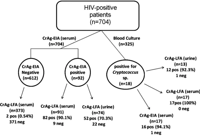
A total of 704 HIV-infected study participants with available specimens were identified and were tested as outlined in Figure 1. Blood culture was performed on 325 patient specimens, with 18 of 325 yieldingC. neoformans. Serum was not available from 1 of the 18 culture-positive patients for analyses in the study; thus the EIA testing was performed on 17 culture-positive sera. Of the 17 serum specimens tested, 16 were positive by EIA and 17 were positive by LFA with a 5-minute incubation time yielding a sen-sitivity of 94% and 100%, respectively. Thirteen of the 18 patients whose blood culture was positive for Cryptococcus also had a corresponding urine specimen, 12 of which were positive by LFA (92% sensitivity).
Sera from all 704 patients were tested using EIA; 92 (13.1%) were positive. No sera tested in this study gave in-determinate results by EIA. Of 91 EIA-positive sera that were available for further analysis, 82 (90.1%) were positive by LFA with a 5-minute incubation time. Discordant results between EIA and LFA were most often observed in serum exhibiting lower EIA values (data not shown). A random subset of 373 serum samples was selected from the 612 EIA-negative sera and tested using LFA: 371 of 373 (99.5%) were LFA negative, and 2 (0.5%) were LFA positive. The resultant
j statistic describing agreement between LFA and EIA was
0.923 (95% CI, .877–.967).
Urine specimens were available from 74 patients whose sera were EIA positive. When these urine specimens were tested by
LFA using a 5-minute incubation, 52 (70.3%) were positive, and 22 (29.7%) were negative. Among urine specimens from 63 patients with LFA-positive sera, 51 (81.0%) were LFA positive, and 12 (19.1%) were LFA negative.
All 91 EIA-positive sera were also tested by LFA using an extended incubation time. When the results of the serum LFA were read after 15 minutes, 5 additional sera became positive (n587; 95.6%). Of these 5 sera, only 1 came from a patient with
blood culture performed, which was negative forCryptococcus spp. All EIA-negative specimens remained LFA negative after the extended incubation period, increasing thejstatistic to
0.959.
DISCUSSION
Resource-limited countries, especially those in sub-Saharan Africa and Southeast Asia, continue to experience a high in-cidence of cryptococcosis; managing this disease is a persistent public health challenge [2]. Although several CrAg tests are currently available for cryptoccoccal diagnosis, these tests are not readily accessible in resource-limited settings, resulting in no diagnosis or underdiagnosis of these often fatal infections. In the present study, a recently developed cryptococcal assay, the LFA, was evaluated for test sensitivity and agreement with other available diagnostic methods using archived serum and urine specimens from HIV-infected patients in Thailand. Results showed that, with serum specimens, the LFA was 100% sen-sitive when compared with the gold standard blood culture.
Figure 1. Flow chart of the specimen testing algorithm. A total of 704 sera were tested by cryptococcal antigen enzyme immunoassay (CrAg-EIA); 91 EIA-positive sera were tested by lateral flow immunoassay (LFA); a subset of CrAg-EIA–negative sera (n5373) was further tested by LFA. Specimens
Additionally, the LFA had a high level of agreement with the cryptococcal EIA. When urine was evaluated, the LFA was found to be very sensitive (92%) when compared with blood culture, and moderately sensitive (70.7%) when compared with EIA-positive samples.
One of the limitations of this study was that the specimens were collected from patients hospitalized with acute respiratory illness for whom complete clinical details were not available, including whether they had meningitis. Patients with more in-vasive infections (ie, meningitis) may have a higher burden of circulating organisms and therefore antigen, and this may im-pact the performance of tests that measure CrAg. Accordingly, discrepant results between serum EIA and LFA were more often observed in serum with lower EIA optical density values, possibly reflecting the presence of low levels of circulating antigen.
This study was performed in a reference laboratory and therefore the performance of the LFA in a field or hospital set-ting is unclear at this time. However, the LFA was simple to perform and did not require any additional laboratory equip-ment. Incubation could be performed at room temperature and the assay itself could be accomplished in 3 easy steps since this method does not require any pretreatment of specimen (to re-move rheumatoid factor). Finally, the LFA yielded results that were unambiguous. Thus, the LFA has many characteristics that may make it a valuable POCT in resource-limited areas. In ad-dition, this study demonstrates that the CrAg LFA satisfies most of the WHO ASSURED criteria [16, 17]: specifically, the assay is sensitive, user-friendly (small specimen volume, simple to use), rapid (10–15 minutes to perform), and equipment-free (including no requirement for refrigeration). The rapid turnaround time will allow diagnoses to be potentially provided during patient visits, allowing treatment to begin immediately if warranted. Recently, Jarvis et al [19] strongly recommended the integration of CrAg screening into national antiretroviral treatment pro-grams in sub-Saharan Africa to reduce the human and economic costs due to the disease [19]. Although not tested in this study, the LFA may also have utility as a screening tool for early diagnosis of cryptococcosis.
Extending the incubation time for sera from 5 to 15 minutes improved the sensitivity of the LFA when compared with EIA, thereby increasing agreement between the EIA and the LFA. Since the time of this study, the manufacturer of the LFA has modified the testing protocol, now recommending testing twice the volume of patient specimen, reducing the recommended specimen:diluent ratio from 1:5 to 1:2, increasing the amount of conjugate in the chromatographic strip, and increasing the recommended incubation time of the LFA to 10 minutes. Cur-rently the LFA has CE marking (a mandatory conformance marking for the European Economic Area) for use in the Euro-pean Union and in countries that use CE approval and has been
submitted to the United States Food and Drug Administration for approval (personal communication, Sean Bauman, IMMY). For POCTs to have maximum value in remote settings, the use of minimally invasive, easily obtained, processing-free specimens is optimal. This study was performed with sera and urine as test specimens in a controlled laboratory setting (reference laboratory); however, in remote areas with insufficient technical expertise or specimen processing capabilities, sera may not be the ideal specimen type. The lower sensitivity of the LFA for urine compared with serum could be due to reduced excretion of CrAg into the urine, compared with the blood. HIV infection [20] and treatment of HIV with the antiretroviral drugs te-nofovir and indinavir [21, 22] have been previously demon-strated to reduce the glomerular filtration rate and, potentially, reduce the amount of CrAg excreted in the urine. Urine has been used for antigen detection in other fungal [23–25] and nonfungal diseases [26, 27]. The kinetics of ex-cretion of CrAg antigen in urine is unclear, and additional studies testing CrAg in urine need to be performed, including studies where urine is collected under controlled conditions. Another minimally invasive specimen type is whole blood from a finger stick. Point-of-care assays for whole blood have been developed for viral and parasitic diseases [8, 9, 12, 13], all of which have been useful in resource-limited countries. Further studies evaluating the LFA using finger-stick blood would en-hance the accessibility of this assay. Additionally the test’s cost, ranging from $1.25 to $2.50 per test (depending on the country and volume of purchase) is based on the World Bank’s list of economies, thereby ensuring affordability to the countries most in need.
In summary, this study demonstrates that the LFA is a sensi-tive test forCryptococcusspp compared with the gold standard culture, and has a high level of agreement with EIA. Given the ease of use, temperature stability, minimal requirements for laboratory infrastructure, and potential low cost of this test, the LFA shows great promise as a POCT for diagnosis of crypto-coccosis. The availability of this assay as a POCT for use in remote locations could have a meaningful impact on crypto-coccal diagnosis.
Acknowledgments
The authors express their gratitude to Apiwat Lapamnouysup for the technical assistance in this study, and acknowledge Immuno-Mycologics, Inc. (Norman, Oklahoma), for the donation of the lateral flow immuno-assay kits for this study.
The findings and conclusions in this article are those of the author(s) and do not necessarily represent the views of the CDC. The use of product names in this manuscript does not imply their endorsement by the US Department of Health and Human Services.
References
1. Ong EL. Common AIDS-associated opportunistic infections. Clin Med
2008; 8:539–43.
2. Park BJ, Wannemuehler KA, Marston BJ, Govender N, Pappas PG, Chiller TM. Estimation of the current global burden of cryptococcal meningitis among persons living with HIV/AIDS. AIDS2009; 23:525–30. 3. Gade W, Hinnefeld SW, Babcock LS, et al. Comparison of the PRE-MIER cryptococcal antigen enzyme immunoassay and the latex ag-glutination assay for detection of cryptococcal antigens. J Clin Microbiol1991; 29:1616–9.
4. Sekhon AS, Garg AK, Kaufman L, et al. Evaluation of a commercial enzyme immunoassay for the detection of cryptococcal antigen. My-coses1993; 36:31–4.
5. Anderson D, Crowe S, Garcia M. Point-of-care testing. Curr HIV/AIDS Rep2011; 8:31–7.
6. Lin YH, Wang Y, Loua A, et al. Evaluation of a new hepatitis B virus surface antigen rapid test with improved sensitivity. J Clin Microbiol
2008; 46:3319–24.
7. Pavie J, Rachline A, Loze B, et al. Sensitivity of five rapid HIV tests on oral fluid or finger-stick whole blood: a real-time comparison in a healthcare setting. PLoS One2010; 5:e11581.
8. Ghanchi NK, Beg MA, Hussain R. Estimation of parasite load using rapid diagnostic test ICT Now Malaria P.f/P.v inPlasmodium falcipa-rummalaria. Scand J Infect Dis2009; 41:597–601.
9. Quintana M, Piper R, Boling HL, et al. Malaria diagnosis by dipstick assay in a Honduran population with coendemicPlasmodium falci-parumandPlasmodium vivax. Am J Trop Med Hyg1998; 59:868–71. 10. Peeling RW, Ye H. Diagnostic tools for preventing and managing maternal and congenital syphilis: an overview. Bull World Health Organ2004; 82:439–46.
11. Mukherjee P, Ghosh S, Ramamurthy T, et al. Evaluation of a rapid immunochromatographic dipstick kit for diagnosis of cholera em-phasizes its outbreak utility. Jpn J Infect Dis2010; 63:234–8. 12. Rocha A, Braga C, Belem M, et al. Comparison of tests for the detection
of circulating filarial antigen (Og4C3-ELISA and AD12-ICT) and ul-trasound in diagnosis of lymphatic filariasis in individuals with mi-crofilariae. Mem Inst Oswaldo Cruz2009; 104:621–5.
13. Weil GJ, Lammie PJ, Weiss N. The ICT Filariasis Test: a rapid-format antigen test for diagnosis of bancroftian filariasis. Parasitol Today1997; 13:401–4.
14. Peeling RW, Mabey D. Point-of-care tests for diagnosing infections in the developing world. Clin Microbiol Infect2010; 16:1062–9. 15. Loubiere S, Moatti JP. Economic evaluation of point-of-care diagnostic
technologies for infectious diseases. Clin Microbiol Infect2010; 16: 1070–6.
16. Mabey D, Peeling RW, Ustianowski A, Perkins MD. Diagnostics for the developing world. Nat Rev Microbiol2004; 2:231–40.
17. WHO. Mapping the landscape of diagnostics for sexually transmitted infections. Key findings and recommendations.: UNICEF/UNDP/ World Bank/WHO, 2004.
18. Olsen SJ, Thamthitiwat S, Chantra S, et al. Incidence of respiratory pathogens in persons hospitalized with pneumonia in two provinces in Thailand. Epidemiol Infect2010; 138:1811–22.
19. Jarvis JN, Wainwright H, Harrison TS, Rebe K, Meintjes G. Pulmonary cryptococcosis misdiagnosed as smear-negative pulmonary tubercu-losis with fatal consequences. Int J Infect Dis 2010; 14(Suppl 3): e310–2.
20. Krawczyk CS, Holmberg SD, Moorman AC, Gardner LI, McGwin G Jr., Group HIVOS. Factors associated with chronic renal failure in HIV-infected ambulatory patients. AIDS2004; 18:2171–8.
21. Campbell LJ, Ibrahim F, Fisher M, Holt SG, Hendry BM, Post FA. Spectrum of chronic kidney disease in HIV-infected patients. HIV Med
2009; 10:329–36.
22. Horberg M, Tang B, Towner W, et al. Impact of tenofovir on renal function in HIV-infected, antiretroviral-naive patients. J Acquir Im-mune Defic Syndr2010; 53:62–9.
23. Durkin M, Witt J, Lemonte A, Wheat B, Connolly P. Antigen assay with the potential to aid in diagnosis of blastomycosis. J Clin Microbiol
2004; 42:4873–5.
24. Swartzentruber S, Rhodes L, Kurkjian K, et al. Diagnosis of acute pulmonary histoplasmosis by antigen detection. Clin Infect Dis2009; 49:1878–82.
25. Wheat LJ. Current diagnosis of histoplasmosis. Trends Microbiol
2003; 11:488–94.
26. Dirven K, Ieven M, Peeters MF, van der Zee A, De Schrijver K, Goossens H. Comparison of three Legionellaurinary antigen assays during an outbreak of legionellosis in Belgium. J Med Microbiol
2005; 54:1213–6.
27. Neuman MI, Harper MB. Evaluation of a rapid urine antigen assay for the detection of invasive pneumococcal disease in children. Pediatrics