Manual for
Treatment and Control
of Lameness in Cattle
Manual for
Treatment and
Manual for
Treatment and
Control of Lameness
in Cattle
By
The University of Tennessee College of Veterinary Medicine retains copyright for all medical illustrations drawn by Deborah K. Haines, MFA, CMI, FAMI, Medical Illustrator at The University of Tennessee College of Veterinary Medicine.
Blackwell Publishing Professional
2121 State Avenue, Ames, Iowa 50014, USA
Orders:1-800-862-6657 Office:1-515-292-0140 Fax:1-515-292-3348
Web site:www.blackwellprofessional.com
Blackwell Publishing Ltd
9600 Garsington Road, Oxford OX4 2DQ, UK Tel.:+44 (0)1865 776868
Blackwell Publishing Asia
550 Swanston Street, Carlton, Victoria 3053, Australia Tel.:+61 (0)3 8359 1011
Authorization to photocopy items for internal or personal use, or the internal or personal use of specific clients, is granted by Blackwell Publishing, provided that the base fee of $.10 per copy is paid directly to the
been granted a photocopy license by CCC, a separate system of payments has been arranged. The fee codes for users of the Transactional Reporting Service are ISBN-13: 978-0-8138-1418-6
ISBN-10: 0-8138-1418-9/2006 $.10.
First edition, 2006
Library of Congress Cataloging-in-Publication Data
van Amstel, S. R. (Sarel Rens), 1942– Manual for treatment and control of lameness in cattle / by Sarel R. van Amstel &
Jan Shearer. — 1st ed. p. cm. Includes index.
ISBN-13: 978-0-8138-1418-6 (alk.paper) ISBN-10: 0-8138-1418-9 (alk.paper) 1. Lameness in cattle. I. Shearer, Jan K. II. Title.
SF967.L3V36 2006 636.2′089758—dc22
2006008604
Contents
1 An Introduction to Lameness in Cattle 1
2 Horn Formation and Growth 16
3 Nutrition and Claw Health 31
4 Biomechanics of Weight (Load) Bearing and Claw Trimming 42
5 Laminitis 127
6 Pain Management 141
7 Upper Leg Lameness 147
8 Infectious Claw Diseases 165
9 Cattle Behavior, Cow-Friendly Facilities, and Proper Handling 181 10 Footbaths for the Management of Infectious Skin Disorders
of the Foot and Hoof knife sharpening 191
Index 207
An introduction to lameness in cattle
Lameness is one of the single most important health problems in cattle. In dairy cat-tle, there are few conditions as common or as costly as those affecting locomotion. Cows suffering lameness disorders have reduced milk yield, lower reproductive per-formance, and decreased longevity. In large herds, seriously affected animals must endure extreme pain and discomfort in the simple process of walking to and from the feed bunk, milking parlor, water trough, etc. Consequently, lameness represents an important animal welfare issue. In feedlot cattle, lameness reduces feed conver-sion and weight gain. In cow–calf operations lameness reduces the cow’s ability to forage or graze, thereby decreasing her milk production and body condition, which limits her ability to properly care for her calf or become pregnant. For these and many other reasons, prompt recognition and treatment of lame cows should be a high priority in all cattle operations.
Once lameness has been identified and treatment initiated, the next step is to try to understand its underlying causes. Even a cursory investigation of herd lameness proves that it is a complex multifactorial problem. It may be related to feeding and nutrition, housing conditions, environmental factors, management practices, or a combination of any or all of these. Narrowing it down to the most significant factors or causes in any given situation requires information and an understanding of the relationship of each of these to lameness.
Prevalence of lameness
The determination of prevalence or incidence of lameness is commonly used for the purposes of making comparisons or estimating economic losses. These calculations may also be used for the establishment of benchmarks and for monitoring progress or change that may justify the need for intervention. Throughout this manual, ref-erence to these epidemiologic terms and concepts may be used for any and all of the above. However, our objective in the following is to establish some sense of how common lameness is and what it costs.
A prevalence rate is a snapshot in time evaluation, and therefore has limita-tions as a precise indicator or predictor of the amount of lameness that may have been experienced previously, or that which may be experienced in the future. How-ever, one study found that a single measure of prevalence was well correlated with mean prevalence over time, and may therefore be useful as a tool to assess the extent of lameness in a herd, or as a means to determine the effect of lameness
2 Manualfortreatmentandcontroloflameness in cattle
intervention strategies. As with incidence, prevalence is also dependent upon the sensitivity or level of detection. In herds where the level of detection is extremely sensitive, prevalence or incidence may be quite high or possibly even overestimated. Where tolerance for lameness is greater, prevalence or incidence may be underes-timated. Therefore, some degree of misclassification is unavoidable where there is no standardization of the assessment criteria. Locomotion scoring is the tool most commonly applied when conducting assessments of lameness prevalence. These techniques are described elsewhere in this manual. Readers are advised to review those sections for additional information on detection of lameness in dairy cattle (Chapter 4).
Wells et al. conducted an epidemiologic investigation of the prevalence of lame-ness in 17 dairy herds in Minnesota and Wisconsin. Cows from 14 herds were housed in stanchions or tie stalls, whereas cows from 3 other herds were housed in either free stalls or dry lot. Two investigators evaluated the locomotion of cows during visits to the farm during the summer and spring. The scoring system proved itself to be reliable with agreement between the two observers at 92.7% and 91.3%. The prevalence of clinical lameness as detected by the trained observers (trial investi-gators) was 13.7% (117/853) during the summer and 16.7% (134/801) during the spring visits. These prevalence rates were 2.5 times higher than those estimated by the herd managers. A more recent study of 30 Wisconsin herds by Cook found slightly higher prevalence rates. Similar to the study by Wells et al., 15 herds were housed in free stalls, 13 herds in stanchions and tie stalls and the remaining 2 herds had access to free stalls or tie stalls. In this study, the trial investigator assumed responsibility for locomotion scoring during both the summer and winter visits. A locomotion scoring system of 1–4 was used whereby cows scoring either 3 or 4 were considered clinically lame. Cows with a locomotion score of 1 (no gait abnormality) were 54.9% and 55.9% during the summer and winter visits, respectively. Overall herd prevalence for lameness was 21.1% during the summer compared with 23.9% during the winter. Prevalence of lameness was also significantly related to type of housing and stall surface in this study. Free stall herds had an elevated prevalence of lameness during the winter months, whereas there were no seasonal differences observed for tie-stall herds. Further, within the herds housed in free stalls, there were no seasonal differences in lameness prevalence rate for cows in sand-bedded stalls. Free-stall-housed cows bedded with materials other than sand had higher lameness prevalence rate.
those classified as a clinically lame for the purposes of calculating the prevalence of lameness. Results indicated that the mean prevalence of lameness as identified by trained observers was 22.11% (range 0–50%). The mean prevalence of lameness as estimated by the dairymen was 5.73% (range 0–35%), indicating that dairymen usually underestimate the prevalence of lameness. Similar prevalence rates of 37 farms in England and Wales were observed in a study by Clarkson et al. Researchers used a five-point scale and found a mean annual prevalence of lameness of 20.6% (range 2.0%–53.9%) for the entire study period . The mean prevalence of lameness during the summer and winter was 18.6% and 25%, respectively.
Incidence of lameness
Incidence rates are usually calculated on an annual basis from herd records of in-dividual animal treatments. These data must be scrutinized carefully when used for determining the true or actual incidence of lameness. For example, in some oper-ations records are kept only on those cases requiring antibiotic treatment for the purposes of residue avoidance. Others report only those lameness conditions that may require treatment by a veterinarian. These studies invariably underestimate the incidence of lameness. Information reported by claw trimmers may be a better source of data for a calculation of lameness incidence. But, these data are often not recorded because they do not conform to the farm’s record-keeping system, or the terminology used may not be consistent or easily interpreted by the dairymen. As a consequence, there is wide variation in the incidence rates reported. Vermunt cites incidence rates for veterinary-treated lameness as low as 2.5% to data collected from farm records that show rates of 55% or more.
4 Manualfortreatmentandcontroloflameness in cattle
Economic loss associated with lameness
The economic loss incurred as a result of disease arises primarily from the conse-quences of disease and not the cost of treatment. British researchers estimated that sole ulcers were responsible for the greatest economic loss ($627/case, converted to US dollars assuming the value of the British pound at 1.6 to 1 US dollars), followed by digital diseases such as white line disease and sole abscess which accounted for losses of $257/case. Digital dermatitis and foot rot accounted for smaller, but sig-nificant losses at $128/case. Lower milk yields, reduced reproductive performance, higher involuntary culling rates, discarded milk, and the additional management effort required to care for lame cows accounted for the majority of economic loss. Guard reports similar but slightly lower rates of economic loss based on clinical observation and records of lameness in New York dairy herds. Based on an incidence rate of 30 cases/100 cows/year, a fatality rate of 2%, an increase in days open of 28 days, and costs for treatment and additional labor of $23/case, he estimated a cost of $9000/100 cows/year. Cost per clinical case in Guard’s example is $300/lame cow, or $90/cow in the herd. The estimates of loss per cow are similar for both studies. The difference in costs per cow in the herd is largely a function of the incidence. Clearly, lameness is one of the most costly of health problems affecting dairy cattle.
Lameness as a cause for reduced performance and culling
Lameness is reported to be the third most common cause of culling or premature removal from the herd, behind reproduction and mastitis. Depending upon one’s definition of culling, this may be a bit confusing. For example, cows that leave the herd by way of sale for dairy purposes or those that leave due to low production are removed for “voluntary” (at the will of the dairymen) reasons. Those that leave the herd due to reproductive failure, disease and injury, death, mastitis, or due to feet and legs problems are involuntarily lost from the herd. Since the loss of animals for such reasons is not at the discretion of the dairymen, they are termed “involuntary.” In the strictest sense, culling is a voluntary procedure applied to eliminate cows with low milk-producing ability.
Lameness severely limits milk production and reproductive performance. Lame cows do not go to pasture, spend little time at the feed bunk, and prefer to lie down most of the time. If the cow does not eat, she would not be able to maintain milk production or body weight. Under these conditions she becomes a cull for reasons of low production. A study reported by Warnick et al. observed that lame cows produced less milk 2 weeks before and 3 weeks after the diagnosis of lameness was made. A Florida study found that cows affected with foot rot in the early postpar-tum period produced 10% less milk during lactation as compared with unaffected controls. And, a recent study reported by Juarez et al. found that milk production decreased as locomotion score increased.
conception rates (17.5% versus 42.6%), lower overall pregnancy rates (85.0% ver-sus 92.6%), and a higher incidence of cystic ovarian disease (25.0% verver-sus 11.1%). These researchers also observed that culling rates for lame and nonlame cows be-fore the start of breeding (95 days) were 30.8% and 5.4%, respectively. Another study by Garbarino et al. observed a direct effect of lameness on ovarian activity within the first 60 days postpartum. Lameness resulted in a 3.5 times greater likeli-hood of delayed cyclicity. Researchers also found that the interval to the first luteal phase was prolonged in lame cows as compared to nonlame cows (36 days versus 29 days).
The direct effects of lameness are estimated to account for 15% of the cows culled in the United States (National Animal Health Monitoring System). However, con-sidering the impact of lameness on milk production and reproductive performance, it has been estimated that indirect effects of lameness could easily account for an additional 49% of culling in US dairy herds. This suggests that the significance of lameness is easily underestimated when considering its effects on culling rate. A greater emphasis on record keeping may help producers better understand the true incidence of lameness and potentially improve their awareness of its effects on performance and profitability.
Finally, British surveys indicate that cattle sold to slaughter as a result of lame-ness have carcasses worth only one half as much as those sold to slaughter for other reasons. Lame cows spend less time eating, more time lying down and lose weight rapidly. Reduced value of cows culled for beef purposes represents an important cause of economic loss that is often times overlooked. Cows with serious or com-plicated foot problems need to be evaluated and treated carefully. The objective should be to relieve pain and suffering so that the animal can either be returned to service or prepared for eventual movement to slaughter. Whenever pain cannot be adequately controlled to achieve either of these objectives, euthanasia should be considered.
Animal welfare considerations
Lameness causes significant pain and discomfort for affected animals. Furthermore, despite prompt care and treatment, the recovery period may be quite prolonged. One study found the average duration of lameness to be 27 days. Another study observed that animals were clinically lame on average for about 8 weeks and that gait was affected for as long as 3 months or more. Add to this the fact that lame cows often go undetected until late in the course of the disease, and it is not hard to see why lameness is considered to be one of the most important animal welfare issues.
6 Manualfortreatmentandcontroloflameness in cattle
Also, the treatment of certain lameness disorders can be time-consuming, ultimately requiring the veterinarian to charge a fee that some dairymen find excessive. The combination of these problems results in situations where veterinarians are inclined to avoid working on foot problems, and producers are inclined to avoid seeking their assistance, even when veterinary expertise may be necessary. Complicated foot problems where surgical intervention may be required are not brought to the attention of the veterinarian. Instead, the dairyman may be forced to trust his own experience or that of a trimmer for treatment of these conditions. The result may be ineffective or inappropriate treatment, additional treatment delays, and/or failure to provide veterinary care when necessary, which only increases animal suffering. Complicated foot problems, described elsewhere in this manual, require veterinary examination. In some cases, treatment is not indicated. Movement to slaughter or euthanasia may be the better options from an animal welfare perspective.
The influence of genetic factors in lameness
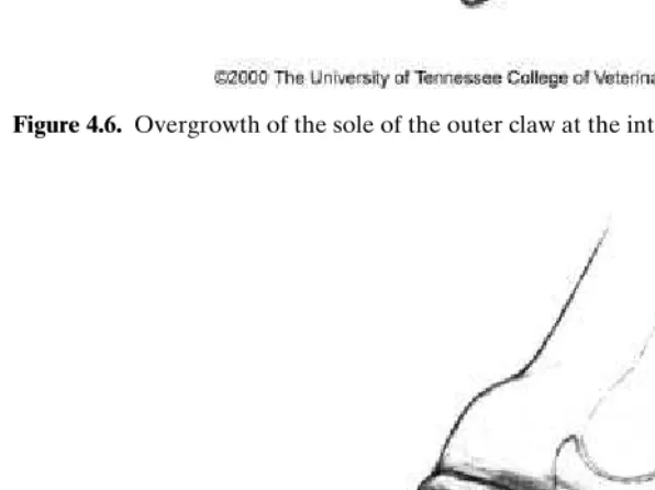
Genetic factors have a significant influence on feet and leg traits in dairy cattle. Specific traits scored include foot angle, legs–side view, and legs–rear view. Her-itability values tend to be low (particularly for legs–rear view and foot angle) as scores can vary significantly depending on the cow’s stance at the time of scoring. Simply moving the cow forward a few steps can make major differences in scoring of feet and leg traits. Other factors, which markedly influence posture and stance, are overgrown claws or pain associated with foot disorders.
Heritability estimates for feet and leg traits on Holstein cows range from about 0.08 to 0.16, which means that single scores from an individual cow are not a reliable measure of that cow’s genetic merit for a specific trait. However, where scores from multiple offspring are available, the breeding value of a specific bull or cow can be reliably estimated. Successful genetic improvement requires selection based on information from progeny tests of bulls and not on the evaluation of a specific individual animal.
Generally speaking, cow legs should be sturdy with a strong pastern and good flexibility in the hock. Abnormally straight hocks, weak pasterns, sickle hocks, splay toes, or overlapping toes are associated with an increase in the incidence of lameness. The ideal conformation of the cow’s foot should be short, steeply angled, high in the heel, and even clawed. Some suggest that the ideal hoof angle is 50◦–55◦for
front feet and 45◦–50◦for rear feet.
Nutrition and feeding management
disorders. A primary goal in feeding is to maximize dry matter intake, and thus op-timize performance yet avoid those conditions, which might lead to rumen acidosis and laminitis. Feedlots emphasize keeping feed available at all times and encourage intake numerous times daily in order to avoid rumen acidosis. Dairies apply similar feeding strategies to encourage consistent feed intake and minimize production losses.
Total mixed rations containing high-quality forages have been one of the strate-gies used to lower the risk of rumen acidosis and laminitis. Proper formulation and mixing of feed ingredients helps achieve optimal success. It is often recommended that hay and ensiled feeds be chopped as coarse as possible and not mixed exces-sively to the extent that effective fiber attributes are lost. However, if forages are too coarse, cows may sort or select for concentrates rather than consume a balanced diet containing both feed grains and forage. Because of social hierarchy issues, most rec-ommend housing mature cows separately from heifers. In all cases animals should be introduced to the milking herd ration gradually, preferably through the use of a properly formulated transition ration. Transition is a critical period of adjustment for animals and has important links to the pathogenesis of laminitis.
In the southern United States, the nutritionist’s challenge is to maintain feed in-take and avoid feeding-related health problems during periods of hot weather. One strategy is to increase the nutrient density of rations and thus maintain an accept-able rate of dry matter intake. This strategy can be troublesome if not monitored carefully, in part because heat-stressed cattle tend to eat less frequently (feeding during cooler times of the day only) but proportionally more at each feeding. The combined effect of these types of rations and feeding patterns increases the risk for rumen acidosis. However, add to this the fact that heat-stressed cattle often have a lowered rumen pH due to decreased salivary buffering, and it is easy to understand how acidosis becomes a major feeding challenge during the summer months.
Housing, environment, behavior, and management
Despite research and clinical observation highlighting its significance in limiting the performance and profitability of dairy cattle, lameness remains a prominent health disorder in dairy farms throughout the world. In countries such as the United States where economic incentives have encouraged producers to expand herd size, there has been a gradual change from pasture-based to confinement-type housing systems. Properly designed confinement systems offer the advantages of improved protection of animals from inclement weather conditions. For example, confinement conditions offer convenience for the implementation of cow cooling measures in hot weather and the provision of wind blocks in cold conditions. It also creates facilities for improved access to feed and water, and a comfortable place for the cow to lie down and rest.
8 Manualfortreatmentandcontroloflameness in cattle
ulcers and white line disease. In conditions where concrete floors are also abrasive, there is excessive wear on the claw’s weight-bearing surface. Excessive wear of the sole results in the development of thin soles, and frequently separation of the white line, especially at the toe (claw zone regions 1 and 2). Confinement conditions also limit cows to a smaller area, thereby increasing exposure of the cow’s foot to manure slurry and moisture. This increases potential for the development of infec-tious skin disorders of the foot (digital and interdigital dermatitis) and heel horn erosion.
Housing considerations
The dairy cow is a land animal. Its foot was not designed for prolonged standing on hard abrasive surfaces. But, as herd size continues to increase so must the amount of surface area of concrete, otherwise earthen floors become a combination of manure, urine, and moisture ridden soil that soon becomes a quagmire of muck and mud. In today’s modern housing systems, out of necessity cows must spend a majority, if not all, of their time on concrete. Options for resting are limited to a free stall, alleyway, or, in the best-case scenario, a well-groomed drylot. It should not surprise anyone that lameness has become a major health problem in the dairy industry.
Concrete
Concrete, depending upon how it is formulated and mixed, is capable of creating an extremely abrasive surface for cows’ claws. New concrete is more abrasive than old one, and wet concrete is up to 83% more abrasive than dry concrete. Claws may wear more than they grow during the first two months on concrete. Animals housed on wet concrete suffer doubly: first, because of the increased abrasiveness associated with wet concrete, and secondly, because moisture softens the claw horn, thereby permitting an increased rate of wear. A further cause of increased claw wear occurs from poor handling procedures where crowding or rushing cattle results in increased wear from twisting and turning on rough abrasive flooring surfaces. For this reason, the proper design of facilities, which incorporates ideas for easing cow movement thereby reducing rotational forces on claws, is an important housing consideration.
slippery when covered by manure slurry. On the other hand, a brush or broom-type finish may result in a surface that is too abrasive.
Smooth concrete reduces wear and may contribute to claw horn overgrowth that may require more frequent trimming of claws. Smooth surfaces are also slippery and predispose to injury, usually of the upper leg from falling. Grooving the sur-face of smooth concrete floors increases traction and reduces injuries from falling. Most recommend grooving a parallel or diamond pattern in the floor to maximize traction. Grooves should be 3/8 to 1/2 in. wide and 1/2 in. deep. When grooves are wider than 1/2 in., the floor is less comfortable because support at the weight-bearing surface is less uniform. For the same reason, it is advised that the floor area between the grooves be kept flat and uniform as well. Grooves in walkways that run in a parallel pattern should be 2–3 in. apart, whereas grooves on a di-amond pattern may be slightly wider at 4–6 in. on center. The didi-amond pattern is considered to be particularly useful in high-traffic areas. As much as possible, avoid orienting grooves at right angles to the direction of the manure scraper travel.
In recent years some operations have incorporated rubber belting along feed mangers and in alleys or walkways to and from the milking parlor. Observation of cow behavior indicates that cows prefer the softer surface offered by the rubber belting. In fact, in some situations cows may find the rubber flooring more comfort-able than the adjoining stall. When this happens, cows may block access to the feed manger. Rubber belts can also be slippery walking surfaces when wet. Grooving the belts (only belts without reinforcing wires) helps reduce slipping injuries. Guide-lines for grooving rubber belts are essentially the same as those described above for concrete. Primary problems with rubber belting are related to manure handling and securing them to the underlying floor. For example, in flush barns where rub-ber may not be properly secured, manure and other debris may become entrapped beneath the rubber. In barns that scrape manure, depending upon how the rubber is secured to the floor, scraping may result in frequent displacement of the rubber. Rubber flooring must be secured in such a way as to make it resistant to displace-ment from either the twisting or turning action of the wheels or the scraper itself. Despite these drawbacks, rubber belting is a flooring surface modification that ap-pears to improve cow and foot comfort, but more research is needed to confirm this observation. Furthermore, it is not a substitute for a poorly designed stall. In herds where belting does not work well, it may be due to other cow comfort issues (poor stall design, heat stress, etc.) that have not been properly addressed.
10 Manualfortreatmentandcontroloflameness in cattle
dirt or grass lots can reduce the mechanical impact of hard surfaces on feet and legs, but maximum benefit in some areas is seasonal.
Free-stall design and comfort
Proper design of stalls for cows should consider a cow’s resting behavior and normal lying positions as described by Kammer and illustrated in a video by Dr. Neil Anderson and others from Canada. They suggest that resting areas for cows provide six basic freedoms: (1) freedom to stretch their front legs forward, (2) freedom to lie on their sides with unobstructed space for their head and neck, (3) freedom to rest their heads against their sides without hindrance from a partition, (4) freedom to rest with their legs, udders, and tails on a platform, (5) freedom to stand or lie without fear or pain from neck rails, partitions, or supports, and (6) freedom to rest on a clean, dry, and soft bed. Cows are clearly very adaptable creatures considering that these freedoms are rarely accomplished in most modern housing systems.
According to a least one study the incidence of lameness is much higher in free stalls (35%) compared with straw yards (8%). However, similar observations have been made by Ward and others who found that large herds with free-stall housing experienced more lameness compared to large herds where cows were housed in straw yards. A comfortable stall encourages resting, thereby improving cow comfort and overall performance. Some British as well as US recommendations for Holstein cattle advise construction of a free stall 8 ft long (7 ft 6 in. for two facing rows) by 4 ft wide with a brisket board (15 in. high) located 5 ft 8 in. from the stall curb. Excessive curb height (over 6 in. high), inadequate bedding of the free stall, and insufficient lunge space have all been related to an increase in herd lameness.
A study by Faull and Hughes suggests that the above recommendations on stall design dimensions are insufficient. Their observation of Holstein–Friesian cows at pasture indicates that they need 95 (240 cm)×47 (120 cm) in. of living space and an additional 24 in. (60 cm) of lunge space for rising. In other words, they recommend that the stall be slightly longer than 9 ft 8 in. (300 cm). Few barns in the United States have been constructed with stall dimensions approaching these recommendations. Clearly, some feel that this is excessive stall size and cost prohibitive. Others suggest that these recommendations may be appropriate for larger framed cattle but not for the average Holstein cow in the United States. In view of the requirements of cows for normal resting, and in light of the high rates of lameness in free-stall-housed cattle, it is possible that further study of stall design is warranted to maximize cow comfort.
Environment
which predisposes to softer claw horn, greater wear rates, and claw disorders result-ing from excessive thinnresult-ing of the sole. Thin soles have become a major complication in confinement housed dairy cattle contributing to separation of the sole from the white line in zones 1 and 2 (the abaxial toe region). These lesions tend to have serious consequences resulting in toe abscesses that may become chronic problems in affected animals.
In North America and other areas affected by periods of hot and humid or hot and dry weather conditions, heat stress contributes to major problems with lameness. Increased rates of lameness tend to coincide and/or follow periods of intense hot weather. In North America, heat-associated lameness tends to peak during the late summer and early fall months (July through October). Heat stress predisposes to rumen acidosis from reduced salivary buffering and respiratory alkalosis from increased respiratory rates. The result is rumen acidosis despite being fed rations that normally would not predispose to acidosis conditions. The abatement of heat stress during such periods is critical to performance as well as health in dairy cattle.
Cow behavior
Social interactions
Cows are moved from pen to pen for a variety of reasons. In many cases, it is done to give the cow access to a diet that is better designed to meet her needs based on age or stage of lactation. Cows are also moved for management purposes such as when a cow is moved from a close-up pen to a maternity pen at the time of calving. Depending upon her physiological or health status, it may be necessary to locate her in a pen where she can be monitored frequently and assisted as needed. The movement of cows to new groups, however, is not without risk. In fact, recent observations suggest that the frequent movement of cows to new groups results in significant social turmoil. Research suggests that with every pen move, cows require 2–5 days to readjust and reestablish social rank. During this time subordinate cows may reduce feed intake by as much as 25%. The result is depressed performance and in some cases development of disease. The same occurs when cows are pulled from the main herd for lameness and reassigned to a lame or sick cow group. Forcing her into a “new group situation” when she is already feeling vulnerable to social interaction may complicate her recovery. It is particularly important in these situations to avoid overcrowding. Efforts to make these animals comfortable with adequate facilities to manage heat and/or cold stress, a well-designed stall, and plenty of bedding are essential.
12 Manualfortreatmentandcontroloflameness in cattle
in alleys and walkways. It is for these reasons that most recommend that barns be designed and managed to provide for a larger number of stalls than cows. Most dairy operations in the United States are guilty of overcrowding their barns. When stall numbers are equivalent to or less than the total number of animals in the barn, timid heifers may have less opportunity to rest. Recommendations that call for at least 10% more free stalls than cows make sense to permit more choice and encourage lying time.
Herdsmanship or management
Cow handling and herding
Cow-handling strategies are also important to minimizing foot problems. A study by Clarkson et al. found that farmers who allowed their cattle to walk in single file had less lameness compared to farmers that pushed their cows to the parlor and back. Clackson and Ward found that rushing cattle over rough flooring surfaces led to a greater potential for damage to the corium and a greater incidence of lameness. Cows should be allowed to move at their own pace over hard and rough surfaces. Movement at the herdsman’s pace increases foot problems and injuries from falling or slipping. In herds, cows are sometimes moved to and from milking facilities on horseback, with dogs or small four-wheel vehicles. While this may be a more convenient way to move animals, the tendency is to move animals too quickly, which encourages feet and/or leg injury. Although there may not be a completely satisfactory solution to this situation, by caring for animal walkways and making personnel aware of this concern, one can limit lameness disorders occurring in this way.
Dr. Neil Chesterton, a veterinarian from New Zealand, makes the point so effec-tively in a video he has produced on cattle handling. He demonstrates how a cow walking on a rock-covered concrete pad will avoid deliberately tramping on stones by carefully watching where she places each of her front feet while walking. When this cow is placed in a group of cows and rushed across a similar rock-covered concrete pad, cows place their feet indiscriminately on the flooring surface and thus stomp all over the rocks, which is a very simple, but very effective example of how cattle-herding practices in combination with cattle tracks or floors may be an important predisposing cause of lameness.
Standing or lying time
Figure 1.1. Poor stall design will decrease lying down and resting time.
time ruminating or “cud-chewing.” When cud-chewing time is reduced, the natural buffering of rumen contents by saliva is decreased.
14 Manualfortreatmentandcontroloflameness in cattle
hours per day. Indeed, it is not uncommon for herds to experience an increase in lameness by making this seemingly insignificant change. Overcrowding, poor stall design, lack of bedding or poor bedding management, and heat stress are just a few of the complications that should be considered when cows fail to spend the appropriate amount of time at rest.
Knowledge, training, and awareness of lameness
Finally, a study by Mill and Ward found that knowledge, training, and awareness of lameness by dairymen significantly influenced the amount they experienced. The farmers who were the most aware of lameness disorders, had the most training, and consulted with their veterinarians about foot problems tended to have the least problems. Clearly, it is important that dairymen understand the problem of lameness to enable them to recognize and deal with it properly.
Eye of the master
There is no substitute for the careful observation of an owner or manager. The phrase “eye of the master” is quoted as that quintessential factor that makes all the difference in the successful care and management of dairy cattle. Several years ago, a large dairy in Florida hired an experienced dairyman from the Midwest to simply observe each and every cow in the dairy, each and every day. It was this person’s specific responsibility to identify sick cows, lame cows, cows in heat, and cows that just did not look right. He did, and the dairy prospered greatly from his efforts. The point is that no matter how large our herds or how great our technology, cows are individuals and must be cared for and managed accordingly. Every herd needs people who truly care about the animals with which they work if it is to be successful.
Bibliography
Anderson, N: Observation on cow comfort using 24-hour time lapse video. Proceedings of the 12th International Symposium on Lameness in Ruminants. Orlando, FL, January 9–13, 2002, p. 27–34.
Clarkson, MJ, DY Downham, WB Faull, JW Hughes, FJ Manson, JB Merritt, RD Murray, WB Russell, JE Sutherst, and WR Ward: Incidence and prevalence of lameness in dairy cattle. Vet Rec, 1996, 138:563–567.
Cook, NB: Prevalence of lameness among dairy cattle in Wisconsin as a function of housing type and stall surface. JAVMA, 2003, 223(9):1324–1328.
Esslemont, RJ and EJ Peeler: The scope for raising margins in dairy herds by improving fertility and health. Br Vet J, 1993, 149:537–547.
cattle: The influence of cubicles and indoor and outdoor walking surfaces. Vet. Rec. 1996. 139:130–136.
Garbarino, JA, Hernandez, J, Shearer, JK, Risco, CA, and Thatcher WW: Effect of Lameness on Ovarian Activity in Post-Partum Holstein Cows. J. Dairy Sci. 2004, 87:4123–4131. Guard, C: Laminitis in dairy cattle: Recognition of the disorder and management of the
causative factors. Proceedings of the American Association of Bovine Practitioners, January 1996, 28:71–74.
Guard, C: Recognizing and managing infectious causes of lameness in cattle. In: The AABP Proceedings, January 1995, No. 27, pp. 80–82.
Guard, C: Laminitis in dairy cattle: Recognition of the disorder and management of the causative factors. In: The AABP Proceedings, January 1996, No. 28, pp. 71–74.
Juarez, S. T., P. H. Robinson, E. J. DePeters, and E. O. Price. 2003. Impact of lameness on behavior and productivity of lactating Holstein cows. Appl. Anim. Behav. Sci. 83:1–14. Leonard, F. C., J. M. O’Connell, and K. J. O’Farrell. Effect of overcrowding on claw health
in first-calved Friesian heifers. Br. Vet. J. 1996. 152:459–472.
McDaniel, BT: “Management and housing factors affecting feet and leg soundness in dairy cattle”. Proceedings of the American Association of Bovine Practitioners, 1983, 14:41–49. Melendez P, Bartolome J, Archbald LF, Donovan A. The association between lameness,
ovarian cysts and fertility in lactating dairy cows. Theriogenology. 2003, 59:927–937. Phillips, CJC: Cattle Behaviour and Welfare, 2nd edition. Blackwell Science Ltd., Osney
Mead, Oxford, 2002.
Tranter, WP and RS Morris: A case study of lameness in 3 dairy herds. NZ Vet J, 1991, 39:88–96.
Vermunt, J: Herd lameness—A review, major causal factors, and guidelines for prevention and control. In: Proceedings of the 13th International Symposium and 5th Conference on Lameness in Ruminants, Maribor, Slovenia, 2004, pp. 3–18.
Warnick, LD, CL Guard, and YT Grohn: The Effect of Lameness on Milk Production in Diary Cattle, Proceedings of the American Association of Bovine Practitioners, 1998, 31:182.
Wells, SJ, AM Trent, WE Marsh, and RA Robinson: Prevalence and severity of lameness in lactating dairy cows in a sample of Minnesota and Wisconsin herds. JAVMA, 1993, 202(1):78–82.
Chapter 2
Horn formation and growth
Structure and function
The horn-producing germinal layer of the epidermis and its supporting dermal structure, the corium, consists of four different regions, each producing a structurally different type of horn. Perioplic horn overlying the perioplic corium is found just below the skin horn junction and extends to the back of the claw to include the heel horn. Horn of the wall is produced in the area of the coronary corium and is situated between the perioplic corium and the sensitive laminae. Horn of the white line (WL) (laminar horn) is produced by the epidermis overlying the laminar corium (sensitive laminae). The solar horn overlies the solar corium and is situated between the laminar horn of the WL and the perioplic horn of the heel. The four different regions of the corium are shown in Figures 2.1 and 2.2. The corium consists of a rich vascular network, which terminates in dermal papillae or vascular pegs. Side and secondary papillae are common in the coronary corium, and small secondary papillae are present on the terminal papillae of the laminar corium. A vascular peg consists of a main arteriole, which connects directly to a venule, at the tip (Figure 2.3).
Between the arteriole and venule is an extensive capillary network. In the corium of horses there are several vascular shunts between the arteriole and venule. These shunts may open during laminitis, thus cutting off blood supply to the tip of the vascular peg, which will adversely influence formation of horn cells. Recent work suggests that these shunts are relatively uncommon in the corium of cattle until after the damage to the vascular system has occurred as in the case of laminitis.
The basement membrane (BM) is situated between the epidermis and corium and is a key structure bridging the cytoskeleton of the keritinocytes in the basal layer of the epidermis via the corium to the connective tissue of the distal phalanx. The BM consists of a complex lattice composed of both collagen and glycoprotein (laminin, fibronectin, amyloid P, entactin, and heparin sulfate proteoglycan). It provides both anchorage and orientation to keratinocytes in the basal layer, and damage of the BM leads to loss of organized structure. Keratinocyte proliferation and differentiation are controlled by cues from the BM. Pathological changes within the BM can result in hyperproliferation of keratinocytes and an abnormal keratinocyte growth pattern and expression of keratin protein subtypes.
The basal layer of the epidermis consists of keratinocytes undergoing active proliferation and differentiation. Neighboring keratinocytes are tightly bound by desmosomal intracellular junctions and are imbedded in a lipid-rich extracellular matrix known as intercellular cementing substance (ICS). Keratinocytes produce
Figure 2.1. Regions of the corium.
metalloproteinases (MMPs) and cytokines in response to polymorphonuclear cells (PMNs) resulting in degradation of extracellular matrix during wound healing. MMPs may play a role in the pathogenesis of laminitis.
Keratinocytes become large, polygonal, and flatter upon transition into the Stra-tum spinosum (StraStra-tum medium), and cell contents are replaced with keratin pro-teins (keratinization). These cells then undergo programmed death in the final stages of keratinization and differentiation (cornification) (Stratum corneum). The plasmalemma (cell membrane) of cornified cells remains permeable to water and solutes, but not to large molecules such as protein.
18 Manualfortreatmentandcontroloflameness in cattle
Figure 2.2. Longitudinal section of claw showing anatomical structures.
level inside the cell stabilizes the remainder of the intracellular protein matrix. During conditions of low hydration of the epidermis, extensive secondary hydrogen cross-linking through glycine and tyrosine makes the protein matrix less flexible, thus increasing horn hardness. High levels of epidermal moisture result in greater distances between secondary cross-linking, which makes the protein matrix more flexible, thus decreasing horn hardness.
Keratin formation may be regulated by several factors such as epidermal growth factor (EGF), receptors for which have been demonstrated in the bovine claw. Other factors, which may play a role, include the hormone relaxin, which inhibits EGF and prolactin and hydrocortisone, which decrease protein synthesis in bovine claw tissue explants. Glucocorticoids have been shown to be elevated during lactation. Insulin stimulates protein synthesis; however, low concentrations of insulin have been measured in lactating dairy cows.
The epidermal layer overlying the vascular pegs produces horn cells in the form of tubules (tubular horn). Cells within the tubules are arranged in a steep spiral around the center axis. Tubules differ in size, number, and shape in various parts of the claw and are round near the inside of the wall and oval near the surface. There are approximately 80 tubules/mm2in the wall and 20 tubules/mm2in the sole
Figure 2.3. Dermal papillae (vascular peg).
Eighty percent of the horn of the WL consists of laminar horn, which is produced from:
(a) Germinal epithelium overlying the laminar corium just below the coronary corium. This forms the outer zone of the WL and consists of soft nontubular horn.
(b) Germinal epithelium overlying the dermal papillae (also referred to as dermal caps or cap-papillae), which protrudes from the dermal folds on the laminar corium (See Figure 2.4) and produces nontubular cap-horn. This horn repre-sents the middle zone of the WL.
20 Manualfortreatmentandcontroloflameness in cattle
Dermal papillae
Figure 2.4. Dermal fold with dermal papillae (cap papillae).
Because of the high turnover rate the cells are generally less mature and therefore softer and less resistant to wear and other environmental factors.
Horn quality and physical properties
levels of protein, energy, lipids, vitamins A, D, and E, calcium, and phosphorous. Micronutrients such as sulfur containing amino acids like cysteine and methionine are essential for cross-linking of keratin filaments. Trace minerals particularly zinc, copper, and the vitamin biotin have very important roles in the keratinization of horn cells and integrity of the ICS of claw horn.
The external and internal environment of claw horn will affect its moisture con-tent. A hydrostatic force exists between the dermis (corium) and epidermis (horn), moving water toward the outer horn cells. The plasmalemma of cornified ker-atinocytes in the outer layers of the epidermis is highly permeable to the passive movement of water and crystalloids, but not to macromolecules such as protein. This movement of water creates a gradient, in which the outer surface of the horn has a low hydration level while the inner layers adjacent to the dermis maintain a high hydration level. In addition, a variable osmotic gradient is present inside the cell caused by solutes and keratin proteins, which will further regulate the water content of horn cells. Immersion of sole horn in solute-free water resulted in wa-ter uptake as evidenced by a 4% increase in mass afwa-ter 10 days. Maximum weight change, however, occurred within 48 hours. Prolonged water contact of claw horn occurs in many dairy operations because of flush and sprinkler systems commonly in use to clean udders, manage manure accumulation, and reduce heat stress. This may have adverse effects on claw health and may increase the incidence of lameness. The physical properties of horn are commonly expressed in terms of its stiffness, hardness, and fracture toughness, all of which are affected by the hydration status. Stiffness is defined as resistance to deformation, and it relates to the flexibility of horn. By increasing the hydration of horn cells, the spaces between secondary bonding sites of the matrix keratin protein are widened, resulting in increased flexibility. Although a positive relationship between the level of horn moisture and degree of wear has been reported, flexible horn may be more resistant to abrasive wear of concrete because of its ability to expand and contract.
Hardness may be defined as the resistance of a material to penetration by a harder object. An inverse relationship exists between hardness and water content of the horn. The number of horn tubules per unit area may also affect horn hardness since intertubular material can absorb more moisture with fewer tubules per mm2.
The outer wall has more tubules as compared to the sole and therefore represents harder horn. Increased moisture content of horn resulted in an increased rate of wear.
Horn cells and ICS may be affected by certain compounds. For example, high levels of copper sulfate are reported to destroy the ICS, making the claw horn more brittle. Likewise, constant exposure to urine and manure can destroy both the horn cells and ICS, resulting in loss of horn as observed in the condition of heel erosion. Both internal and external factors may act synergistically to produce poor horn quality. For example, changes in blood supply as seen with laminitis will result in production of poor quality horn. Poor quality horn is more susceptible to the effects of environmental influences.
22 Manualfortreatmentandcontroloflameness in cattle
feeder cattle on high planes of nutrition, this growth rate may be increased to as much as 2 1/2 times that of normal. The horn growth rate depends on several factors including breed, developmental abnormalities, nutrition, environmental factors, the integrity of the blood supply through the corium, and the biomechanics of weight bearing. For example, horn growth rate of free stall housed cattle is greater than that of cattle on pasture or in tie stalls. Claw horn proliferation and keratinization are increased in the summer, compared to the winter months.
Bibliography
Baillie, C., C. Southam, A. Buxton, and P. Pavan. 2000. Structure and properties of bovine hoof horn.Adv.CompositesLett., 9(2):101–113.
Bertram, J.E.A., and J.M. Gosline. 1987. Functional design of horse hoof keratin: The mod-ulation of mechanical properties through hydration effects.J.Exp. Biol., 130:121–136. Collins, S.N., B.C. Cope, L. Hopegood, R.J. Latham, R.G. Linford, and J.D. Reilly. 1998.
Stiffness as a function of moisture content in natural materials: Characterisation of hoof horn samples.J.Mater.Sci., 33:5185–5191.
Douglas, J.E., C. Mittal, J.J. Thomason, and J.C. Jofriet. 1996. The modulus of elasticity of equine hoof wall: Implications for the mechanical function of the hoof.J.Exp. Biol., 199:1829–1836.
Greenough, P.R., J.J. Vermunt, J.J. McKinnon, J.J. Fathy, F.A. Berg, P.A. Cohen, and R.D.H. Cohen. 1990. Laminitis-like changes in the claws of feedlot cattle.Can.Vet.J., 31:202– 208.
Hinterhofer, C., Ch. Stanek, and K. Binder. 1998. Elastic modulus of equine hoof horn, tested in wall samples, sole samples and frog samples at varying levels of moisture.Berl.Munch.
Tierarz. Wschr., 11:217–221.
Leach, D.H., and G.C. Zoerb. 1983. Mechanical proper ties of equine hoof wall tissue.Am.
J.Vet.Res., 44(1):2190–2194.
Toussaint Raven, E. 1989. Structure and function. InCattleFoot CareandClaw Trimming, E. Toussaint Raven, ed. Farming Press, Ipswitch, UK, pp. 24–26.
Vermunt, J.J., and P.R. Greenough. 1995. Structural characteristics of the bovine claw: Horn growth and wear, horn hardness and claw conformation.Br.Vet.J., 151:157–180. Wagner, I.P., and D.M. Hood. 2002. Effect of prolonged water immersion on equine hoof
epidermis in vitro. AJVR, 63(8):1140–1144.
Anatomy
The foot
the claws to the carpus or tarsus is named dorsal, while the back or posterior aspect is named palmar in the forelimb and plantar in the hind limb.
The metacarpophalangeal and metatarsophalangeal joints each have two syn-ovial sacs, which communicate at the palmar or plantar aspect at the level of the proximal sesamoid bones between the interosseus muscle and the metacarpal or metatarsal bones. The palmar or plantar pouch extends more proximal than the dorsal pouch and lies deep to the interosseus muscle and the digital flexor tendons. Synovial fluid accumulation in either the palmar or plantar pouch of the joint or the proximal flexor tendon sheath will result in a fluctuant swelling immediately above the dewclaws.
Each digit of the foot consists of three phalanges (proximal, middle, and distal or P1, P2, and P3; Figure 2.5) and the navicular bone (distal sesamoid) and two joints—
proximal interphalangeal (PIP) joint and distal interphalangeal (DIP) joint. P1is
longer than P2 (Figure 2.5). Little longitudinal growth occurs in P1 after birth.
P1 has a distinct marrow cavity. Amputation through the distal end of P1 with
Figure 2.5. Anatomical structures of the digit.
24 Manualfortreatmentandcontroloflameness in cattle
exposure of the marrow cavity will sometimes result in excessive granulation of the exposed bone marrow. The palmar or plantar pouch of the PIP joint is situated deep to the terminal portion of the superficial digital flexor tendon (SFT). Care must be taken not to open this pouch during surgical procedures requiring resection of the SFT. Collateral ligaments and a palmar/plantar ligament support the PIP joint. P3is completely enclosed within the claw horn capsule (Figure 2.5). Its solar
surface is concave. The articular surface of P3 has a steep slope (25◦–30◦ from a
horizontal bearing surface) (Figure 2.5), which complicates the surgical approach to the DIP joint. The extensor process becomes increasingly irregular with advancing age. There is a large vascular channel (axial foramen) on the axial side of P3close
the DIP joint.
The deep flexor tendon is attached to the flexor tuberosity at the back of P3
(Figure 2.5). Because of the concave ventral (solar) surface of P3, it has potentially
two pressure areas, one at the tip and the other at the heel that can cause pressure on the solar corium with sinking, resulting in either a toe or sole ulcer (Figure 2.5). The navicular bone is attached to P3by three small distal ligaments and is also
attached to P2by two collateral ligaments. The navicular bursa is situated between
the navicular bone and the deep flexor tendon and permits movement of the deep flexor tendon over the surface of the navicular bone during extension and flexion of the claw. Although the navicular bursa is well protected by surrounding fibroelastic tissue, it is frequently involved in conditions that can cause suppurative processes within the heel. P3, the DIP joint, navicular bone, and navicular bursa are within
the claw capsule (Figure 2.5).
The DIP joint is formed by the articular surfaces of P2, P3, and the navicular bone.
The joint capsule forms dorsal and palmar/plantar pouches. The dorsal pouch ex-tends to the coronary band and lies deep to the insertion of the common digital extensor tendon. Arthrocentesis can be performed at this level, axially or abaxially to the extensor tendon. The palmar/plantar pouch of the DIP joint is well protected by the navicular bone, its distal ligament, the deep digital flexor tendon, and sur-rounding fibroelastic tissue. However, the palmar/plantar pouch is adjacent to the so-called retroarticular space (Figure 2.6), which is frequently involved in a suppu-rative inflammation following ascending infections of the sole or WL. Extension of the infection from the retroarticular space is one of the ports of entry into the joint. Care should be taken not to open the joint capsule at this point during surgical procedures requiring resection of the deep digital flexor tendon.
Lateral and medial digits are connected by proximal and distal interdigital (cru-ciate) ligaments. The proximal cruciate ligament is situated at the proximal ex-tremities of both the proximal phalanges. The distal cruciate ligament is wider and more superficial than the proximal cruciate ligament and lies just above the palmar/plantar interdigital cleft (Figure 2.7). The origin of the distal cruciate lig-ament is at the distal aspect of the proximal phalanx. The liglig-ament continues su-perficial to the deep flexor tendon and inserts on the axial surface of the navicular bone and P3. The insertion of the distal cruciate ligament forms a significant part
of the structure of the suspensory system of the caudal aspect of P3with some of
of the flexor tendon.
26 Manualfortreatmentandcontroloflameness in cattle
Tendon sheath
The proximal limit of the digital flexor tendon sheath (DFTS) is the distal third of the metacarpus/tarsus (6–8 cm proximal to the proximal sesamoid bones). It ends distally just dorsal to the navicular bone (Figure 2.6). The distal part of the DFTS consists of a single compartment (Figure 2.6). Proximal to this, at the level of the digital annular ligament, the DFTS consists of an inner and outer compartment. The SFT forms a tendinous tube, enclosing the inner proximal compartment of the DFTS. The outer proximal compartment encloses the tendinous tube formed by SFT. The inner compartment extends further proximal to the fetlock than the outer compartment. Puncture of the tendon sheath above the dewclaws usually results in aspiration of fluid from the outer compartment. The tendon sheath is often involved in septic conditions of the claw and should be carefully evaluated in septic conditions involving the deep structures within the claw.
The claws
The purpose of the claw horn capsule is to protect the corium and dissipate the con-cussion forces that occur when the digits impact the ground. It consists of the wall, which can be divided into the abaxial (outside) and the axial (inside). The abaxial wall is further subdivided into dorsal (or front/toe) and lateral (abaxial) aspects. The wall is demarcated from the heel on the abaxial side of the claw by the abaxial groove (Figure 2.8). The wall consists of two types of horns: perioplic and coro-nary (Figure 2.2). Perioplic horn is the softer horn lying just below the coronet at the skin–horn junction. At the back of the foot the periople gradually widens and
Figure 2.9. Area of the white line. (Dotted line)
eventually becomes the horn of the heel. Coronary horn, the hardest horn within the claw capsule, makes up the bulk of the horn of the wall. The wall has faint ridges or rugae, which run horizontally and parallel to each other. Toward the heel these ridges diverge, reflecting a more rapid rate of growth in the heel region due to faster rates of wear. In mature Holstein cattle the length of the dorsal wall should be a minimum of 3 in. from just below the top of the hairless portion of the wall to the weight-bearing surface. Ideal heel height is 1.5 in., measured at the abaxial groove. The sole is produced by the epidermis overlying the solar corium and merges imperceptibly with the horn of the heel at the heel–sole junction. The sole is con-nected to the wall by means of the WL. White line horn is produced by epidermis overlying the laminar corium. It courses forward from the area of the heel on the abaxial side of the claw, around the tip of the toe and about 1/3 of the way back on the axial side of the claw’s weight-bearing surface (Figure 2.9). Where the WL leaves the weight-bearing surface, it courses upward on the axial side of the claw. The WL is a unique and important structure. It is the softest horn within the claw capsule. This permits it to provide a flexible junction between the harder horn of the wall and the softer horn of the sole. On the other hand, because of its softer nature it also represents a weak area on the weight-bearing surface that is vulnerable to damage.
Suspensory system of the third phalanx (P
3)
28 Manualfortreatmentandcontroloflameness in cattle
(a)
(b)
Figure 2.10. Parts of Digital cushion. a) Ventral view b) Complete foot medial claw with saggital section showing parts of digital cushion and P3.
dermal and epidermal components is the interdigitating dermal (sensitive laminae) and epidermal laminae (horn leaflets). The result is that P3hangs within the claw
Figure 2.11. Suspensory system of caudal aspect of P3.
capabilities with respect to mechanical load carried on the claws of cattle vary signif-icantly. In the horse, load bearing is primarily on the wall. Cattle, on the other hand, simply cannot handle the same amount of mechanical load on the walls of their claws. Instead, weight bearing in cattle requires displacement of load to alternate support structures within the sole and heel.
The primary structures within the supportive apparatus of the bovine claw are the solar corium and associated connective tissue, and the digital cushion (Figure 2.5), which consists of loose connective tissue and varying amounts of adipose tissue. The digital cushions are arranged in a series of three parallel cylinders (Figures 2.10). The volume of all three pads in a single claw adds up to∼5.7 ml. All three cushions extend from the skin–horn junction at the heel toward the tip of the third phalanx. The abaxial and middle fat pads are shorter than the axial pad. The shorter pads lie superficial to the deep digital flexor tendon and do not reach further distal than the insertion of the tendon. The axial fat pad courses from the axial border of the heel bulb toward the middle of the sole surface, where it ends in the middle third. Recent studies by Swiss researchers have shown that the amount of fat (and thus cushioning capacity) increases with increasing age. This is believed to have significant implications for animals relative to their susceptibility to claw disorders. The caudal aspect of P3 and the digital cushion is attached to the inside of the
abaxial claw wall, and further support is provided axially by its attachment to the distal interdigital cruciate ligament (Figure 2.11).
Bibliography
Desrochers, A. 2001. Surgical treatment of lameness.Vet.Clin.North Am. FoodAnim.Pract., 17(1): 143–158.
30 Manualfortreatmentandcontroloflameness in cattle
Maierl, J., P. Bottcher, S. Hecht, and H.G. Liebich. 2002. A new method to assess the volume of the fat pads in the bovine bulb. InProceedingsof the 12th InternationalSymposium on Lameness in Ruminants, January 9–13. Orlando, FL.
Stanek, C. 1997. Tendons and tendon sheaths. InLameness in Cattle,P.R. Greenough, ed., WB Saunders Co., Philadelphia, PA, pp. 188–192.
Nutrition and claw health
The health and function of the bovine claw is dependent upon sound nutrition and feeding practices. In this context, the avoidance of rumen acidosis, which is considered to be the predominant predisposing cause of laminitis, is believed to be of paramount importance. Acidosis in its acute form is a life-threatening dis-ease. In its subclinical form, acidosis contributes to decreased performance, poor body condition, and lameness most often due to laminitis and related claw dis-orders. In addition to being the single largest component of the dairy cow’s diet, the one most often incriminated in rumen acidosis and laminitis is carbohydrate. The rapid fermentation rates of certain nonstructural carbohydrates place desirable rumen microbes in jeopardy. Therefore, rations must be carefully formulated and fed to avoid potential problems. Not all studies reported in the literature have been able to demonstrate an association between rumen acidosis and laminitis. These inconsistencies substantiate the view of most people that laminitis is multifactorial and is likely complicated by many other factors. Rumen pH is a balance between the acid produced by carbohydrate fermentation and rumen buffering from saliva. Heat stress contributes to rumen acidosis by altering feeding behavior (encouraging slug feeding) and reducing salivary buffering. Although occasionally questioned as a cause of laminitis, the effect of elevated levels of dietary protein in dairy cat-tle diets has not shown conclusive evidence of contributing to laminitis. Research into the role of vitamins, particularly biotin, suggests significant benefits to claw health. Similar information exists on the role of minerals and trace minerals in dairy cattle diets. A claw healthy diet should include appropriate supplementation of both vitamins and minerals to support the proper growth and development of claw horn. Laminitis results from disrupted blood flow in the corium that leads to damage of the dermal–epidermal junction and the underlying connective tissue matrix of the corium. Inflammation predisposes to the activation of matrix met-alloproteinases (MMPs) that breakdown the strong collagen fiber bundles of the suspensory apparatus of P3. This permits sinking and rotation of P3 and
predis-poses to the ulcers of the toe, sole, and heel. There are, however, alternate the-ories that suggest that hormonal changes associated with calving may be major contributors to weakening of the suspensory apparatus. If these observations are correct, it may help to explain those inconsistencies in the literature and those observed clinically that do not show a clear relationship between laminitis and nutrition.
32 Manual for treatment and controlof lameness incattle
Introduction
The management of feeding and nutrition is the primary area of interest when attempting to reduce lameness problems. This may or may not be the correct ap-proach, depending upon the specific types of lameness experienced. For example, it would be hard to influence the incidence of infectious foot diseases (foot rot, inter-digital dermatitis, or inter-digital dermatitis) by manipulation of the diet alone. Laminitis and claw disorders share a closer relationship to metabolic disease disorders that are often linked to nutrition and/or feeding issues. Cow comfort considerations are also critical factors in sorting out lameness problems and must be thus evaluated in herd problem situations as well. However, for the purposes of this discussion, our attention will be on nutrition and claw health.
Rumen acidosis
Acidosis is generally associated with the ingestion of large amounts of highly fer-mentable carbohydrate-rich feeds, which ultimately result in the excessive produc-tion and accumulaproduc-tion of lactic acid in the rumen. In its acute form, the disease is characterized by severe toxemia, ataxia, incoordination, dehydration, ruminal stasis, weakness, and recumbency. The mortality rate is high. The subclinical form of rumen acidosis (better known as SARA, for subacute rumen acidosis) is far more common than the acute form of this disease. Major clinical manifestations would include variable feed intake, depressed fat test, poor body condition de-spite sufficient energy intake, mild to moderate diarrhea, and occasional cases of epistaxis (nose-bleed) or hemoptysis (the expectoration of blood from the mouth). Conditions such as laminitis or undefined lameness, abomasal disorders, and liver abscesses are generally secondary observations.
Although few studies have been able to establish a direct link between rumen acidosis and laminitis, most assume that the feeding program is a major underlying factor. In reality, much of the information ascribed to cattle is based on information from studies of starch overloading models in horses. Recent work suggests that an oligofructose overload model may be appropriate for the study of acute bovine laminitis. Researchers were able to successfully create classical symptoms of rumen acidosis and laminitis in cows treated with an alimentary oligofructose overload. The following is an attempt to identify some of the more important predisposing factors relative to nutrition and feeding of dairy cattle.
Nutrition and feeding considerations
diet, what ends up in front of the cow is still at the mercy of those responsible for mixing, delivery, and management of the feed bunk. Add to these selective eating or feed-sorting behavior of cows and it is easy to see that there is ample room for error. Equally important are dietary changes that naturally occur during the cow’s lactation cycle. In recent years, nutritionist’s have concentrated their attention on feeding programs during the transition period in an attempt to ease the adjustment of cows to higher energy rations necessary to sustain milk production. A Florida study concluded that large differences in the fiber and net energy content of close-up and early lactation diets can contribute to an increase in incidence of rumen acidosis and subclinical laminitis.
Carbohydrate
Feeding rations high in nonstructural carbohydrates to animals that are not suffi-ciently adapted has the potential to result in a lowered rumen pH. Lowered rumen pH is associated with a change in the rumen microflora from predominantly gram-negative to predominantly gram-positive lactic-acid-producing bacteria. Coincident with this change in rumen pH and microflora is the release of endotoxin from the outer cell walls of dying and disintegrating gram-negative bacteria. Aided by a damaged and dysfunctional rumen mucosa, lactic acid, endotoxin, and possibly his-tamine are absorbed into the blood stream. These products are rapidly dispersed to the microcirculation of the corium, where directly or indirectly (through vasoactive mediators) blood flow is disrupted, leading to the lesions observed in laminitis.
While there is little dispute that rumen acidosis may occur as described above, it is not clear that laminitis will inevitably occur as a consequence. Three different studies observed no correlation between laminitis and the feeding of rations high in carbohydrate. Despite conflicting information in the literature, one would still have to conclude that there seems to be an association (albeit complex) between carbohydrate nutrition, rumen acidosis, and laminitis, but more research is needed to sort out the details of these relationships.
Protein
34 Manual for treatment and controlof lameness incattle
laminitis. In consideration of the above information, one must conclude that there is simply insufficient information to know what effects, if any, protein may have on foot health.
Vitamins
Vitamin deficiencies sufficient to cause obvious disease are relatively rare under modern feeding conditions. More common are those conditions where vitamin levels are sufficient to prevent the occurrence of clinical disease, but are possi-bly insufficient to support optimum growth and performance. For example, rickets from a deficiency of Vitamin D is extremely uncommon, since hay and exposure to sunlight normally provide the cow with ample quantities of this vitamin. On the other hand, sporadic instances of white muscle disease associated with Vitamin E and selenium deficiency occur in unsupplemented animals raised in areas where soils are normally deficient in selenium. Sudden death of calves exhibiting a gener-alized weakness or stiffness of the legs may be observed in animals affected. Vita-min A has important roles in the maintenance of epithelial tissues including claw horn.
B-Vitamins are synthesized by rumen microflora and therefore, until recently, rarely fed to dairy cattle. The one exception in recent time is biotin. Biotin is essential for keratin protein synthesis and the formation of long-chain fatty acids that make up the intercellular matrix of claw horn. Canadian research suggested that cattle fed high grain diets are subject to potential biotin deficiency, since the rumen microbes responsible for biotin synthesis are sensitive to low rumen pH. Since then several feeding trials with biotin supplemented at a rate of 20 mg/day have shown benefits to claw health, including an improvement in the healing rate of sole ulcers, a decrease in the occurrence of vertical wall cracks in beef cattle, an improvement in white line health, a decrease in the incidence of lameness in pastured dairy cattle in tropical Australia, a reduction in incidence of sole hemorrhages, an increase in milk production in biotin-supplemented cows, and an improvement in horn quality and strength. While cost and a lack of scientific information were once reasons to question the value of biotin supplementation, current cost and a growing body of scientific information suggests that biotin is worthy of consideration in the diets of lactating dairy cattle.