Open Access Journal of Ophthalmology
Clinical Pathology of Diabetic Retinopathy and
Macular Edema
Marashi A
*Marashi eye clinic, Syria
*Corresponding author: Ameen Marashi, Retina specialist at Marashi eye clinic, Aleppo, Syria, E-mail: [email protected]
;
www.amretina.tkAbstract
Diabetic retinopathy and macular edema is multifactorial complex disease, VEGF can play central role in non-chronic diabetic macular edema pathogenesis and VEGF blockade agents may improve vision, where in non-chronic diabetic macular edema inflammatory cytokines are the main driver of edema and intravitreal steroids may result in edema resolution, however vascular element is not always the cause of macular thickening and visual loss from non-vascular elements such as vitreomacular abnormalities which needs to be managed surgically, while diabetic retinopathy can be non-proliferative or proliferative in the presence of neovascularization which they managed by pan retinal laser photocoagulation and proliferation can complicate in to tractoinal retinal detachment and vitreous hemorrhage, which may require surgical management in certain cases.
Keywords:
Cytokines; Pericytes; Ischemia; Diabetic retinopathy; Diabetic maculopathyIntroduction
Diabetic retinopathy and macular edema is responsible for vision loss in working age group due to hyperglycemia, when approaching patients with diabetic retinopathy, it is essential to understand the underlying pathological mechanisms in order to individualized treatment as diabetic retinopathy and macular edema is multifactorial complex disease. A lot of agents or procedures are available for targeting various pathological mechanisms, such as VEGF, inflammatory, or vitreomacular abnormality, however optimum treatment results can be achieved by using the right agent or procedure at the right place.
Macular Edema
Macular thickening and cyst formation are due to fluid accumulation because of increased vascular permeability
as a result of inner blood retinal barrier break down after loss of pericytes and thickened basement membrane induced by hyperglycemia, this process is governed by multiple and complex factors and mechanisms such as vascular, inflammatory and biochemical [1].
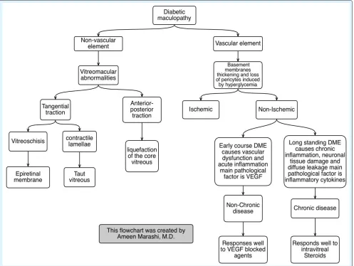
Macular edema can be induced by one or multiple factors at the same time, and it is important to understand that pathogenesis mechanism can be changed from one to another. The best way to targetpathological factors in clinical practice is to understand it mechanism verities, diabetic macular edema can be induced by vascular and non-vascular (vitreomacular abnormality) elements and sometimes mixed where vascular is in term can be presented as ischemic or non-ischemic, where the latter can be as chronic or non-chronic course (Figure 1).
Review Article
Volume 1 Issue 1
Received Date: July 08, 2016
Figure 1:A flow chart created by the author explaining the pathological elements causing the diabetic maculopathy.
Vascular Element
Non-ischemic
Non-chronic disease: When diabetic macular edema starts to develop the main mechanism is vascular dysfunction, and acute inflammation causing hypoxia and thus governed by upregulated vascular endothelial growth factor (VEGF) and other inflammatory cytokines [2] such as IL-1b, IL-6, IL-8, and MCP-1, where in non-chronic disease VEGF may play major role in pathogenesis and targeting it by VEGF blockade agents can cause macular edema resolution. VEGF can be targeted by blocking the VEGF receptor using monoclonal antibodies such as Ranibizumab or Bevacizumab which they inhibit VEGF-A isoforms, or by trapping VEGF using fusion proteins such as Aflibercept, ziv-aflibercept, or conbercept which they inhabit VEGF-A VEGF-B, and PIGF.
Clinical trials have evaluated the safety and efficacy of intravitreal VEGF-blockade agents for diabetic macular edema treatment and compered it head to head and with other treatment modalities such as laser and steroids. The main outcome of these clinical trials is the following:-VEGF blockade agents are safe and effective to use for diabetic macular edema [3] (Figure 2).-VEGF blockade agents are superior to laser treatment alone and to steroids in a long term follow-up [4]. -There is not much deference in visual out come when combining intravitreal VEGF blockade agents with laser treatment in contrast to intravitreal VEGF blockade agents alone [5]. -Patients with central diabetic macular edema that received intravitreal VEGF blockade agents as differed treatment
didn’t gain visual benefits as those who received VEGF
Open Access Journal of Ophthalmology
3
adopted chronic course [6]. -Patients may benefits equally to all VEGF blockade agents when BCVA is good at baseline where Aflibercept showed more efficacies in the 1st 12 months follow up when BCVA is worse at baseline
[7].
Figure 2: Shows thickening of the macula and cyst formation as a result of diabetic macular edema (above) where (bottom) shows resolution of diabetic macular edema after injecting intravitreal VEGF blockade.
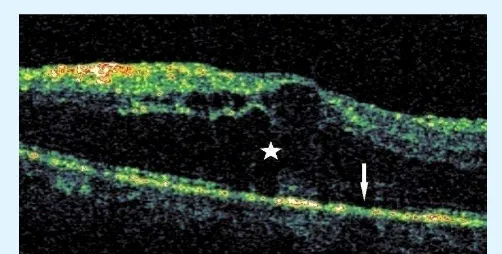
Chronic disease: As the diabetic macular edema becomes long standing the fluid leakage become diffuse and cause photoreceptor loss (Figure 3) inflammation governed by mediators such as MCP-1, TNF-α, IL-1b, IL-6, IL-8, and IP-10 where VEFG may not play a significant role and thus explain the poor response to intravitreal VEGF blockade agents in chronic DME. The process of chronic inflammation itself is not self-resolving leading to tissue stress and it further damage with increased sub retinal microglia accumulation which will cause more fluid leak induced by leukostasis and cytotoxic effect (8).
Figure 3: Chronic macular edema showing diffuse leakage and neural tissue damage (star) and loss of photoreceptor loss (arrow).
This cascade of events can be shot down using intravitreal steroids, commercially intravitreal steroids are available in three forms: Triamcinolone Acetonide, dexamethasone 0, 7 mg biodegradable implant and FluocinoloneAcetonide Implant 0.19 mg non-biodegradable implant.
A lot of trails have studied the safety and efficacy of intravitreal steroids and they concluded the following Intravitreal steroids are safe and effective for diabetic macular edema treatment [9].-Intravitreal steroids can resolve persistent diabetic macular edema which may not respond well to other treatment modalities [10]. Intravitreal steroids induce risk of increased intra ocular pressure and cataract formation [11,12].
Ischemic
traction which can feature either epiretinal membrane due to vitreoschisis, or taut vitreous due to glial cell proliferation or contracted lamellae. These vitreomacular abnormalities are governed by several mechanisms such as non-enzymatically cross linking of vitreous collagen along with glial cells and inflammatory cells infiltration and deposition of glial fibrillary acidic protein and cytokeratin.
Figure 5: Shows macular thickening due to vitreomacular thickening with focal disturbance of inner retinal layers.
The best way to diagnose vitreomacular abnormality is by OCT showing focal disturbance of inner retinal layers (Figure 5) however clinically in the absence of vascular element and presence of vitreomacular abnormalities, treatment with VEGF blockade agents, intravitreal steroids and laser may not reduce macular thickening and improve vision, as this abnormality should by be addressed surgically by performing parsplana vitrectomy with ILM peeling in cases of moderate visual loss [13].
Diabetic Retinopathy
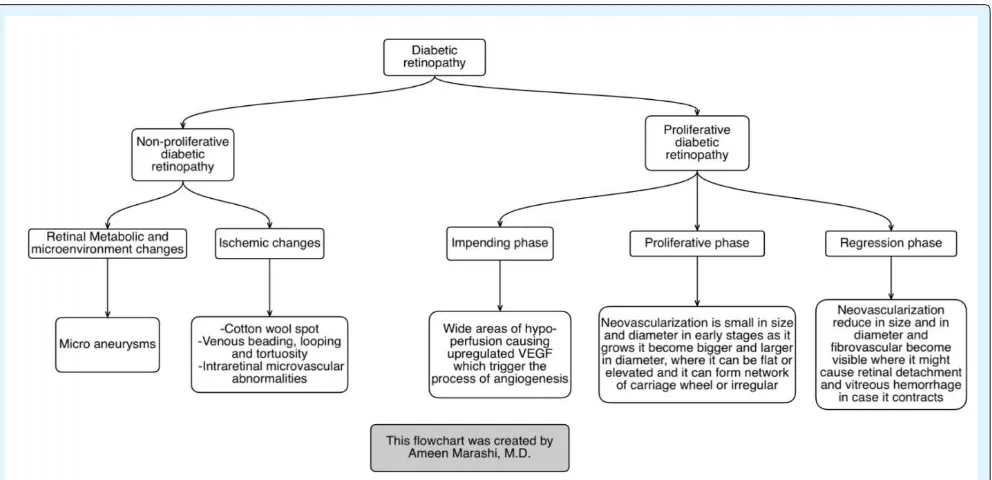
The metabolic and retinal microenvironment causes pericytes, endothelium and capillary damage due to agglutinated erythrocyte and th rombus, all that forms hyper cellular sacs in the capillary wall and thus forms micro aneurisms which is the main feature of non-proliferative stage of diabetic retinopathy as this process progress more micro aneurisms forms and retinal tissue reaches state of relative ischemia and thus will trigger VEGF production and interim will induce neovascularization which is the main feature of proliferative stage (Figure 6) which may lead eventually to vitreous hemorrhage or/ and tractinal retinal detachment and blindness (Figure 6).
Figure 6: A flow chart created by the author explaining the pathological elements causing the diabetic retinopathy.
Non-proliferative stage
In non-proliferative stage the main features are:
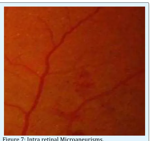
Microaneurisms formed from hyper-cellular sacs in the
Open Access Journal of Ophthalmology
5
Figure 7: Intra retinal Microaneurisms.
Cotton-Wool spots: Ischemia causes cystic bodies changes in the RNFL and interim will cause swelling RNFL ends with neural deposits and thus will form cotton-wool spots (Figure 8) Venous beading, looping and tortuosity, may proceeds proliferative stage as ischemia increases (Figure 9). Intraretinal microvascular abnormalities (IRMA) is a shunt runs from retinal arteriols to venule bypassing capillary bed, usually associated next to retinal ischemia (Figure 10).
Figure 8: Cotton wool spots.
Figure 9: Venous looping.
Figure 10: Intraretinal microvascular abnormalities (IRMA).
Proliferative stage
The proliferation has a cycle of three phases
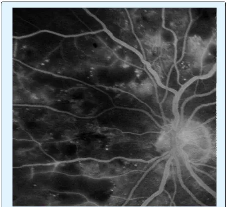
The impending phase: VEGF is upregulated when the retinal tissue reaches the state of relative ischemia and thus initiates the process of angiogenesis, in this stage level of VEGF concentration is high in the vitreous [14], and this can be noted clinically as areas of hypo perfusion on fluorescein angiograms (Figure 11).
Figure 11:Fluorescein angiogram shows areas of hypo perfusion and capillary dropout.
Figure 12: Retinal Neovascularization with hemorrhage.
The regression stage: Neo vessel appears stripped in its early stages as it starts to regress and reduce it diameter (Figure 13),
fibro vascular membrane becomes more visible forming fibro-vascular tissue which may contract causing traction retinal detachment in the areas of fibro-vascular tissue attachment with posterior hyaloid [16].
Vitreous hemorrhage is one of the most common complications of proliferative stage and it is induced by contraction of fibro-vascular tissue or spontaneous bleeding [17].
Clinically non proliferative diabetic retinopathy is monitored by glycemic control while proliferative diabetic retinopathy requires pan retinal photocoagulation treatment in the absence of diabetic macular edema while in the presence of diabetic macular edema, VEGF blockade agents can be introduced to address both macular edema and proliferation and pan retinal photocoagulation treatment can be differed to patients who are hard to follow up or treatment failure [18], however surgical management in cases of proliferative diabetic retinopathy is reserved for cases of tractional retinal detachment involving or threatening the macula and in cases of non-clearing vitreous hemorrhage.
Figure 13: Regressed Neo vascurlaztion.
Conclusion
Pathology of diabetic macular edema and retinopathy is multifactorial, understanding the involving factors is important, to individualize the treatment for every patient by targeting the underlying mechanism, sometimes the one or more mechanism is involving and sometimes the pathology changes the mechanism from one form to another. Diabetic macular edema can be caused by vascular element or vascular element; however non-proliferative diabetic retinopathy features mainly microaneurisms due to metabolic changes while proliferative diabetic retinopathy is caused by upregulated VEGF triggering the process of angiogenesis.