N
europhysiology/pathophysiology content is a frequent source of anxiety for undergraduate students and their instructors. This learning module supple-ments traditional lecture and overhead presentations to offer a novel, nonthreatening, and entertaining introduction to neuropathology. The module is based on a ridiculous analogy between the human brain and the cauliflower. This module has been used with both underclassmen and more advanced health science undergraduate students and has produced enthusiastic student responses while deescalating both student and instructor anxiety.ADV PHYSIOL EDUC 24: 22–29, 2000.
Key words:neuroanatomy; neurophysiology; teaching neurophysiology
Human physiology and pathophysiology coursework is emphasized in diverse undergraduate programs. Students of clinical laboratory science and the allied health professions as well as physical education, pre-medicine, and physiology students enroll in under-graduate physiology classes in increasing numbers every year (3). These students come with disparate levels of interest in, and preparation for, physiology coursework. One commonality among all undergrad-uate physiology/pathophysiology students is the level of anxiety evoked by the seemingly innocuous state-ment, “Next week, we’ll start neuro.” Knowledge acquired during states of anxiety is likely to be con-crete, transient, and result in poor educational out-comes (4).
During the 1998 –1999 academic year, we developed a nonthreatening learning module that has successfully deescalated the student anxiety historically triggered by the neurophysiology content of our undergraduate pathophysiology course. This module reviews
neuro-anatomy and physiology while simultaneously intro-ducing pertinent neurophysiology. Thus novice stu-dents are motivated by an immediate understanding of the clinical relevance of the material rather than being bewildered by a series of seemingly disembod-ied strange-sounding names. This module has en-hanced student learning and allowed us to expand the neuropathology content of our course. Most impor-tantly, it has generated an unprecedented enthusiasm for neurophysiology and neuropathology among our students. The development of the module presented in the following sections of this paper was inspired by the observation of neurologist and neuroanatomist Stephen Goldberg (1) that, “the best memory tech-niques involve the use of ridiculous associations.”
MODULE OBJECTIVES
1. To review the anatomy and physiology of the brain
3. To correlate brain pathophysiology and clinical presentation
MATERIALS
● One large well-proportioned cauliflower
● One large knife with a sharp point
● One large black plastic trash bag
● Handouts/overheads created with Microsoft Word
PREPARATION
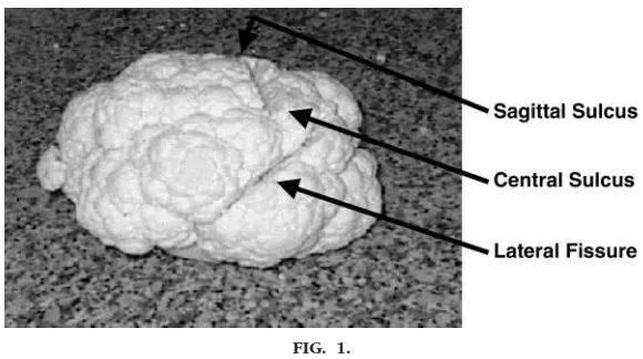
With the use of a sharp knife, trim the green leaves from the cauliflower. Etch the sagittal sulcus, central sulcus (of Rolando) and lateral fissure (of Sylvius) on the surface of the cauliflower (see Fig. 1). Place the cauliflower, core-side down, in the trash bag. Before class, place the trash bag on the podium in a manner that reveals the general shape of its contents.
METHODS
Students are provided with printouts of all overheads that are presented during the lecture. We have found that these handouts free students from the distracting aspects of obsessive note taking. The handouts have ample white space to allow the students to jot down insights that develop during the presentation. The lecture begins with a traditional didactic format. An overhead of the gross anatomy of the brain is dis-played, and the structures that comprise the central
nervous system are identified. This slide is based on similar figures in classic physiology texts (2) but has historically produced a glazed expression in the eyes of many students. As the second or third student nods off, the instructor gestures toward the trash bag and speculates that use of an actual brain may clarify this discussion of brain pathophysiology.
The lecturer briefly explains that several years ago a very selfless patient, a victim of Parkinson’s disease, donated this brain for teaching purposes. This state-ment can be counted on to refocus student attention. The cauliflower is resurrected from the trash bag, and, despite the relieved laughter that it produces, the lecturer never concedes that this “brain” is, in fact, only a vegetable.
CORTICAL STRUCTURES
The lecturer orients the students to the anterior and posterior aspects of the brain and comments that the class is “obviously” viewing the lateral aspect of the left and dominant hemisphere of this brain. First, gyri (elevations), sulci (depressions), and fissures (smaller depressions) on the surface of the brain are noted. Next, the sagittal and central sulci and the lateral fissure are identified (see Fig. 2).
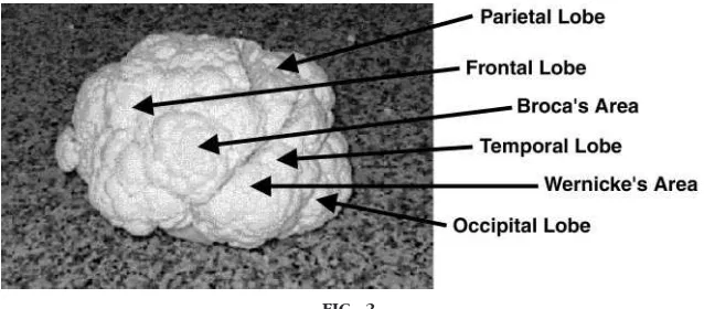
The relationship of these landmarks to the frontal, parietal, temporal, and occipital lobes is demon-strated. However, discussion of the pathophysiology in these areas is deferred. The motor cortex of the frontal lobe and the sensory cortex of the parietal
FIG. 1.
lobe (just anterior and posterior to the central sulcus) are identified. Unless an extremely well-proportioned cauliflower has been selected, the lecturer is reduced to searching out a mostly imaginary cerebellum.
The lecturer locates and, using the knife, resects “Bro-ca’s area” (a small flowerette on the posterior aspect of the inferior temporal gyrus of the motor cortex of the frontal lobe). This introduces the common deficits in the production and/or comprehension of speech (aphasias) that result from specific cortical lesions. While exhibiting the resected Broca’s area to the class, the instructor explains that Broca’s aphasia is a motor aphasia that interferes with the production but not the comprehension of speech. Broca’s aphasia is characterized by halting telegraphic speech (e.g., “Student . . . came . . . class.”). Broca’s apha-sia is sometimes termed nonfluent aphaapha-sia. Similarly, “Wernicke’s area” (a small flowerette on the posterior aspect of the superior temporal gyrus near the audi-tory area in the temporal lobe) is located and re-sected. Wernicke’s aphasia is characterized by an in-ability to comprehend language. Speech production is preserved (e.g., “The student trolley yes sir and base-ball too!”) but meaning is absent. Wernicke’s aphasia is sometimes termed fluent aphasia. It is useful at this time to note the proximity of Broca’s area and Wer-nicke’s area on the cortex. Because both areas are perfused by the middle cerebral artery, isolated dam-age to only one area is rare. Most aphasia is global and has elements of impaired speech production as well as language comprehension.
Throughout the demonstration, the lecturer ostenta-tiously places all resected structures on the podium in
clear view of the class. These structures will be used again later in the demonstration.
MIDLINE STRUCTURES
A complete midline transection is made along the ante-rior-posterior axis of the cauliflower. The lecturer dem-onstrates that this cut, along the sagittal plane, bisects the brain into a right and left hemisphere. Students can be oriented to the frontal and coronal planes at this time. The instructor sets the “right” hemisphere of the brain aside. An overhead illustrating a prototype neuronal pathway is displayed. Similar illustrations are found in all classic physiology texts (2). The instructor notes that the neuron, the basic functional of the brain, has evolved to transmit information and produce behavior. The com-ponents of a neuronal pathway are briefly reviewed (i.e., dendrites, cell body, axon, synaptic buttons, synapse, neurotransmitters, and receptors). The production of the neurolemma by oligodendroglioma cells and the purpose of myelinization and saltatory conduction are explained. A pathway is defined as a series of neurons. A tract is defined as a bundle of pathways. The student is reminded that a tract is the central nervous system equivalent of a nerve in the peripheral nervous system.
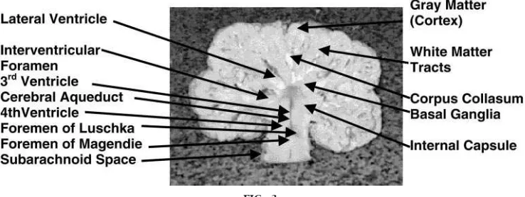
The organization of neuronal cell bodies in the cortex and the myelinated neuronal axons in the deeper white matter tract of the cerebrum is reviewed. The instructor exhibits the medial aspect of the “left” hemisphere to the class. Elaborating on the analogy between the brain and the cauliflower, the instructor draws the students’ attention to the clustering of neuronal cell bodies in the gray matter of the cortex (i.e., the small buds on the surface of the cauliflower
FIG. 2.
flowerettes) and the convergence of the myelinated axon pathways to form deeper white matter tracts (the stalks of the flowerettes; see Fig. 3). The instruc-tor comments that the white matter tracts from sen-sory and motor areas of the cortex come together as they pass through the internal capsulae of each hemi-sphere and proceed caudally to the brain stem and ultimately the spinal cord. The instructor notes that, because many tracts converge in the internal capsule, lesions of the internal capsule cause massive neuro-logical deficits and have profound clinical conse-quences. At this point, the instructor also points out the corpus callosum and notes that it is the site of most interhemispheric information transfer.
The “basal ganglia” is resected next, and the lecturer allows the class to examine the structure. The instruc-tor states that lesions of the basal ganglia result in involuntary and unexpected motor movements along with akinesia (decreased voluntary movement). The instructor reminds the class that this brain was do-nated by a victim of Parkinson’s disease. The class is instructed to pay special attention to the color of the substantia nigra, a nucleus of the basal ganglia. The substantia is normally dark due to the pigmentation of dopamine-producing cells. However, in Parkinson’s disease the dopaminergic cells are lost, and the sub-stantia takes on the pale color that they observe. Loss of dopamine leaves Parkinson’s patients with a rela-tive excess of acetylcholine. Parkinson’s disease is characterized by resting tremor, rigidity, and postural instability. It is useful at this point to contrast move-ment disorders associated with pathology of the basal ganglia with movement disorders associated with
le-sions of the cerebellum. Cerebellar lele-sions produce intentional tremors, difficulty coordinating fine motor movements, and gait ataxia (lack of coordination dur-ing voluntary movement).
Next, the ventricular system is introduced. Explaining that the brain is really a hollow tube with enlarged, cerebral spinal fluid (CSF)-filled ventricles, the instruc-tor uses the knife to identify and carve out the left hemisphere’s lateral ventricle, the interventricular fo-ramen (of Monro), the third ventricle, the cerebral aqueduct (of Sylvius), the fourth ventricle, the fora-men of Luschka, and the forafora-men of Magendie. (see Fig. 3). The three meningeal membranes are identi-fied: innermost the pia mater, the middle arachnoid membrane, and the outermost dura mater that clings to the periosteum of the skull. The instructor defines each area as it is carved out and explains that CSF circulates through the ventricular system to the sub-arachnoid space and is reabsorbed by the sub-arachnoid villi in the superior sagittal sinus. The instructor ex-plains that an obstruction anywhere in the system, an increase in the production of CSF, or a decrease in reabsorption of CSF will increase pressures in the ventricular system. The Monroe-Kellie hypothesis, the pathophysiology of increased intracranial pressure, and herniation syndromes can be introduced at this point if this content is consistent with the course objectives.
CEREBRAL CIRCULATION
At this point, the left hemisphere, mangled by dissec-tion and resecdissec-tion, is set aside, and the instructor
FIG. 3.
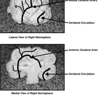
displays the right hemisphere to the class. This hemi-sphere is used to describe cerebral circulation, the functional and behavioral capabilities of the cerebral lobes, and the clinical manifestations of specific cor-tical lesions. The expectation is that, at the comple-tion of this seccomple-tion, students will be able to predict the pathophysiology associated with the occlusion of specific cerebral arteries.
The brain’s blood supply is provided by two arterial systems: the internal carotid system and the vertebral system. The instructor traces the paths of these two systems on the surfaces of the brain. The bifurcation of the internal carotid artery into the anterior and
middle cerebral artery is illustrated. The anterior ce-rebral artery supplies blood to ;80% of the midline surface of the cerebrum. The middle cerebral artery supplies blood to ;80% of the lateral surface of the cerebrum. The vertebral system supplies blood to the cerebellum and the remaining 20% of the lateral and midline surfaces of the posterior cerebrum, including the occipital lobe (see Fig. 4).
MOTOR SENSORY DEFICITS
The instructor uses the right hemisphere to visually remind students that the motor and sensory cortexes are located in the frontal and parietal lobe,
respec-FIG. 4.
tively, anterior and posterior to the central fissure and continue from the Sylvian fissure up the lateral aspect of the cortex to the sagittal sulcus and continue downward on the midline structures. The instructor maps the motor and sensory tracts on the surface of the right hemisphere and emphasizes that midline lesions of either the motor or sensory tract impair movement and sensation in the contralateral lower extremities (in this case the left leg), whereas lesions of the motor or sensory tract on the lateral aspect of either the cortex or sensory tract impair movement of the contralateral right upper extremities (in this case, the left arm). In contrast, cerebellar lesions affect move-ment on the side of the body ipsilateral to the lesion.
BEHAVIORAL DEFICITS
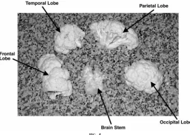
The previous discussion leaves students with the im-pression that discrete areas of the brain control spe-cific functions. It is important to advise students that the brain, in fact, functions as an integrated whole. However, certain areas of the brain are more involved in some functions than others (see Fig. 5).
The frontal lobe.The instructor breaks off an area of the brain roughly corresponding to the right frontal
lobe and explains that, in addition to motor function, the frontal lobe is involved in planning, problem solv-ing, personality, inhibition of impulses, and the initi-ation of behavior. Thus a patient with a frontal lobe lesion may have problems dressing, reacting appro-priately in social situations, and in starting or com-pleting a task. The instructor can reinforce Broca’s aphasia at this point by reminding the class that le-sions of the right hemisphere are not associated with motor aphasia.
The parietal lobe.Next, the parietal lobe is broken off. In addition to perception of sensory input, the parietal lobe is involved in interpretation of visual information, right/left discrimination and the aware-ness of body parts, and the orientation of the body in space. Patients with parietal lobe lesions tend to ig-nore the side of the body contralateral to the lesion, have problems realizing the relationship of their con-tralateral side to potential hazards, and do not receive accurate sensory input from the side of the body contralateral to the lesion. These deficits put them at serious risk for injury.
FIG. 5.
ranging from cortical blindness (complete loss of vi-sion without injury to the eye or optic nerve) to field cuts (visual loss in selected portions of the visual field).
The brain stem. After all of the lobes have been resected and carefully placed on the desk, the instruc-tor is left holding the brain stem. The instrucinstruc-tor char-acterizes the brain stem as the most primitive part of the brain controlling respiration, heart beat, alertness, and sense of balance. The vomiting center (the floor of the 4th ventricle) and the midbrain reticular acti-vating system (consciousness) are also identified. Le-sions of the brain stem are often associated with vertigo. Lesions of the brain stem threaten survival by interfering with breathing and heart beat.
WHAT TO DO WITH THE LEFTOVERS
The similarity between the mammalian brain and the cauliflower is a useful device for achieving the objec-tives of this module. Whereas the association is truly ridiculous, it facilitates review of the anatomy and physiology of the brain (objective 1) while simulta-neously introducing students to common pathologi-cal alterations in brain physiology (objective 2) in a manner that eliminates anxiety as a barrier to learn-ing. We have presented the module as a monologue. However, during the presentation, we take every op-portunity to engage the student: questions are asked and elicited, clinical states are dramatized, and dia-logue is encouraged. The final objective of the mod-ule is the correlation of brain pathophysiology with clinical presentation (objective 3). The ground work for this objective was laid during the presentation, and the objective is met by reviewing the presenta-tion content. The instructor uses the resected areas of the brain, carefully and conspicuously arranged on
tor and sensory deficits is acceptable. If necessary, the instructor will cue the students by retrieving the “in-ternal capsule” from the leftovers on the podium and carefully replacing it in the remains of the left hemi-sphere.
Describe the deficits associated with occlusion of the left middle cerebral artery.
Students must correlate knowledge of the cerebral circulatory system and the functional properties of the cerebral lobes. A response that includes paralysis/ paresis and sensory loss in the right upper extremity and aphasia is acceptable. Also the behavioral deficits associated with lesions of the frontal, parietal, and temporal should be included in the student re-sponses. If necessary, the instructor will cue the stu-dents by retrieving Broca’s area and Wernicke’s area, asking students to replace them in the frontal and temporal lobes, and by reviewing anterior cerebral circulation. The appropriate lobes of the brain can be displayed to remind students of the behavioral deficits associated with occlusion of the left middle cerebral artery.
Describe the signs and symptoms of occlusion of the vertebral arterial system.
DISCUSSION
The learning module presented in this paper repre-sents a typical presentation for our undergraduate pathophysiology course and amply fills our 65-min class period. However, the content of the module can be modified to meet the needs of specific groups of students and the time available. We have tailored this presentation to meet the requirements of more ad-vanced undergraduate clinical courses and graduate pathophysiology courses. It is our observation that the ridiculous nature of the analogy between the brain and the cauliflower effectively engages students and demystifies neuroanatomy and pathophysiology content. Over the past year, student evaluations of this module have confirmed this observation. The following three responses typify student comments:
“I really enjoyed your (neuropathology) lecture. Great technique with the head of cauliflower. It helped in making your points clear, and it helped me visualize the areas you were discussing. You definitely held my attention throughout the class.”
“Your lecture was wonderful. I found it extremely interes-ting . . . the brain exhibit was great. Your lecture burned the topic into my brain!! Excellent learning tool.”
effect) and the anatomy of a large white protuberance of the mustard family is an effective device for intro-ducing undergraduate health-care students to neuro-pathology. From the instructor’s point of view, this lecture is simply fun to do. It is hard to tell whether we or the students have been more astonished and delighted by their reactions to this learning module. However, we would like to add a word of caution to others preparing this recipe—a little ham greatly en-hances the flavor.
Address for reprint requests and other correspondence: J. Masters, 408 Robinson Hall, College of Nursing, Northeastern Univ., Boston, MA 02115.
Received 8 February 2000; accepted in final form 8 August 2000
REFERENCES
1. Goldberg S.Clinical Neuroanatomy Made Ridiculously Sim-ple. Miami, FL: MedMaster, 1988.
2. Guyton AC.Textbook of Medical Physiology(9th ed). Philadel-phia: Saunders, 1996.
3. McCleary VL, Aasen G, and Slotnick HB.Predictors of suc-cess in undergraduate human physiology.Am J Physiol Advan Physiol Educ22: S119 –S126, 1999.