Brain Research 881 (2000) 73–76
www.elsevier.com / locate / bres
Short communication
Differential Schwann cell migration in adult and old mice: an in vitro
study
a a b
Arnaldo R. Santos Jr. , Maria Lucia F. Wada , Francesco Langone , Alexandre
c ,
*
L.R. Oliveira
a
Department of Cell Biology, State University of Campinas–UNICAMP, CP 6109, CEP 13089-970, Campinas, SP, Brazil
b
Department of Physiology and Biophysics, State University of Campinas–UNICAMP, Campinas, SP, Brazil
c
Department of Anatomy, State University of Campinas–UNICAMP, Campinas, SP, Brazil
Accepted 8 August 2000
Abstract
The influence of aging on Schwann cell (SC) proliferation, migration and viability was studied in vitro. SCs were cultured in Ham F-10 medium enriched with 20% fetal calf serum (FCS), 40% FCS or collagen I gel plus 20% FCS. The migration of adult mice derived SCs was stimulated with FCS and collagen. With aging, SC migration, multiplication and viability decreased, indicating that ideal culturing conditions should be adjusted. 2000 Elsevier Science B.V. All rights reserved.
Theme: Development and regeneration
Topic: Glia and non-neuronal cells; Aging process
Keywords: Schwann cell; Cell culture; Collagen; Cell migration; Aging; Mouse
Peripheral nerve regeneration occurs as a result of a were used. After dissection, the nerves were reduced into series of events, which involve axonal regeneration and fragments about 1 cm long and washed in Ham F-10 reorganization of the extracelullar microenviroinment medium (Sigma) supplemented with 20% fetal calf serum [2,4,5,11]. After nerve lesion, Wallerian degeneration takes (FCS) and 100 mg / ml of gentamicin (Schering-Plough). place distally to the lesion. This process is characterized by The fragments were then cut in smaller, 2 mm long pieces macrophage invasion and Schwann cell (SC) multiplica- and cultured in culture plates with six wells (Corning / tion, mainly into the distal stump [1,2,8]. Such newly Costar) at 378C for 20 days. Three different experimental produced SCs organize themselves within the basal lamina conditions were used: (1) Ham F-10 medium sup-left by degenerated axons, originating the so-called ‘bands plemented with 100mg / ml of gentamicin and 20% FCS,
¨
of Bungner’. (2) Ham F-10 medium supplemented with 100 mg / ml of
Taking into account that with aging, nerve regeneration gentamicin and 40% FCS, or (3) Ham F-10 medium is less successful [6,10] and considering the importance of supplemented with 100 mg / ml of gentamicin and 20% the SC for the development of this process, the aim of this FCS in well culture plates coated with 1 ml of collagen I study was to investigate in vitro its multiplication and gel. The collagen was extracted according to the method migration capacity with aging. Also, we have investigated described by Schor [9] and was prepared with 0.9 ml of the importance of collagen and serum factors for SC collagen solution, 0.05 ml of 4% NaHCO and 0.05 ml of3
migration and multiplication. 10 times concentrated Ham F-10 medium (Sigma). Within
For this study, sciatic nerves from adult (8 months old, the incubation period, the total number of migrant cells n55) and old (2 years old, n55) C57BL / 6J male mice was evaluated on days 1, 2, 4, 6, 8 and 10 in all experimental conditions. On the 20th day, the culture medium was collected and the non-adherent cells were
*Corresponding author. Tel.: 155-19-788-7391; fax.
155-19-289-counted with an Olympus IX-50 inverted microscope with
3124.
E-mail address: [email protected] (A.L.R. Oliveira). a phase contrast system.
74 A.R. Santos et al. / Brain Research 881 (2000) 73 –76
After 15 or 20 days of culture, the samples were fixed in with anti-S-100 antibody showed a bipolar morphology 10% formalin for 1 h and washed in phosphate buffered with thin- and long-cell prolongations, except in the saline (PBS) 0.1 M in pH 7.2 at 378C. In order to block nuclear region (Fig. 2). SC counting revealed that explants nonspecific staining, the specimens were incubated for 1 h from adult animals cultured with 20% FCS plus collagen I
with 1% bovine serum albumin (BSA, Sigma) in PBS, gel or 40% FCS displayed an increase of SCs when
washed and the monoclonal anti-S-100 antibody (dilution compared to samples cultured only with 20% FCS. On the 1:300) was applied. The samples were rinsed and the other hand, the explants from old animals showed a
anti-rabbit CY-3 secondary antibody was added. decreased SC number in relation to adult animals. An
In all experimental conditions, cells from adult or old important finding was that the number of SCs derived from animals were able to migrate from the explants. The cell old animals did not increase substantially when the migration pattern from adult or old animals was very medium was enriched with FCS or collagen I (Fig. 1A). similar except that in general, the cells from adult animals Schwann cell autograft produced in vitro has been showed a more intense migration rate than those from old reported as a novel method for repairing long gaps [3,5] animals. Also, an increase of migrating cells was propor- following extensive peripheral nerve lesions. However, tional to the increase of FCS concentration. The cells that with aging, there is evidence that SCs decrease in their grew on collagen I gel showed the highest migration rate capacity to multiply and produce neurotrophic factors,
(Fig. 1). basal lamina components and myelin [12]. Considering the
After 15 days of culture, we observed SCs migrating relative importance of SCs for nerve regeneration and from explants to the culture plate or to collagen I gels in taking into account that a percentage of peripheral nerve all experimental conditions. The Schwann cells labeled lesions occur in middle age or elderly individuals, we have investigated the behavior of SCs obtained from old mice, submitted to different experimental conditions. Under these experimental conditions, it was possible to determine if extracellular stimuli would be able to increase the survival, migration and proliferation rates of the SCs.
With regard to the migration of SCs, both adult and aged cells started migrating around the fourth day, but with different rates. Basically, migration in the old mouse-derived cells (OMDC) and adult mouse-mouse-derived cells (AMDC), when cultured with FCS 20 and 40%, was similar up to the sixth day. On the other hand, culturing with collagen stimulated SC migration only in the AMDC. These findings show that the SCs retain its migratory ability with aging but the capacity to respond to extracellu-lar stimuli may be reduced. This fact can be related with the capacity to synthesize and express receptors for matrix components, such as collagen, fibronectin and basal lamina elements, which are essential for cell migration [7,11].
In this context, we observed that the viability of the SCs in culture was greatly increased when the medium was supplemented with FCS 40% or with collagen plus FCS 20% (Fig. 1B). Such results reinforce the hypothesis that the absence of extracellular stimuli as well as the relation with other cell types which synthesize trophic substances, as well as cytokines, may be a strong factor in changing the behavior of SCs when in vitro. With regard to the SCs from old mice, the viability assay showed that these cells are even more sensitive and almost all of them detached from the plate after 20 days of culture. Interestingly, when the medium was supplemented with FCS 40%, cell detach-ment was considerably reduced. The results were even better when collagen was added. Taken together, our results reinforce the fact that cultured SC behavior is
Fig. 1. (A) Migration of cells from the sciatic nerves explants from adult
altered compared to that shown during in vivo Wallerian
or old animals in the different experimental conditions. (B) Counting of
A.R. Santos et al. / Brain Research 881 (2000) 73 –76 75
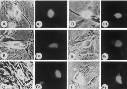
Fig. 2. Phase contrast and immunocytochemical labeling (S-100) of the Schwann cells cultured in the different experimental conditions. (A–C) represent adult derived cells and (D) to (F), old mouse-derived cells. (A), (A1), (D), (D1), cultured with 20% FCS. (B), (B1), (E), (E1), cultured with 40% FCS. (C), (C1), (F), (F1), cultured on collagen I gel with 20% FCS. Scale bar525mm.
with extracellular matrix components as well as trophic References substances may increase cellular viability and make the
cell behavior closer to the in vivo conditions. Also, with [1] W. Beuche, R.L. Friede, The role of non-resident cells in Wallerian degeneration, J. Neurocytol. 13 (1984) 767–796.
aging, all the behavioral alterations are sharper and cellular
[2] L.B. Dahlin, Prevention of macrophage invasion impairs
regenera-migration as well as multiplication and viability in culture
tion in nerve grafts, Brain Res. 679 (1995) 274–280.
are greatly reduced. These facts should be taken into
[3] H. Fansa, G. Keilhoff, G. Forster, B. Seidel, G. Wolf, W. Scheneider,
account and studied further in order to define ideal Acellular muscle with Schwann-cell implantation: An alternative culturing conditions for SCs at different donor ages, which biologic nerve conduit, J. Rec. Microsur. 15 (1999) 531–537.
will be crucial for the development of nerve repair [4] S.M. Hall, Regeneration in cellular and acellular autografts in the peripheral nervous system, Neuropathol. Appl. Neurobiol. 12 (1986)
techniques employing cultured SCs.
401–414.
[5] T.W. Hudson, G.R.D. Evans, C.E. Schmidt, Engineering strategies for peripheral nerve repair, Clin. Plas. Surg. 26 (1999) 617–628. [6] E. Kerezoudi, P.K. Thomas, Influence of age on regeneration in the
Acknowledgements peripheral nervous system, Gerontology 45 (1999) 301–306.
[7] A.D.O. Levi, P. Guenard, P. Aebischer, R.P. Bunge, The functional characteristics of Schwann cells cultured from human peripheral
´
We are grateful to Prof. Dr. Aureo T. Yamada for
nerve after transplantation into a gap within the rat sciatic nerve, J.
providing the secondary antibody used in this study. The Neurosci. 14 (1994) 1309–1319.
authors are also thankful to Prof. Dr. Mary Anne H. Dolder [8] L. Lubinska, Patterns of Wallerian degeneration of myelinated fibers
76 A.R. Santos et al. / Brain Research 881 (2000) 73 –76
phrenic nerve. Interpretation of the role of axoplasmatic flow of the [11] K. Torigoe, K. Hashimoto, G. Lundborg, A role of migratory trophic factor, Brain Res. 233 (1982) 227–240. Schwann cells in a conditioning effect of peripheral nerve regenera-[9] S.L. Schor, Cell proliferation and migration on collagen substrata in tion, Exp. Neurol. 160 (1999) 99–108.
vitro, J. Cell Sci. 41 (1980) 159–175. [12] D.E. Weinstein, The role of Schwann cells in neural regeneration, [10] K. Tanaka, H.D. Webster, Myelinated fiber regeneration after crush Neuroscientist 5 (1999) 208–216.