Synthesis of 60- and 72 kDa heat shock proteins
in early porcine embryogenesis
Y.T. King
a, W.C. Lee
a, M.S. Gao
c, J.L. Wang
a,
C.F. Tu
b, S.C. Wu
b, Y.H. Kuo
b,∗aDepartment of Comparative Medicine, Pig Research Institute Taiwan,
P.O. Box 23, Chunan, Miaoli 35099, Taiwan, ROC
bDepartment of Applied Biology, Pig Research Institute Taiwan,
P.O. Box 23, Chunan, Miaoli 35099, Taiwan, ROC
cDepartment of Life Science, National Tsing Hua University, Hsinchu, Taiwan, ROC
Received 20 July 1999; received in revised form 2 May 2000; accepted 16 June 2000
Abstract
Proteins of selected embryonic stages were metabolically labeled with [35S]-methionine and
analyzed by two-dimensional SDS-polyacrylamide gel electrophoresis (2-D PAGE) to study protein expression from 4- to 8-cell to blastocyst stage of porcine embryos. Two proteins with molecular weights of 60 and 72 kDa were de novo synthesized during the 4- to 8-cell stage were the earliest that were detected. They were identified as HSP60 and HSP72 according to their locations on 2-D autoradiography and the immunoblotting result of anti-HSP 60 and HSP 72 antibodies of 1-cell stage of porcine embryos. In protein translation in early pig embryogenesis the timing of their synthesis suggests that HSP60 and HSP72 play significant roles as chaperones. © 2000 Elsevier Science B.V. All rights reserved.
Keywords: Pig-embryology; Embryogenesis; Heat shock protein; Autoradiography; Western blotting
1. Introduction
Heat shock proteins (HSP) are highly conserved stress proteins, and are recognized as molecular chaperones (Craig, 1993). According to their molecular weights they are divided into five families 110, 90, 70, 60 kDa, and the small HSP. The HSP70 family was categorized into a constitutive and inducible form (Lee et al., 1991). HSP72 is expressed constitutively and in living cell HSP70 is heat-inducible (Craig, 1993). Inducible HSPs are expressed when living cells are subjected to stressful elements such as hyperthermia and heavy metals.
∗Corresponding author. Tel.:+886-37-672352; fax:+886-37-660104.
E-mail address: [email protected] (Y.H. Kuo).
While accompanying ATP hydrolysis, both HSP60 and HSP70 can complete the rena-turing process (Craig, 1993). Following the ATP hydrolysis, only the HSP70 family binds to unfolded polypeptides and release renatured proteins. HSP60 is formed by several sub-groups, as well its chaperone function also requires ATP hydrolysis (Hunter, 1974).
Although HSP70 family synthesis occurs in the early stage of mouse embryogenesis (Bensaude et al., 1983; Dvork et al., 1995; Christians et al., 1997), the role of HSP60 and HSP70 in porcine ontogeny has not yet been established. Therefore, only the profile of the de novo synthesis of porcine embryo from 4- to 8-cell stage to blastocyst was studied.
During embryogenesis the [35S]-methionine labeling method and 2-D PAGE were used to analyze the protein profiles. Pig embryonic proteins that occur in the early stage of embryogenesis were labeled metabolically. The autoradiography of 2-D protein profile was compared with that of the silver stain of porcine smooth muscle cell (SMC) and the Western blot from 1-cell stage pig embryos. Those results indicated that in the early stage of porcine embryogenesis, HSP60 and HSP72 were the first detectable set of the de novo synthesized proteins to develop.
2. Materials and methods
2.1. Synchronization and superovulation
Landrace gilts of 7–8 months old, with a body weight of 100–150 kg, was selected as the embryo donor. All pigs were fed 1.2 kg commercial feed twice daily, and water was provided ad libitum. The gilts were synchronized by a 15 days morning feeding of Regumate® (con-taining 4% alterenogest, 20 mg per day; Hoechst, France) that was mixed with commercial feed. Twenty-four hours after the last feeding of Regumate®, superovulation was induced by an injection of PMSG (2000 IU, i.m., China Chem. and Pharm., Taiwan) and an injection of hCG (1750 IU, i.m., China Chem. and Pharm., Taiwan), which subsequently occurred 76–78 h after the PMSG injection. The gilts were then artificially inseminated with frozen semen 24 h after receiving a hCG injection.
2.2. Embryo collection
Before the embryos were collected, the pigs were made to fast overnight. Gilts were calmed by an injection (i.m.) of stresnil (2 mg/kg, Janssen Pharmaceutical, Belgium) and atropine sulfate (0.04 mg/kg, China Chem. and Pharm., Taiwan), and then were initially anesthetized by an injection of sodium pentothal (10 mg/kg, Abbott Australasia Pty Ltd., Australia) into an ear vein. Anesthesia was maintained in an oxygen gas mixture throughout the operation via a closed-circuit system using 4% halothene (Zeneca Ltd., UK). Pig em-bryos of different stages were flushed from the Fallopian tubes or the uterus horn into dishes with Dulbecco’s PBS (Gibco/BRL, USA) containing 0.4% BSA (Fraction V, Sigma, USA).
2.3. Metabolic labeling
At different developmental stages, fresh embryos were cultivated in a 37◦C incubator
1991). Two embryos were mouth pipetted into100ml of Dulbecco’s PBS labeling media
and cultured at 37◦C for 3 h. The labeling media contained 1.67 mCi/ml [35S]-methionine (specific activity<1000Ci/mmol, Amersham, USA). After isotopic labeling, the embryos were washed three times with Dulbecco’s PBS containing 0.4% BSA solution, and they were then lysed in a 20ml lysis buffer for 2-D PAGE (Lee et al., 1991).
2.4. Gel electrophoresis
The methods of 2-D PAGE on the embryonic proteins were performed according to those specified by Laemmli (1970), O’Farrell (1975) and Lee et al. (1996). Approximately 30ml
of embryonic protein was loaded onto the isoelectrofocusing (IEF) gel and electrophoresised at 400 V for 16 h and then at 800 V for 1 h. Subsequently, the IEF gels were laid onto 9% SDS polyacrylamide slab gels with 4.75% stacking gel in the second dimension (30 mA per gel for 4 h). The slab gels were then vacuum dried on a piece of 3 M filter paper by a gel drier, and then exposed to the intensification screen. The profiles of 4- to 8-cell and morula were exposed for 16 h and while the others were exposed for 72 h. The 2-D gel images were captured by a phosphoimager (Phosphoimager 445SI, Molecular Dynamics, USA). The images were then analyzed by ‘The Software Package for the Analysis of 2-D Gel image’ (Pdi, 1995).
2.5. Immunoblot analysis
The proteins from 15 1-cell stage embryos were pooled for immunoblot analysis. The pro-tein spots on the 2-D PAGE were transferred onto the nitrocellulose membrane (Hybond-C extra, Amersham, USA) by a semi-dry blotter (OWL Scientific Plastics Inc., USA). This membrane was incubated for 1 h with 3% gelatin containing TTBS (20 mM Tris–HCl, pH 7.4, 500 mM NaCl, 0.05% Tween 20) and then rinsed with TTBS. Subsequently, the membrane was incubated with monoclonal antibodies to HSP70/72 and HSP60 (1:500 di-luted in TTBS containing 1% gelatin, Stressgene, USA) at room temperature for 1 h. After three TTBS washes, the membrane was reacted with goat-anti-mouse antibody conjugated with alkaline phosphatase (diluted 1:1000 in TTBS containing 1% gelatin, Sigma, USA) at room temperature for 30 min. The membrane was again rinsed three times with TTBS and developed at room temperature in developing buffer (14 mg of nitro blue tetrazolium, 0.7% N,N-dimethylformamide, 30 mg of 5-bromo-4-chloro-3-indolul phosphate per 100 ml, 1 mM MgCl2, and 100 mM NaHCO3, pH 9.8).
3. Results
during the early stage of porcine embryogenesis, a 3 h autoradiographical labeling period and a 16–72 h exposure time is required.
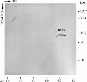
Two proteins, with molecular weights of 60 and 72 kDa were the first set of de novo synthesized proteins found at the 4- to 8-cell stages (Fig. 1). Fig. 1 also illustrates the fact that the number of proteins increased throughout the different embryogenic stages. Selected proteins 18 out of 20 were successfully located in SMC 2-D PAGE (Fig. 2). A Western blot of 1-cell stage embryos identified HSP60 and HSP72 (numbered 1 and 2, respectively) (Fig. 3). It also illustrated that HSP60 and HSP72 existed before the first embryonic segregation (Fig. 3). A unique protein numbered 12 (Fig. 2) was only detected in early blastocyst and deserves more study. HSP60 and HSP72 were confirmed as the de novo synthesized proteins at porcine 4- to 8-cell stage. These results imply that these two HSPs play vital roles in porcine embryogenesis.
4. Discussion
Hunter (1974) chronicled the development of porcine embryogenesis. As well the protein metabolism of mammalian oocyte has been intensively studied (Morange et al., 1984). The studies of animal embryos indicate that the blocking stages vary among animals. For the pig, for example, it was reported to occur at the 4-cell stage for pig (Davis, 1985). Edwards and Hansen (1997) indicated that in bovine embryogenesis HSP70 cognates were synthesized from the two-cell to hatched blastocyst stage (Edwards and Hansen, 1997). However, the protein profile that occurs during the early stage of porcine embryogenesis was unavailable. The study of Muller et al. (1985) indicated neither the HSPs nor the thermotolerance could be induced by heat shock (Muller et al., 1985). The expression of mouse embryonic protein have indicated that HSP72 is spontaneously expressed at the onset of zygotic genome activity, which is one of the leading genes expressed in the mouse embryo (Chastant et al., 1996). The constitutive HSP70 (i.e. HSP72 in this report) in mouse embryo is found in the 1-cell to blastocyst stage (Dvork et al., 1995). Moreover, their response to environmental changes is not expressed until the morula or early blastocyst stage (Christians et al., 1997). Crosby indicated that in the early developmental stages, especially during the 1- to 4-cell stages of ovine embryo, the protein synthesis remains high. However, it decreased to 95% during 4- to 8-cell stage, then increased at the fifth mitosis (Crosby et al., 1988). Edwards and Hansen indicated that in a bovine embryo the HSP70 family expression appeared as early as during the 2-cell stage (Edwards and Hansen, 1997). After fertilization, both HSP72 and HSP70 are synthesized in the mouse zygotic genome (Bensaude et al., 1983). However, the HSP 60, 70, and 90 families are constitutively expressed in 8-cell or blastocyst of a mouse embryo (Mirkes, 1997). The de novo synthesis of HSP60 in porcine embryogenesis was reported for the first time by our study.
Fig. 1. Stage specific protein synthesis in porcine embryo. Porcine embryonic proteins were labeled by [35S]-methionine at different developmental stages. Each gel contains protein samples from two embryos. The
Fig. 3. Western blotting of HSP60 and HSP72 in pig 1-cell stage oocyte. This 2-D PAGE result was obtained from 15 of the 1-cell stage porcine embryos. Locations of HSP60 and HSP72 are as labeled.
embryogenesis, in which the developmental program must be linearly and chronologically prepared. The evidence clearly implicates HSP in the protection of embryos from stress (Lindquist and Craig, 1988). Furthermore, Edwards and Hansen indicated that in the thermal resistance of early embryogenesis heat inducible HSP is prominent (Edwards and Hansen, 1997). The functional study of these proteins during the early developing mouse embryo
b
indicates that HSP60 and HSP72 were significant to the embryonic development. A retar-dation of mouse embryogenesis occurred if their functions were blocked by the specific antibodies (Neuer et al., 1998). The effect of HSP60 on mice embryogenesis is detected as early as on the third day (2 days earlier than that of HSP70 in embryogenesis) and contin-ued for the entire preimplantation period (Neuer et al., 1998). Although the timing of the initiation of embryonic development is different among animals, HSP60 and HSP72 are putatively the protein chaperones for embryogenesis of mammalian embryos. Our study in-dicates that after the blocking stage in porcine embryogenesis HSP60 and HSP72 were the two foremost synthesized de novo chaperones. Their maternal oriented resources probably important for the embryo before the blockage.
5. Conclusions
The simultaneous expression of both HSP60 and HSP72 in early porcine embryogenesis has been performed in this study for the first time. The acquisition of these two chaperones after the blocking stage implies their necessity for porcine embryogenesis before the be-ginning of further embryonic segregation. The result adds more information of the possible function of HSP60 and HSP72 on early porcine embryogenesis.
Acknowledgements
The authors would like to thank our colleagues at The Pig Research Institute, Taiwan. Drs. K.H. Lee, C.Y. Liu and S.H. Liu are also thanked for their valuable opinions.
References
Bensaude, O., Babinet, C., Morange, M., Jacob, F., 1983. Heat shock proteins, first major products of zygotic gene activity in mouse embryo. Nature 305, 331–333.
Craig, E.A., 1993. Chaperones: helpers along the pathways to protein folding. Science 260, 1902–1903. Chastant, S., Christians, E., Campion, E., Renard, J.P., 1996. Quantitative control of gene expression by
nucleocytoplasmic interactions in early mouse embryos: consequence for reprogrammation by nuclear transfer. Mol. Reprod. Dev. 44, 423–432.
Christians, E., Michel, E., Renard, J.P., 1997. Developmental control of heat shock and chaperone gene expression. HSP70 genes and heat shock factors during preimplantation phase of mouse development. Cell. Mol. Life Sci. 53, 168–178.
Crosby, I.M., Gandolfi, F., Moor, R.M., 1988. Control of protein synthesis during early cleavage of sheep embryos. J. Reprod. Fertil. 82, 769–775.
Davis, D.L., 1985. Culture and storage of pig embryos. J. Reprod. Fertil. Suppl. 33, 115–124.
Dvork, P., Dvorakova, D., Yoshiki, A., Ohashi, T., Kitamura, K., Kusakabe, M., 1995. Expression of paternal and maternal mitochondrial HSP70 family, hsc74, in preimplantation mouse embryos. Int. J. Dev. Biol. 39, 511–517.
Edwards, J.L., Hansen, P.J., 1997. Differential responses of bovine oocytes and preimplantation embryos to heat shock. Mol. Reprod. Dev. 46, 138–145.
Hunter, R.H.F., 1974. Chronological and cytological details of fertilization and early embryonic development in the domestic pig, Sus Scrofa. Anat. Rec. 178, 169–186.
Lee, W.C., Lin, K.Y., Chiu, Y.T., Lin, J.H., Cheng, H.C., Huang, H.C., Yang, P.C., Liu, S.K., Mao, S.J.T., 1996. Substantial decrease of heat shock protein 90 in ventricular tissues of two sudden-death pigs with hypertrophic cardiomyopathy. FASEB J. 10, 1198–1204.
Lee, W.C., Lin, K.Y., Chen, C.M., Chen, Z.T., Liu, H.J., 1991. Induction of heat-shock response and alterations of protein phosphorylation by a novel topoisomerase II inhibitor, with angulatin A, in 9L rat brain tumor cells. J. Cell. Physiol. 149, 66–76.
Lindquist, S., Craig, E.A., 1988. The heat-shock proteins. Annu. Rev. Genet. 22, 631–677.
Mirkes, P.E., 1997. Molecular/cellular biology of the heat stress response and its role in agent-induced teratogenesis. Mutat. Res. 396, 163–173.
Morange, M., Diu, A., Bensaude, O., Babinet, C., 1984. Altered expression of heat shock proteins in embryonal carcinoma and mouse early embryonic cells. Mol. Cell. Biol. 4, 730–735.
Muller, W.U., Li, G.C., Goldstein, L.S., 1985. Heat does not induce synthesis of heat shock proteins or thermotolerance in the earlist stage of mouse embryo development. Int. J. Hyperthermia 1 (1), 97–102. Neuer, A., Mele, C., Liu, H.C., Rosenwaks, Z., Witkin, S.S., 1998. Monoclonal antibodies to mammalian heat
shock proteins impair mouse embryo development in vitro. Hum. Reprod. 13, 987–990.
O’Farrell, P.H., 1975. High resolution two-dimensional electrophoresis of proteins. J. Biol. Chem. 250, 4007–4021. Pdi, 1995. 2-D The Software Package for the Analysis of 2-D Gel image. Pdi Inc. Huntington Station, New York,
USA.
![Fig. 1. Stage specific protein synthesis in porcine embryo. Porcine embryonic proteins were labeled by[35S]-methionine at different developmental stages](https://thumb-ap.123doks.com/thumbv2/123dok/3157385.1385711/5.612.85.422.91.515/specic-synthesis-porcine-embryonic-proteins-methionine-different-developmental.webp)