Research Article
Potato Cyst Nematode in East Java: Newly Infected Areas and Identification
(ж)Nematoda Sista Kentang di Jawa Timur: Daerah Sebaran Baru dan Identifikasi
Happy Cahya Nugrahana1)*, Siwi Indarti2), & Edhi Martono2) 1)Agricultural Quarantine Major Service of Surabaya
Jln. Ir. H. Juanda, Semambung, Gedangan, Sidoarjo, East Java 61253
2)Department of Crop Protection, Faculty of Agriculture, Universitas Gadjah Mada
Jln. Flora 1, Bulaksumur, Sleman, Yogyakarta 55281
*Corresponding author. E-mail: happy_cahya@rocketmail.com
ABSTRACT
Potato Cyst Nematodes (PCN), Globodera rostochiensishas noted to be a devastated pest on potato in Indonesia. It is listed as the A2 pest by Plant Quarantine of Republik Indonesia, and it was also being a highly concerned plant parasitic nematode species worlwide. Therefore, both intensive and extensive surveys should be done to monitor the spread of PCN, especially in East Java as one of the centre of potato plantations in Indonesia. The aim of this study was to study the distribution of PCN in four potato plantations in East Java, i.e. Batu, Magetan, Probolinggo, and Pasuruan which were located between 1,205 to 2,063 m above the sea level. Extraction and isolation of cysts from soil samples was done using Baunacke method, and it was followed by identification of the nematodes using morphological and molecular approaches according to Baldwin and Mundo-Ocampo. The results showed that PCN was found on all sampling sites, i.e. Batu (Sumber Brantas, Jurang Kuali, Tunggangan, Junggo, Brakseng); Magetan (Dadi, Sarangan, Singolangu); Probolinggo (Tukul, Pandansari, Ledokombo, Sumberanom, Wonokerto, Ngadas), Pasuruan (Wonokerto, Tosari, Ledoksari, Ngadiwono). Magetan and Pasuruan were noted as new infested areas in East Java. Both morphological and molecular methods showed that the species found on all sites was Globodera rostochiensis.
Keywords: East Java, Globodera rostochiensis, identification, newly infected area, potato
INTISARI
Nematoda Sista Kentang (NSK),Globodera rostochiensis telah tercatat sebagai hama yang menghancurkan tanaman kentang di Indonesia. NSK terdaftar sebagai Organisme Pengganggu Tumbuhan Karantina golongan A2 oleh Badan Karantina Pertanian Republik Indonesia, dan juga merupakan spesies nematoda parasit tanaman yang sangat merugikan di seluruh dunia. Oleh karena itu, baik survei intensif maupun ekstensif harus dilakukan untuk memantau penyebaran NSK, terutama di Jawa Timur sebagai salah satu sentra tanaman kentang di Indonesia. Tujuan dari penelitian ini adalah untuk mempelajari distribusi NSK pada empat daerah sentra penanaman kentang di Jawa Timur, yaitu Batu, Magetan, Probolinggo, dan Pasuruan yang terletak antara 1.205 sampai 2.063 m di atas permukaan laut. Ekstraksi dan isolasi sista NSK dari sampel tanah dilakukan dengan metode Baunacke, dan dilanjutkan dengan identifikasi secara morfologi dan molekuler menurut Baldwin dan Mundo-Ocampo. Hasil penelitian menunjukkan bahwa NSK ditemukan di semua lokasi pengambilan sampel, yaitu Batu (Sumber Brantas, Jurang Kuali, Tunggangan, Junggo, Brakseng); Magetan (Dadi, Sarangan, Singolangu); Probolinggo (Tukul, Pandansari, Ledokombo, Sumberanom, Wonokerto, Ngadas), Pasuruan (Wonokerto, Tosari, Ledoksari, Ngadiwono). Magetan dan Pasuruan tercatat sebagai daerah sebaran baru di Jawa Timur. Hasil identifikasi secara morfologi dan molekuler menunjukkan bahwa spesies yang ditemukan di semua lokasi adalah Globodera rostochiensis.
Kata kunci: daerah sebaran baru, Globodera rostochiensis, identifikasi, Jawa Timur, kentang
INTRODUCTION
Plant parasitic nematode was one important factor reducing the productivity of potato in Indonesia, which only ranged from 13.4 to 15.3 tons/ha (Hadisoeganda, 2006). The most notorious nematode on potato is the
Potato Cyst Nematodes (PCN), including Globodera
rostochiensisand Globodera pallida.The nematode causes economic loss on solanaceae crops due to plant chlorosis, stunting, and general poor growth (Luc et al.,
1995), while the level of damage caused by nematode can reach 50−75% (Hadisoeganda, 2006).
Submitted May 31, 2017; accepted July 27, 2017
The PCN was first discovered in 2003 in Bumiaji, Kota Batu, East Java Province, and the species is confirmed as Globodera rostochiensis(Mulyadi et al.,
2003; Indarti et al., 2004). Nurjanah et al. (2016) mentioned that this species was found in Probolinggo and Batu, but the exact infested locations are still unconfirmed.
The PCN has ability to settle in one area, and difficult to be eradicated completely. The PCN can spread locally through farming activities in the area. Meanwhile, remote deployment might be happened through planting of potato seeds, moving of plants, and usage of infected soil and water (Wang et al.,2001, Ibrahim
et al.,2003). The usage of agricultural equipment and vehicles can also be potentially spread the PCN (EPPO, 2004; Brodie, 1993). The importance of PCN as pest on potato need to be cautioned, especially on the potential to be spread into uninfested areas. Therefore, surveilance and monitoring to track movement of PCN should be done. Then, this study was aimed to trace and ascertain the possibility of the massive spread of PCN in East Java area. The molecular technique was also used to support the morphological technique to ensure the correct identification of PCN species (Bulman & Marshall, 1997).
MATERIALS AND METHODS
PCN Cyst Collection
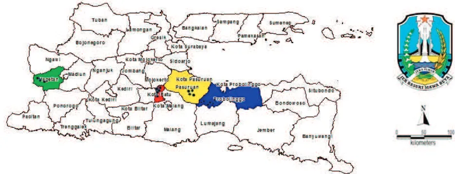
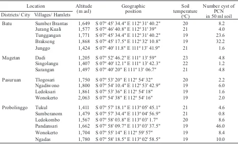
PCN soil sampling. Eighteen soil samples were collected from potato plantation in East Java (Figure. 1), which were determined by altitudes and soil temperature. Soil samples were collected using the purposive sampling method, that was taking soils from rhizo-sphere of the potato plants allegedly infected by PCN, about 0 to 20 cm deep for as much as 500 ml soil (Shurtleff & Averee, 2000).
Cyst extraction. Mature cyst of PCN were isolated by sieving and extracting 50 ml of soil samples using Baunacke Method (Benzooijen, 2006). Cyst were slightly dried and collected into the tube, then were kept at 4 oC.
PCN population analysis. Number of PCN cyst in each of the soil samples taken were counted.
Identification
Morphological characterization.Morphological observation was done on juveniles hatched from the cyst, which were then fixed with FAA (Dropkin, 1992).
Morphological differentiation between Globodera
rostochiensis and G. pallida was done by observing differences of knob stylet of the second juveniles (20 specimens/sampling location) and perineal pattern of the cyst (10 specimens/sampling location).
Photo micrographs of cyst vulval area and semi permanent fixed larvae were made with an automatic camera Olympus DP 21 attached to a Olympus CX 31 compound microscope; and measurements were made with an ocular micrometer on the same micro-scope. Specimens were morphologically identified with recent taxonomic keys based on combination of second-stage juveniles and cysts characters including body length, body width, stylet length, tail and hyaline part of tail length, Granek’s ratio (the distance from anus to the nearest edge of vulval basin divided vulval basin diameter), and the number of cuticular ridges between vulva and anus (Fleming & Powers, 1998).
Molecular characterization.DNA extraction was performed following methods described by Fullaondo
et al. (1999) with some modifications. Sixty PCN cysts were crushed in Eppendorf tubes using plastic micro-pestles in 300 µl of lysis buffer and 5 µl of
proteinase K. The mixture was incubated at 65oC for
The regression analysis showed that the number of PCN cysts increased when the soil temperature declined. From the pattern shown on Figure 2, a relationship model between soil temperature and the number of PCN cysts could be made using the following equation:
Total of PCN cyst = 20.545-0.0457 × Soil Temperature
That model could be used to interpret the impact of temperature on the number of cyst. In the range of 19−23°C, an every one degree Celcius of soil tem-perature decrement could add 0.0457 PCN cyst.
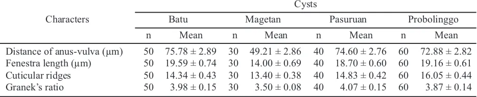
Morphological measurement of second-stage juveniles and cysts are shown in Table 3 and 4. The body of second-stage juveniles were vermiform, head rounded, and slightly set off with prominent cephalic sclerotization. Stylet was short, well developed, basal knobs rounded without forward projection (Figure 3C and 3D). Tail tapered uniformly with hyaline, and ended with a finely rounded to pointed terminus (Figure 3E−J). Second-stage morphological values from the
1 hour and then incubated at 95oC for 10 minutes.
300 µl of chloroform and isoamylalcohol (24:1) was added. The homogenate was sentrifuged at 11,000 rpm for 10 min. The supernatant was transferred to a new tube and 1.5 time (supernatant total volume) binding buffer was added with the supernatant, and
stored for 20 min at -20 oC. This mixture was
sentrifuged at 11,000 rpm for 10 min. The pellet was precipitated with 500 ml 70% ethanol, then sentrifugated at 14,000 rpm for 10 min. The pellet was dried in laminar air flow and dissolved in 20 µl nuclease free water.
The fragment of ITS region was amplified using the species-specific primers PITSr3 (AGCGCAGA
CATGCCGCAA for G. rostochiensis, amplicon size
434 bp) and PITSp4 (ACAACAGCAATCGTCGAG for G. pallida, amplicon size 265 bp) in combination with common primer ITS5 (GCAAGTAAAAGTC GTAACAAGG) (Bulman & Marshall, 1997; White et al.,
1990) in a multiplex reaction. The PCR amplification was done in a 25 µl reaction volume containing beads ready to go, 1 µl primer, 19 µl nuclease free water and 3 µl of DNA template. The amplification temperature profile was: 2 min initial denaturation at 94oC and 7
min final extension at 72oC with the intervening 35
cycles of 30 s at 94oC, 30 s primer annealing at 60oC
and 30 s primer extension at 72oC. All PCR products
were run in electrophoresis at 50 V for 1 h in 0.8% agarose gels and visualized on UV ilumination.
RESULTS AND DISCUSSION
This research found two new infested areas infected by PCN, i.e. Pasuruan and Magetan. Before 2015, the distribution area of PCN was only in Batu and Probolinggo, but after 2016, the PCN has spread onto those two new locations which was marked with the establishment of cyst in soil (Table 1).
The number of cyst in Pasuruan and Magetan was fairly low, ranging from 1.2 to 6.0 cysts/50 ml soil. This suggests that PCN in those two districts were still in the process of development. Meanwhile, the highest number of cyst was found in Pandansari, Probolinggo (46.0 cysts/50 ml soil), and the lowest number was found in Sumberanom, Probolinggo (0.8 cysts/ 50 ml soil) (Table 2).
Temperature is a limiting factor that regulates the activity of nematodes including ability to extend its distribution range (Kaczmarek et al., 2014). According to Wallace (1964), the lowest temperature that inhibits
the activity of G. rostochiensisis 5−10 oC, the temperature
optimum for emergence is 21oC, for development is
18−24°C, and for reproduction is 21−27°C. Temperature that inhibits the emergence of G. rostochiensis is
30oC, inhibits the activity of PCN is 30−35°C, and
hampered its development is 29−32°C. According to Mulyadi (2009) larval activity takes place at 10°C and stops at 40°C. The optimum temperature for the
development of G. rostochiensison host plants range
between 18−24°C and inhibited at 29−32°C. The
optimum temperature for G. rostochiensis growth at
20°C (Nurjanah et al., 2016), range between 15.7− 23.1oC (Trifonova, 1999). The results showed that the cysts were present in all sampled areas, with soil tem-peratures ranged between 19−23°C, and the altitude ranged between 1,205 to 2,063 m above the sea level. (Table 2). Thus, soil temperature at all locations
seemed to be suitable for the development of G.
rostochiensis.
Location Globodera rostochiensis Before 2015 2016 to 2017
Batu + +
Magetan - +
Pasuruan - +
Probolinggo + +
Figure 2. The relationship between the soil temperature and the number of Globodera rostochiensiscysts in East Java
Location Altitude
(m asl)
Geographic position
Soil temperature
(oC)
Number cyst of PCN in 50 ml soil Districts/ City Villages/ Hamlets
Batu Sumber Brantas 1,649 S 07º 45' 34.4" E 112º 31' 40.2" 20 8.2 Jurang Kuali 1,577 S 07º 46' 40.8" E 112º 31' 39" 21 4.0 Tunggangan 1,771 S 07º 45' 34.4" E 112º 31' 40.2" 19 23.6 Brakseng 1,868 S 07º 45' 17.5" E 112º 32' 10.8" 19 32.2 Junggo 1,424 S 07º 40' 11.8" E 111º 13' 41.9" 21 1.6
Magetan Dadi 1,205 S 07º 52' 46.2" E 111º 13' 59" 23 4.8 Singolangu 1,407 S 07º 40' 12.1" E 111º 13' 42.3" 22 1.2 Sarangan 1,497 S 07º 40' 20" E 111º 13' 06.7" 21 4.8
Pasuruan Tlogosari 1,750 S 07º 53' 20" E 112º 54' 32" 20 2.2 Ngadiwono 1,800 S 07º 54' 10.4" E 112º 53' 42.9" 19 6.0 Ledoksari 1,861 S 07º 53' 36" E 112º 54' 18" 19 1.6 Wonokerto 2,063 S 07º 54' 38" E 112º 54' 16" 19 2.0
Probolinggo Tukul 1,411 S 07º 57' 18.1" E 113º 05' 45.1" 21 4.6 Sumberanom 1,479 S 07º 57' 34.4" E 113º 04' 56.9" 21 0.8 Ledokombo 1,567 S 07º 58' 03.8" E 113º 03' 1.7" 20 8.6 Pandansari 1,662 S 07º 58' 09.7" E 113º 03' 37.5" 19 46.0 Wonokerto 1,704 S 07º 55' 14" E 112º 59' 57" 19 8.4 Ngadas 1,780 S 07º 58' 18.5" E 113º 02' 58.5" 19 10.0 Table 2. Distribution of PCN cyst in potato producing areas in East Java
analysed populations were very close with observa-tion by Skandar et al.(2011).
The first stage in identification of PCN generally involves the spherical with an anterior necklike
projections (Globodera); perform (elongated-ovoid),
the length of the body being about 1.5 times its width, with a protruding neck and no vulval cone (Punctodera) from lemon-shape cysts, with both anterior and posterior
projections (Heterodera). Cactodera cysts are less
commonly found in agricultural soil samples and
range in shape from ‘Globodera-like’ spheroidal to
lemon-shape, with a small posterior cone, the vulva is terminal, becoming a circum-fenestrate opening in the cyst. Identifications of cysts samples are generally made on the basis of cyst shape and fenestration (Fleming & Powers, 1998).
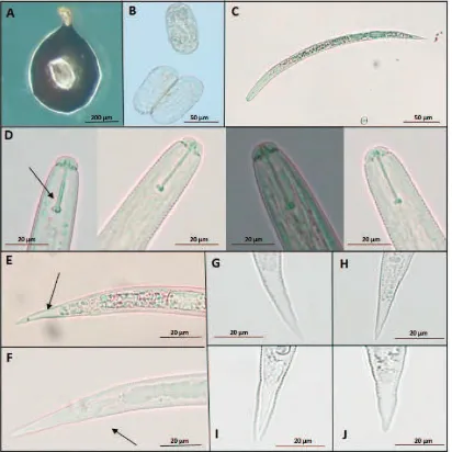
vulval basin. Vulval cone could not be found. Bullae-like structures were noted in few specimens (Figure 4C and D). Cyst wall pattern was ridge-like with heavy punctuations (Figure 4A and E). Cysts morphological values from the analyzed populations were very close with taxonomic keys (Fleming & Powers, 1998).
On average, the population from Probolinggo has the shortest second-stage juveniles (441.90 ± 2.63 µm), while the population of Batu has the longest (463.35 ± 5.61 µm). The population of Batu has the broadest body (22.45 ± 0.24 µm) compared to the population of Probolinggo (19.99 ± 0.22 µm). The stylet of Globodera sp. is slighty, stout, and heavily sclerotized, with well-developed, rounded basal knobs that slope backward (except in second-stage juveniles of G. pallida, in wich the knobs project forward and the stylet is about 23 µm, and the basal knobs are more rounded and the stylet is typically shorter about 21 µm for G. rostochiensis) (Shurtleff & Averee, 2000). Specimens from Probolinggo have the longest stylet (20.72 ± 0.06 µm), and the lowest were from Magetan (20.53 ± 0.10 µm). The results of this study indicate that the length of the stylet is smaller than the literature. Knob stylet of all samples is rounded. The juvenile tail tapers to pointed tip with a clear terminus (hyaline) (Figure 3E). The specimens from Batu have the longest tail (49.91 ± 0.36 µm), and the shortest are from Probolinggo (47.74 ± 0.28 µm). Magetan has the longest hyaline tail terminal length (27.92 ± 0.34
Characters Table 3. Morphological characters of Globoderasp. second-stage juveniles
Characters Table 4. Morphological characters of Globoderasp. cysts
µm), and the shortest are from Pasuruan (26.54 ± 0.31 µm). Granek’s ratio (the distance form the anus to the nearest edge of fenestra, divided by the length of the fenestra) and a number of cuticular ridges between anus and fenestra can be used for identification.
Fleming and Powers mentioned that G. rostochiensis
had Granek’s ratio > 3 and number of cuticular ridges
between anus and fenestra are 12−31, and G. pallida
had Granek’s ratio < 3 and number of cuticular ridges between anus and fenestra are 8−20. Furthermore, the population of Magetan has the lowest mean value of Granek’s ratio (3.50) while the population of Pasuruan has the highest (4.07). Finally, the population of Magetan has the lowest mean number of cuticular ridges between vulva-anus (13.40), while the population of Probolinggo has the highest value (16.05).
The vulva is near the terminus and anus, not on a terminal valvular cone (Figure 4E and F). It lies between finely papillated crescentic areas occupying most of the vulval slit and surrounded by a single, circular fenestra. In most species, all the eggs are retained within the mature cyst, but a few species also lay eggs in an external gelatinous matrix (Shurtleff & Averee, 2000).
Eggs of Globodera sp. (Figure 3B) remain viable
in the soil for a long period and encysted eggs are stimulated to hatch by root exudates. Juveniles (Figure
3C) become active at 10oC and maximum root invasion
Figure 3. Photomicrographs of cyst, eggs, and second-stage juveniles of Globodera rostochiensis: cyst (60× magnification) (A); eggs (400×magnification) (B); whole juvenile (100× magnification) (C); stylet variations of G. rostochiensisfrom: Batu, Magetan, Pasuruan, Probolinggo, narrow show anterior regions showing stylet and knob (D); tail showing hyaline (E); arrow showing anal (F); tail variations of G. rostochiensis from: Batu (G), Magetan (H), Pasuruan (I), Probolinggo (J); D−J photographed at 400× of magnification
rupture the root tissue but remain attached to the root by their heads and protruding necks which stay inserted in the root tissue. The fertilized females become large and subspherical and go through a sequence of the color change prior to dying and becoming cysts. PCN generally complete one generation during a growing season. Eggs will remain viable in cysts for over 20 years in soils under severe environmental stress. They
withstand temperatures of extreme cold (-15oC) and
soil desiccation for long periods. A large portion of eggs will hatch only if they are stimulated by potato root exudates (Luc et al., 1995).
However, essential morphological and morpho-metrical character, such as the juvenile stylet length,
the stylet knobs anteriorly flattened to rounded, without forwarding projections, the number of cuticular ridges and Granek’s ratio indicated that the specimens were
G. rostochiensis.When compared with G. pallida, the stylet of the second-stage juvenile was slightly shorter. The stylet knob is rounded. Cysts have a smaller Granek’s ratio and more cuticular ridges between the anus and vulva.
Molecular identification with multiplex PCR
determined all samples from four locations as G.
rostochiensis(Figure 5).Using two sets of primers in the same reaction enables species identification in only one step without subsequent analysis. The PCR
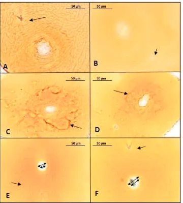
Figure 4.Photomicrographs of terminal areas of Globodera rostochiensiscysts: anal-vulval regions with arrows showing anal areas (A and B); bullae-like structures (C and D); cyst wall pattern (E); anal areas with V-shape (F); double arrows show vulva (E and F); A−F photographed at 400× magnification
This primer amplified an approximately 434 bp DNA fragment for all the sample. However, PITSp4 primer
for identified G. pallida nematode species did not
display amplification. The PCR-based methods used in the present study shall play an important role for future surveys and routine identification G. rostochiensis,in order to contain the nematode to its current distribution area. In addition, the use of species-specific primers in PCR multiplex reactions, which allows the simultaneous identification of both species in the same sample, will
eventually help to detectG. pallida.
CONCLUSION
PCN were found in potato planting areas in East Java: Batu, Pasuruan, Probolinggo, and Magetan. Pasuruan and Magetan are new area distributions of
G. rostochiensis in East Java. Based on the morpho-logical and molecular characters, the potato cyst
nematode was determined as G. rostochiensis.
ACKNOWLEDGEMENT
We would like to thank to Indonesian Agricultural Quarantine Agency for partial funding to conduct this study; the farmers in Batu, Pasuruan, Probolinggo, and Magetan for their help; and laboratory of Agricultural Quarantine Major Service of Surabaya.
LITERATURE CITED
Baldwin, J.G. & M. Mundo-Ocampo. 1991. Heteroderinae, Cyst and Non Cyst Forming Nematodes. p.
275−362. InW.R. Nickle (ed.) Manual of
Agri-cultural Nematology. Marcel Dekker, New York.
Benzooijen. 2006. Methods and Techniques
for Nematology Revised Version, Wageningen. 118 p.
Brodie. 1993. Probability of Globodera rostochiensis
Spread on Equipment and Potato Tubers.Journal
of Nematology25: 291−296.
Bulman, S.R. & J.W. Marshall. 1997. Differentiation of Australasian Potato Cyst Nematode (PCN) Populations
Using the Polymerase Chain Reaction (PCR). New
Zealand Journal of Crop and Horticultural Science
25: 123−129.
Dropkin, V.H. 1992. Introduction to Plant Nematology
(Pengantar Nematologi Tumbuhan, Alih Bahasa Supratoyo). Gadjah Mada University Press, Yogyakarta. 366 p.
EPPO. 2004. Globodera rostochiensisand Globodera
Pallida. Bulletin EPPO34: 309−314.
Fleming, C.C., & T.O. Powers. 1998. Potato Cyst Nematode Diagnostics: Morphology, Differential
Hosts and Biochemical Techniques. p. 91−108. In
R.J. Marks & B.B. Brodie (eds.), Potato Cyst
Nematodes-Biology, Distribution, Control. CAB International, Wallingford.
Fulloando, A., B. Enrique, V. Miguel, B. Imanol, S. Azucena, & R. Enrtique. 1999. Identification of
Potato Cyst Nematode Species Globodera
rostochiensis and G. pallida by PCR Using Specific
Primer Combinations. Nematology1: 157−163.
Hadisoeganda, A.W.W. 2006. Nematoda Sista Kentang: Kerugian, Deteksi, Biogeografi, dan Pengendalian Nematoda Terpadu [Potato Cyst Nematodes: Loss, Detection, Biogeography, and Integrated
Nematode Control]. Monografi29: 1−52.
Ibrahim, S.K., H.A. Saad, & N. Mousa. 2003. Potato
Cyst Nematodes Globodera spp. in Libanon
-Occurence and Distribution. Lebanese Science
Journal5: 25−36.
Indarti, S., Mulyadi, B. Rahayu, & B. Triman. 2004.
First Record Of Potato Cyst Nematode Globodera
rostochioensis in Indonesia. Australasian Plant Pathology33: 325−326.
Kaczmarek A., K. MacKenzie, H. Kettle, & V.C. Blok. 2014. Influence of Soil Temperature on
Globodera rostochiensisand Globodera pallida. Phytopathologia Mediterranea53: 396−405.
Luc, M., R.A. Sikora & J. Bridge. 1995. Plant Parasitic Nematodes in Subtropical and Tropical Agriculture Second Edition. CAB international, Wallingford, UK. 629 p.
Mulyadi, B. Rahayu, B. Triman, & S. Indarti. 2003.
Identifikasi Nematoda Sista Kuning (Globodera
rostochiensis) pada Kentang di Batu, Jawa Timur [Identification of Yellow Cyst Nematode (Globodera rostochiensis) on Potato in Batu, East
Java]. Jurnal Perlindungan Tanaman Indonesia
9: 46−53.
Mulyadi, S. Indarti, B. Rahayu T.P., & B. Triman. 2014. Molecular and Pathotype Identification of
Potato Cyst Nematodes. Jurnal Perlindungan
Tanaman Indonesia18: 17−23.
Mulyadi. 2009. Nematologi Pertanian[Agricultural
Nematology]. Gadjah Mada University Press, Yogyakarta. 339 p.
Nurjanah, Y.A. Trisyono, S. Indarti, & S. Hartono. 2016. Identification, Distribution and Genetic Diversity of The Golden Potato Cyst Nematode (Globodera rostochioensis) in Java Indonesia. In
T.R. Nuringtyas, R. Root, A. Widyaparaga, M. Mahardika, A. Kusumaadmaja, & N. Hadi (eds.),
Proceeding of Technology 1st International Conference on Science and Technology 2015 (ICST-2015). AIP Conference Proceeding. AIP Publishing, Melville, New York.
Nurjanah, Y.A. Trisyono, S. Indarti, & S. Hartono. 2016. The Effect of Temperature, Potato Varieties, and the Origin of Cyst on the Reproductive Biology of Globodera rostochiensis. Jurnal Perlindungan Tanaman Indonesia20: 84−88.
Shurtleff, M.C. & C.W. Averee. 2000. Diagnosing
Plant Diseases Caused by Nematode. APS Press. The American Phytopathological Society, Minnesota. 143 p.
Skandar, A.M., Z.A. Handoo, I.A. Zasada, R.E. Ingham, L.K. Carta, & D.J. Chitwood. 2011. Morphological and Molecular Characterization of Globodera
populations From Oregon and Idaho. Phytopathology
101: 480−491.
Trifonova, Z. 1999. Temperatur Influence on the Potato
Cyst Nematode (Globodera rostochiensisWoll.
1923) Development. Bulgarian Journal of
Agricultural Science5: 863−866.
Wallace, H.R. 1964. The Biology of Plant Parasitic
Nematodes. St, Martin’s Press Inc, New York. 280 p.
Wang, J., C.S. Johnson, & J.D. Eisenback. 2001. Effects of Host Resistance and Temperature on
Development of Globodera tabacum solanacearum.
Journal of Nematology33:132–136.