www.elsevier.com / locate / bres
Research report
Effect of extracellular long-time microperfusion of high concentrations
of glutamate and glycine on picrotoxin seizure thresholds in the
hippocampus of freely moving rats
*
˜
´
´
German Sierra-Paredes , Aida Senra-Vidal, German Sierra-Marcuno
Neuroscience Division, Department of Biochemistry and Molecular Biology, School of Medicine, University of Santiago, San Francisco 1, 15705 Santiago de Compostela, Spain
Accepted 19 September 2000
Abstract
The effect of high concentrations of glutamate and glycine on picrotoxin seizure thresholds was investigated by perfusion through microdialysis probes in the hippocampus of freely moving rats. Microperfusion of glutamate at concentrations up to 1 mM, produced no changes in behavior or basal EEG recordings, but microperfusion of 200mM glutamate was sufficient to lower the picrotoxin seizure threshold down to 50% in 60% of the animals studied and produced an increase of 180623% in seizure duration. Microperfusion of 1 mM glutamate reduced seizure threshold in all animals, and markedly prolonged seizure duration (230630%). Microperfusion of 200mM or 1 mM glycine lowered picrotoxin seizure thresholds down to 50% in 70% of the animals and lengthened seizure duration up to 176643%. Continuous microperfusion of the antagonist for the glycine binding site in NMDA receptors 5,7-dichlorokynurenic acid (100
mM) reversed the effect of both glutamate (1 mM) and glycine (1 mM) and suppressed seizures completely in 90% of the animals. These results indicate that although neurotoxicity is not achieved by perfusing glutamate and glycine at concentrations as high as 1 mM, neuronal excitability is modified by altering extracellular glutamate and glycine concentrations, and they suggest that glutamate-induced neuronal hyperexcitability is induced through mechanisms different from excitotoxicity. 2001 Elsevier Science B.V. All rights reserved.
Theme: Disorders of the nervous system
Topic: Epilepsy: basic mechanisms
Keywords: Extracellular amino acid; Seizure; NMDA receptor; Hippocampus; Microdialysis; Extrasynaptic receptor
1. Introduction concentrations of excitatory and inhibitory amino acids have been investigated during different types of epileptic The cellular mechanisms underlying the initiation and seizures in humans and several animal models as indirect spread of epileptic seizures have been related to both evidence of neurochemical modifications on the synaptic increased glutamatergic excitation and decreased GABAer- cleft. Human studies have shown an increase in several gic inhibition [6]. Based on findings of several experimen- amino acids during epileptic seizures [8,15], but some tal models of epilepsy, increasing GABAergic transmission animal experiments have provided conflicting results. and decreasing glutamatergic transmission have been Depending on the method used seizures may be correlated proposed as promising antiepileptic strategies with po- with an increase in the extracellular concentrations of tential advantages compared to current therapies [22,24]. excitatory amino acids [47], but other experiments show Glutamate excitotoxicity has been implicated in the no modification [21,25] or a decrease [40,41] in glutamate pathophysiological process of epilepsy [31]. Extracellular and aspartate during epileptic seizures. These results are difficult to evaluate in terms of the excitatory effect on the postsynaptic receptors, because changes in glutamate con-*Corresponding author. Tel.: 11-43-981-582-382; fax: 1
1-43-981-centration within the synaptic cleft are not reflected by 587-801.
E-mail address: [email protected] (G. Sierra-Paredes). modifications in the extracellular fluid [40].
Direct application of excitatory neurotransmitters such chased from Sigma (St Louis, MO, USA). 5,7-DKA was as glutamate [28], or allosteric positive modulators such as obtained from TOCRIS (Bristol, UK).
glycine [29], failed to induce seizures or excitotoxicity in animals when directly infused into the brain. Glutamate
2.2. Surgical procedure excitotoxicity is more evident in stroke [9] and
autoim-munity [45], but excitotoxicity seems to be a more
Male Sprague–Dawley rats, initially weighing 250–300 complex phenomenon than a single increase in
extracellu-g were used. They were housed in extracellu-groups of three under lar glutamate. Furthermore, most of the chronic models of
controlled environmental conditions (ambient temperature epilepsy seem to need the conjunction of two or more
21618C, humidity 50–60%, 12:12 h light / dark cycle) with factors. Electrically-induced kindling and kainate
injec-free access to food and water except during testing. Rats tions produce both a loss of inhibitory GABAergic neurons
were obtained from the animalary of the University of and excitatory synaptic changes in the rat hippocampus
Santiago. All experiments were performed in a laboratory [5,44], and human studies indicate that both phenomena
under controlled environmental conditions and at the same are also observed in the epileptic brain tissue [12]. In spite
time in the morning in order to avoid circadian variations. of this, glutamate and glycine perfused in extracellular
All efforts were made to minimize animal suffering, and fluid may not be enough to induce seizures, but could
our chronic animal protocols were designed to reduce the result in modified neuronal excitability.
number of animals used [43]. Animal care complied with Changes in the extracellular compartment do not seem
Spanish legislation on Protection of Animals Used in to be involved directly in fast synaptic transmission, but
Experimental and Other Scientific Purposes, and with the are likely to have widespread effects mediated by
ex-European Union regulations. The rats were anesthetized trasynaptic receptors. Amino acids released in the
extracel-with pentobarbital (40 mg / kg, injected intraperitoneally) lular fluid do not easily reach synaptic NMDA and AMPA
and placed in a stereotaxic instrument (D. Kopf, Tujunga, receptors [17,37], but prolonged high concentrations of
CA, USA). Under aseptic conditions, two stainless steel glutamate and glycine may result in the activation of
microscrews to be used as electrodes for EEG recording extrasynaptic NMDA receptor function [36,40], which may
were positioned in the skull above the frontal and occipital lead to the sustained hyperexcitability observed in epileptic
areas of each hemisphere; one screw, used as a reference tissue.
electrode, was anchored in the mid-line, 7–9 mm rostrally In this study we have investigated the effect of
long-to the coronal suture. The intracerebral guide for the term microperfusion of glutamate and glycine in the
microdialysis probe (CMA / 12, CMA / Microdialysis AB, hippocampus of freely moving rats, and how it affects
Stockholm, Sweden) was sterilized with 70% ethanol, picrotoxin seizure thresholds. We also studied the effect of
rinsed in sterile saline and was implanted vertically into the antagonist for the glycine binding site in NMDA
the ventral hippocampus. Stereotaxis coordinates derived receptors 5,7-dichlorokynurenic acid. For this purpose we
from the atlas of Paxinos and Watson [32] were 5 mm have used a new experimental design on a new
whole-posterior, 4.8 mm lateral and 4 mm ventral for the tip of animal model [43] in which partial seizures can be elicited
the cannula relative to bregma and dural surface. Wires repeatedly on different days without changes in threshold
from the microscrews were soldered to a miniature plug or seizure patterns. Picrotoxin seizure thresholds remain
(Cannon MD1-9SL1, USA) and fixed firmly to the skull constant in the same animal in repeated experiments [43]
with dental cement. After surgery, the rats were placed in for time periods as long as 6 months, thus providing a
individual cages and received intramuscular amoxicillin good model to study possible modifications in neuronal
therapy (10 mg / kg every day) for 4 to 5 days. excitability. Picrotoxin, amino acids and the receptor
antagonist were dialyzed through the probe to avoid
possible dynamic effects imposed by the blood–brain 2.3. Experimental procedure barrier on amino acids and some systemic administrated
drugs [1]. Our method is completely reversible, thus The experiments were carried out on conscious, freely permitting the independent study in the same animal of moving rats 10 days after surgery. From the fourth day the each of the separate roles of excitatory and inhibitory animals were placed for 3 h daily in the experimental unit neurotransmitters that may contribute to epileptic seizures. for habituation and EEG control of wakefulness and sleep A preliminary report of some of the findings has been activity. Bipolar cortical EEGs were recorded on magnetic published in abstract form [42] tapes using a Holter–EEG system (Oxford-Medilog 9200, Oxford, UK), and also with a Minihuit electroencephalog-raph (Alvar Electronic, Paris, France).
2. Materials and methods During an experimental session recording time was distributed as follows:
2.1. Drugs and reagents (a) A 15 min reference EEG was recorded before every
probe introduction.
period was chosen to let the animal recover from possible modification had been induced in the duration or number local modifications induced by the tip of the probe. of seizures.
(c) A 60 min post picrotoxin microperfusion control. At the end of the experiments rats were anesthetized All habituation and experimental sessions were recorded with Nembutal and killed by decapitation. A probe was on videotape using a standard camera in order to relate introduced and perfused with Sudan Black to localize behaviorally observed seizures with the EEG recordings. easily the position of the probe. The brain was then In our experiments, seizure threshold was defined as the removed and placed in 4% phosphate-buffered formalde-lowest picrotoxin concentration which produced a specific hyde solution. A week later 50 mm coronal sections were EEG pattern and / or behavioral seizures after 5 min cut and stained with Cresyl Violet, and the position of the perfusion through the rat hippocampus. Only one picrotox- probe was checked under light microscopy.
in dose was perfused in each experimental session. The
lowest picrotoxin concentration used was 100mM, and the 2.4. Data analysis dose was slowly increased (125 mM each step) in each
animal in successive experimental sessions at 3 to 4 day EEG records were analyzed using the Medilog 9200 intervals until an EEG–behavioral seizure was induced. software, version 7.2. Wakefulness, somnolence, and sleep This picrotoxin concentration was the threshold dose. EEG activity (sleep spindles and slow wave sleep) were Seizure types and rest periods between experimental measured as a percentage of total time in the control sessions were described previously in detail [43]. Sub- record. Spike and wave discharge duration, seizure dura-threshold picrotoxin doses (50% of dura-threshold dose, which tion, and seizure onset and offset times were evaluated did not induce EEG or behavioral modifications) were used after picrotoxin and glutamate, glycine and DKA adminis-to investigate the effect of glycine and glutamate. tration. Statistical significance of the difference in duration, We used a CMA / 120 system for freely moving animals total time of seizures and seizure onset and offset times (CMA / Microdialysis AB, Stockholm, Sweden) and CMA / was determined by Student’s paired t-test.
12 microdialysis probes with 4 mm of membrane length. The probe was connected via polyethylene tubing to a
syringe selector (CMA / 111), and to 1 ml syringes 3. Results mounted on a micro-injection pump (CMA / 100). Before
starting each experiment, the probe was perfused with 3.1. Seizure thresholds ethanol and distilled water. After checking the integrity of
the probe under light microscopy, it was perfused with a Our results on individual seizure thresholds and seizure sterile Ringer solution (NaCl 147 mM, KCl 4.0 mM, types were consistent with those described previously [43]. CaCl 2.4 mM) for 10 min, and then introduced into the2 Seizure threshold among animals varied between 100 and rat hippocampus through the chronically implanted in- 300 mM, although it remained unchanged in repeated tracerebral guide. Between re-use, the probe was main- between day experiments within individual rats.
tained in distilled water, and before every introduction it
was sterilized and the integrity of the dialysis membrane 3.2. Effect of glutamate microperfusion was checked. A detailed description of the whole-animal
model and the method to induce seizures is presented Three hours microperfusion of glutamate at
concen-elsewhere [43]. trations up to 1 mM did not produce any changes in
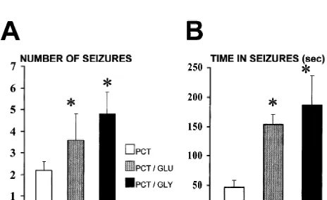
For the control experiments, Ringer solution was per- behavior or basal EEG recordings. Microperfusion on 200 fused at a constant flow rate of 2 ml / min during 2 h. mM glutamate decreased picrotoxin seizure threshold down Picrotoxin dissolved in Ringer solution was perfused at the to 50% of the picrotoxin concentration needed to induce same rate for 5 min. After picrotoxin administration, the seizures in 60% of the animals studied (Fig. 1). The same perfusion of Ringer solution continued for one more hour. concentration of glutamate produced an increase of In order to test the effect of glutamate and glycine, each 180623% in seizure duration (Fig. 2B), and in the number was dissolved in Ringer solution at concentration 200mM of seizures induced after picrotoxin administration (Fig. and 1 mM. Each solution was perfused continuously 2A). In 40% of the animals seizure severity was also throughout the experiment in all the animals on different increased. Microperfusion of 1 mM lowered seizure thres-days, following the same protocol for Ringer solution and hold in all the animals (Fig. 1), and had a significant effect picrotoxin administration in the control experiments. In on seizure duration (230630%) with several animals order to test the effect of 5,7-DKA, it was added to developing recurrent seizures.
glutamate–Ringer solution or glycine–Ringer solution at a
concentration 100 mM and also perfused continuously 3.3. Effect of glycine microperfusion during the experiment in all the animals, administering
Fig. 3. Effect of glycine continuous microperfusion (200mM and 1 mM) on picrotoxin thresholds. The significant decrease (*P,0.01, by Student’s Fig. 1. Effect of glutamate continuous microperfusion (200mM and 1
paired t-test) in seizure threshold induced by glycine microperfusion was mM) in the hippocampus on picrotoxin thresholds. The significant
also prevented by 5,7-DKA (100mM) Data are mean6S.E.M. (n512). decrease in seizure threshold (*P,0.01, by Student’s paired t-test)
induced by glutamate microperfusion was prevented by 5,7-DKA (100 mM). Data are mean6S.E.M. (n512).
4. Discussion of the animals (Fig. 3) and seizure duration increased up to
In this present study we report the effect of high 176643% after continuous perfusion of 200 mM glycine
concentrations of glutamate and glycine on picrotoxin-(Fig. 2B). The total number of seizures was also increased
induced seizure thresholds on freely moving rats. Our (Fig. 2A). Perfusion of 1 mM glycine had the same effect
results show that high extracellular glutamate and glycine on seizure threshold, increasing seizure severity (40% of
levels during prolonged microperfusion do not lead to the rats developed recurrent seizures) and duration (an
excitatory neuronal damage or paroxysmal activity in the increase of 240658%).
living rat brain. This is in agreement with previous reports where raising extracellular glutamate concentrations by 3.4. Effect of 5,7-DKA
microperfusion [28], or long-time pretreatment with gluta-mate transporter inhibitors [7,23,27,40], failed to produce Continuous microperfusion of the antagonist for the
seizures or neuronal damage. For glycine, Obrenovitch et glycine binding site in NMDA receptors 5,7-DKA (100
al. [29] have also shown that no toxic effects are observed mM) reversed the effect of both glutamate and glycine.
after perfusing concentrations up to 10 mM through 5,7-DKA suppressed seizures completely in 90% of the
microdialysis probes implanted in the striatum of animals (Figs. 1 and 3).
halothane-anesthetized rats.
We have found, however, that elevating glutamate and glycine concentrations in the hippocampus significantly increases neuronal excitability. Glutamate and glycine acting alone are not enough to induce epileptiform sei-zures, but their continuous action may produce metabolic changes or receptor rearrangement which facilitate seizure onset when GABAergic inhibition is impaired. The fact that glutamate and glycine produce almost the same effect indicates that their action is likely to be mediated by NMDA receptors. This is also confirmed by the fact that 5,7-DKA, a competitive inhibitor of the glycine binding site in the NMDA receptor, completely prevents the effect of both glutamate and glycine.
The epileptiform synaptic potentials in the hippocampus Fig. 2. Effect of increased glutamate and glycine extracellular con- are clearly dependent on the vesicular glutamate pool centrations on picrotoxin-induced seizures. Continuous microperfusion of
[33,48]. The effects observed could be explained by glutamate (1 mM) or glycine (1 mM) increased significantly: (A) The
overactivation of synaptic NMDA receptors, however, it is number of seizures and (B) mean seizure duration (*P,0.01, by
and glycine concentrations based only on its synaptic Evidence of extrasynaptic glutamate receptors has been
effect. reported [2,20], and a comparison of some properties of
Astroglial cells encapsulate synapses, and astroglial synaptic and non-synaptic NMDA receptors has been glutamate carriers have sufficient capacity to remove all published [10]. Some data suggest that the extracellular glutamate released from the presynaptic site [19]. Glial glutamate and aspartate may act on neuronal NMDA non-cells respond to synaptically released glutamate by activa- synaptic receptors [17,36,37,40], as a possible mechanism tion of electrogenic transporters, which generate a current of neuronal synchronization [40].
that is directly proportional to the amount of glutamate Although there is not direct evidence that extrasynaptic released [14]. The high capacity and affinity for glutamate NMDA receptors participate in the development of sei-uptake by astrocytes in the synaptic region enables synap- zures, the hypothesis described above seems the best suited tic transmission for long periods of time with a high to integrate our data with the majority of previously
signal-to-noise ratio [34]. published experimental research.
The need for astroglial cells working at the synaptic The increase in extracellular glutamate and glycine does region as an effective system for clearance of the pre- not produce neuronal damage or seizure activity. However, synaptically released glutamate may be clearly understood it may potentiate both when associated with other altera-by taking into account the role of the glutamate–glutamine tions, such as the loss of inhibitory neurons. In many brain cycle in both the presynaptic region and the glial cells [38]. injuries, both phenomena are likely to occur.
Upon release into the synaptic cleft, the glutamate diffuses Furthermore, many drugs acting on NMDA receptors away, and the glial glutamate transporters internalize the would bind easily to non-synaptic receptors, preventing the extracellular glutamate. Inside the glial cells, glutamate is potentiation effect which may lead to epileptogenesis. metabolized into glutamine, which is transported out of the NMDA extrasynaptic receptors thus remain a good target glial cells and into the synaptic terminal for subsequent for new antiepileptic drugs.
resynthesis of glutamate. Since glutamine does not act on the glutamate receptor, this release does not interfere with
synaptic transmission [39]. Also, using knockout rats [35], Acknowledgements it was shown that extracellular glutamate levels were
unmodified in the animals which had lost neuronal gluta- The authors gratefully acknowledge the work of the mate transport, while those animals which had lost the laboratory of Neuroanatomy (Director: Prof. J.L. Laban-glutamate transport in glia showed an increase in extracel- deira) who performed the histological study and Prof. J.L. lular glutamate concentrations, suggesting a major role for Otero-Cepeda (Department of Statistics) for statistical glial glutamate transporters in the clearance of extracellular analysis. This work was supported by grant XUGA
glutamate. 20801B97 from the Consellerıa de Educacion e Or-´ ´
This special role of the astrocytes at the synaptic region denacion Universitaria, Xunta de Galicia, Galicia, Spain.´ may be explained by the astroglial heterogeneity [49]. We
also need to take into account the morphological [13] and the pharmacological [11] evidence of synaptic barriers.
References Taken together, all these data suggest that the epileptiform
synaptic potential is generated by the glutamate synaptic
´
[1] E. Aguilar-Veiga, G. Sierra-Paredes, J. Galan-Valiente, R. Soto-pool without the participation of the extracellular gluta- Otero, E. Mendez-Alvarez, G. Sierra-Marcuno, Correlation between´ ˜
mate pool. ethosuximide brain levels measured by high performance liquid
Recent research suggests that the brain extracellular chromatography and its antiepileptic potential, Res. Com. Chem. Pathol. Pharmacol. 71 (1991) 351–364.
environment appears to be an homeodynamic entity which
[2] C. Aoki, C. Venkatesan, C.G. Go, J.A. Mong, T.M. Dawson, Cellular maintains a steady intercellular milieu with regard to ions
and subcellular localization of NMDA-R1 subunit immunoreactivity and small molecules, critical for neuronal function, under in the visual cortex of adult and neonatal rats, J. Neurosci. 14 (1994)
1
the control of neurons and glial cells [26]. Na -dependent 5202–5222.
glutamate transporters of the blood–brain barrier pre- [3] P. Bach-y-Rita, Thoughts on the role ofmen transmission in normal and abnormal mass sustained functions, in: K. Fuxe, L.F. Agnati sumably assist in keeping the glutamate concentration low
(Eds.), Volumen Transmission in the Brain, Raven Press, New York, in the extracellular fluid of the brain [30]. Analysis of our
1991, pp. 489–496.
results is simplified if we also take into account the [4] P. Bach-y-Rita, Nonsynaptic diffusion neurotransmission (NDN) in concept of non-synaptic transmission [3,4,18,46] as a the brain, Neurochem. Int. 23 (1993) 297–318.
complementary mechanism to the glutamate action on [5] Y. Ben-Ari, M. Gho, Long-lasting modification of the synaptic properties of rat CA3 hippocampal neurones induced by kainic acid, synaptic transmission [40].
J. Physiol. 404 (1988) 365–384. Fagni et al. [16] suggested that theL-glutamate added to
[6] H.F. Bradford, Glutamate, GABA and epilepsy, Prog. Neurobiol. 47 the bath stimulated different glutamate receptors to those (1995) 477–511.
cultured cerebellar granule cells, J. Neurochem. 72 (1999) 2181– [29] T.P. Obrenovitch, A.M. Hardy, J. Urenjak, High extracellular glycine does not potentiate N-methyl-D-aspartate evoked
depolariza-2190.
tion in vivo, Brain Res. 746 (1997) 190–194. [8] A.G. Chapman, Microdialysis, in: J.Jr. Engel, T.A. Pedley (Eds.),
˜
´ ´
[30] R.L. O’Kane, I. Martınez Lopez, M.R. DeJoseph, J.R. Vina, R.A. Epilepsy: A Comprehensive Textbook, Lippincott-Raven Publishers,
1
Hawkins, Na -dependent glutamate transporters (EAAT1, EAAT2, Philadelphia, 1997, pp. 1067–1072.
and EAAT3) of the blood–brain barrier, J. Biol. Chem. 274 (1999) [9] D.W. Choi, Glutamate neurotoxicity in diseases of the nervous
31891–31895. system, Neuron 1 (1988) 623–634.
[31] J.W. Olney, R.C. Collins, R.S. Sloviter, Excitotoxic mechanisms of [10] B.A. Clark, M. Farrant, S.G. Cull-Candy, A direct comparison of the
epileptic brain damage, in: A.V. Delgado-Escueta, A.A.Jr. Ward, single-channel properties of synaptic and extrasynaptic NMDA
D.M. Woodbury, R.J. Porter (Eds.), Basic Mechanisms of the receptors, J. Neurosci. 17 (1997) 107–116.
Epilepsies: Molecular and Cellular Approaches. Advances in Neuro-[11] D.R. Curtis, R.M. Eccles, The effect of diffusional barriers upon the
logy, Vol. 44, Raven Press, New York, 1986, pp. 857–877. pharmacology of cells within the central nervous system, J. Physiol.
[32] G. Paxinos, C. Watson, The Rat Brain in Stereotaxic Coordinates, (Lond.) 141 (1958) 446–463.
3rd Edition, Academic Press, London, 1997. [12] N.C. de Lanerolle, J.H. Kim, M.L. Brines, Cellular and molecular
[33] M.A. Rogawski, Excitatory amino acids and seizures, in: T.W. Stone alteration in partial epilepsy, Clin. Neurosci. 2 (1994) 64–81.
(Ed.), CNS Neurotransmitters and Neuromodulators: Glutamate, [13] E. De Robertis, Some new electron microscopical contribution to the
CRC Press, New York, 1995, pp. 219–237.
biology of neuroglia, in: E. De Robertis, R. Carrera (Eds.), Biology [34] L. Ronnback, E. Hansson, Does astroglial network perform quali-¨ of Neuroglia. Progress in Brain Research, Vol. 15, Elsevier, New tative modifications of neuronal messages, in: E. Hansson, T. York, 1965, pp. 1–11. Olsson, L. Ronnback (Eds.), On Astroglia and Glutamate Neuro-¨ [14] J.S. Diamond, D.E. Bergles, C.E. Jahr, Glutamate release monitored transmission, Springer– R.G. Landes, New York, 1997, pp. 155–
with astrocyte transporter currents during LTP, Neuron 21 (1998) 187.
425–433. [35] J.D. Rothstein, M. Dykes-Hobert, C.A. Pardo, L.A. Bristol, L. Jin, [15] M.J. During, D.D. Spencer, Extracellular hippocampal glutamate in R.W. Kuncl, Y. Kanai, M.A. Hediger, Y.F. Wang, J.P. Schtelke, D.F. spontaneous seizures in the conscious brain, Lancet 341 (1993) Welty, Knockout of glutamate transporters reveals a major role from 1607–1610. astroglial transport in exocitotoxicity and clearance of glutamate, [16] L. Fagni, M. Baudry, G. Lynch, Classification and properties of Neuron 16 (1996) 675–686.
excitatory amino acid receptors in the hippocampus. I. Electro- [36] R. Sattler, Z. Xiong, W.Y. Lu, J.F. MacDonald, M. Tymianski, physiological studies of an apparent desensititazion and interactions Distinct roles of synaptic and extrasynaptic NMDA receptors in with drugs which block transmission, J. Neurosci. 3 (1983) 1538– excitotoxicity, J. Neurosci. 20 (2000) 22–33.
1546. [37] G. Seifert, C. Steinhauser, Glial cells in the mouse hippocampus¨ 21
[17] C.R. Farb, T. Aoki, J.E. LeDoux, Differential localization of NMDA express AMPA receptors with an intermediate Ca permeability, and AMPA receptor subunits in the lateral and basal nuclei of Eur. J. Neurosci. 7 (1995) 1872–1881.
amygdala: a light and electron microscope study, J. Comp. Neurol. [38] A. Schousboe, Transport and metabolism of glutamate and GABA 362 (1995) 86–108. in neurons and glial cells, Int. Rev. Neurobiol. 22 (1981) 1–45. [18] K. Fuxe, L.F. Agnati, Two principal modes of electrochemical [39] A. Schousboe, G. Svenneby, L. Hertz, Uptake and metabolism of
communications in the brain: volume versus wiring transmission, glutamate in astrocytes cultured from dissociated mouse brain Adv. Neurosci. 1 (1991) 1–9. hemispheres, J. Neurochem. 29 (1977) 999–1005.
´
[19] I. Hertz, Functional interactions between neurons and astrocytes. I. [40] G. Sierra-Paredes, J. Galan-Valiente, M.D. Vazquez-Illanes, E. ˜
Turnover and metabolism of putative amino acids transmitters, Prog. Aguilar-Veiga, G. Sierra-Marcuno, Effect of ionotropic glutamate Neurobiol. 13 (1979) 277–323. receptors antagonists on the modifications in extracellular glutamate
´
[20] D.M. Kullman, G. Erdemli, F. Asztely, LTP of AMPA and NMDA and aspartate levels during picrotoxin seizures: a microdialysis study receptor-mediated signals: evidence for presynaptic expression and in freely moving rats, Neurochem. Int. 37 (2000) 377–386.
´
extrasynaptic glutamate spill-over, Neuron 17 (1996) 461–474. [41] G. Sierra-Paredes, J. Galan-Valiente, M.D. Vazquez-Illanes, E. ´
[21] A. Lehman, H. Hagberg, I. Jacobson, A. Hamberger, Effect of status Aguilar-Veiga, R. Soto-Otero, E. Mendez-Alvarez, G. Sierra-Mar-˜
epilepticus on extracellular amino acids in the hippocampus, Brain cuno, Extracellular amino acids in the rat hippocampus during Res. 359 (1985) 47–151. picrotoxin threshold seizures in chronic microdialysis experiments,
¨
[22] W. Loscher, Pharmacology of glutamate receptor antagonists in the Neurosci. Lett. 248 (1998) 53–56.
˜
kindling model of epilepsy, Prog. Neurobiol. 54 (1998) 721–741. [42] G. Sierra-Paredes, A. Senra-Vidal, G. Sierra-Marcuno, Effects of ´
[23] L. Massieu, A. Morales-Villagran, R. Tapia, Accumulation of glutamate and glycine intrahippocampal microperfusion on the extracellular glutamate by inhibition of its uptake is not sufficient picrotoxin seizure threshold in freely moving rats, Epilepsia 40 for inducing neuronal damage: an in vivo microdialysis study, J. (Suppl. 7) (1999) 5.
˜
Neurochem. 64 (1995) 2262–2272. [43] G. Sierra-Paredes, G. Sierra-Marcuno, Microperfusion of picrotoxin [24] B.S. Meldrum, The role of glutamate in epilepsy and other CNS in the hippocampus of freely moving rats through microdialysis disorders, Neurology 44 (Suppl. 8) (1994) 14–23. probes: a new method of induce partial and secondary generalized [25] M.H. Millan, T.P. Obrenovitch, G.S. Sarna, S.Y. Lok, L. Symon, seizures, J. Neurosci. Meth. 67 (1996) 113–120.
B.S. Meldrum, Changes in rat brain extracellular glutamate con- [44] R.S. Sloviter, Decreased hippocampal inhibition and selective loss centration during seizures: an in vivo dialysis study with on line of interneurons in experimental epilepsy, Science 235 (1987) 73–76. enzymatic detection, Epilepsy Res. 9 (1991) 86–91. [45] R.E. Twyman, S.W. Rogers, L.C. Gahring, N.G. Carlson, S.D. [26] C.H. Nicholson, Extracellular space as the pathway for neuron–glial Donevan, Antibodies to glutamate receptors: a role in excitatory cell interactions, in: H. Kettermann, B.R. Ransom (Eds.), Neuroglia, disregulation of central nervous system, in: A.V. Delgado-Escueta, Oxford University Press, Oxford, 1995, pp. 387–397. W.A. Wilson, R.W. Olsen, R.J. Porter (Eds.), Jasper’s Basic Mecha-[27] T.P. Obrenovitch, J. Urenjak, E. Zilkha, Evidence disputing the link nisms of the Epilepsies, 3rd Edition, Advances in Neurology, Vol. between seizure activity and high extracellular glutamate, J. Neuro- 79, Lippincott Williams and Wilkins, Philadelphia, 1999, pp. 535–
chem. 66 (1996) 2446–2454. 541.
[47] M.C. Walker, P.T. Galley, M.L. Errington, S.D. Shorvon, J.G.R. potentiation in the hippocampus, in: H. Wheal, V.A. Thomson (Eds.), Jefferys, Ascorbate and glutamate release in the rat hippocampus Excitatory Amino Acids and Synaptic Transmission, Academic after perforant path stimulation: a ‘dialysis electrode’ study, J. Press, New York, 1991, pp. 239–264.
Neurochem. 65 (1995) 725–731. [49] G.P. Wilkin, D.R. Marriot, A.J. Cholewinsky, Astrocyte hetero-[48] H.V. Wheal, L. Simpson, S. Phelps, E. Stockley, Excitatory amino geneity, Trends Neurosci. 13 (1990) 43–46.