DIFFERENTIATING DISORDERS
OF ECF VOLUME/ NA CONTENT REGULATION
VERSUS DISORDERS OF TOTAL BODY FLUID
OSMOLARITY / WATER REGULATION
Susan P. Bagby and William M. Bennett
Portla nd Vetera ns Affa irs Medica l Center, Portla nd 97207; a nd Division of Nephrology/ Hypertension, Oregon Hea lth Sciences University, Portla nd, Oregon 97201
W
hen one is teaching students who are new to concepts of body fluid and electrolyte regulation, a major challenge is to convey the separate but interactive nature of the two systems that respectively regulate extracellular fluid (ECF) volume/Na content and total body fluid osmolarity/water. We have developed a series of strategies/tools designed to both optimize conceptual understanding and translate concepts to clinical practice. These include1) clear delineation of the distinct differences between the two homeostatic systems, reinforced in instructional objectives, lectures, and small group sessions; 2) anticipation and direct confrontation of the common ‘‘Na content5 Na concentration’’ error, soliciting student participation in ousting this misconception;3) modification of terminology to clarify body fluid reality;4) facilitation of active problem-based learning in small group sessions focused on clinical cases of increasing complexity (50% of course hours);5) use of whole body/single-eyeful graphics to convey essential details within a clinically meaningful context;6) standardiza-tion of diagnostic algorithms and pathophysiological graphs across lecturers and course components; and7) provision of hands-on instruction/practice in physical examination of ECF volume in parallel with conceptual learning, thus emphasizing the importance of the bedside exam in detecting disorders of ECF volume/Na content. These approaches require an enthusiastic and well-prepared faculty committed to a high level of consistency and are designed for second-year students with a solid basic science background.AM. J. PHYSIOL. 275 (ADV. PHYSIOL. EDUC. 20): S169–S184, 1998.
In our collective years of teaching medical students, no concept has been more challenging to convey than thesepa ra te-but-intera ctivenature of the two distinct homeostatic systems that regulate extracellular fluid (ECF) volume/ECF Na content on one hand versus total body fluid (TBF) osmolarity/water on the other hand. Few conceptual distinctions are so crucial to safe clinical management of body fluid disorders. Teaching this topic in the second year, and seeing
little evidence of useful retention by students in the third year, convinced us that fresh approaches were needed.
GOAL
regulation and leads to appropriate treatment of these disorders, whether present alone or in combination.
By choice, we have focused on homeostasis at the level of the whole organism and have, over the years, deliberately omitted myriad important details at cellu-lar and molecucellu-lar levels. On the other hand, our approach provides a framework for ready incorpora-tion of such details and can facilitate their conceptual integration in courses designed for different levels of complexity.
THE WORKING REALITY
Body-fluid osmolarity is regulated by hypothalamic sensors, which detect osmolarity, and by effectors, which modify water handling (intake and excretion). Because water moves freely throughout the body fluids (crossing vascular and cellular boundaries readily), this system regulates intracellular fluid (ICF) and ECF compartments as a single unit, the TBF compartment. Thus,a t stea dy sta te,the osmolarity of ICF and ECF are always equal. When disorders of TBF osmolarity/water regulation occur, ICF and ECF osmo-larity will both be altered.
Despite this dependable concordance of TBF osmolar regulation, the specific solute molecules that consti-tute the ICF versus ECF osmoles are distinct, and it is the energy-dependent separation of these solutes at the cell membrane that creates the ICF/ECF boundary. This solute-based subcompartmentalization of the TBF space creates the possibility that the ICF and the ECF compartments may change their respective volum es either concordantly (when water or a noncompartmen-talized solute is in excess or deficit) or discordantly (when a compartmentalized solute is in excess or deficit). For example, if water is ingested in excess (or an evenly distributed solute such as urea is ingested in excess, triggering water retention), the increase in TBF water will be distributed to the ICF and ECF compartments in proportion to their initial sizes; thus their respective volumes will change proportionately (i.e., by a similar percent increase) with two-thirds of the water excess going into the ICF and one-third into the ECF (Fig. 1, upper pathway).
In contrast, in response to the addition of a solute that is restricted to one of the two compartments, homeo-statically retained water—after equilibration—will
re-side in the compartment containing the extra solute. To understand the impact of this aspect of osmoregu-lation on the ECF compartment, it is helpful to consider what would happen to the ECF Volume after NaCl ingestion if no ECF volume/Na content regula-tory system were operative. When NaCl, which is confined to the ECF, is ingested without water (Fig. 1, lower pathway), ECF hypertonicity transiently draws water from the ICF and also activates thirst/water retention. If water ingestion is prevented and osmotic equilibration via water shifts proceeds to completion, the resulting TBF osmolarity is above normal but lower than the ECF osmolarity immediately after salt ingestion. Furthermore, ECF volume is now increased, whereas ICF volume is decreased by the volume of water shifted to the ECF. This state exists until water ingestion occurs; water will then be homeostatically retained in a volume sufficient to restore the osmolar-ity of both ICF and ECF to normal. To achieve this, the water must be retained in sufficient quantity to re-place the volume of ICF water initially shifted to the ECF (thereby normalizing ICF volume and osmolarity) and must further increase the ECF water by an amount sufficient to normalize ECF osmolarity. If there is still no ability to regulate ECF volume or Na content, the ingested NaCl would persist as excess ECF NaCl content. The osmoregulatory system would thus re-tain sufficient water to dilute an elevated ECF Na concentration (and osmolarity) to normal, mandating that the total ECF water content be increased. Thus, compared with the status before NaCl ingestion, the entire volume of ‘‘new’’ water gained in our hypotheti-cal system would exactly equal the volume of water retained within the ECF by the osmotic action of the compartmentalized NaCl; there has been nonetchange in the ICF volume or osmolarity.
Fortunately, operating in parallel with TBF osmoregu-lation, the body is equipped with a separate regulatory system that limits both ECF volume expansion and contraction, thereby protecting the hemodynamically critical intravascular compartment from unopposed
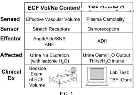
osmoregulation. ECF volume is regulated by intravas-cular sensors, which detect mechanical stretch (thus vascular volume) and by effectors which modify ECF Na content. In fact, altering Na content of the ECF compartment can effect a change in ECF volume only FIG. 1.
Nested r elationship of ex tracellular fluid (ECF) volume/ Na content r egulation within domain of total body fluid osmor egulation: why separate ECF volume r egulation is mandatory. The ECF (inner box containing vascular ring) is located within the TBF and is subject to TBF osmor egulation. Concor dant r esponses of ECF and intracellular fluid (ICF) osmolarity and volume in r esponse to water ingestion (upper pathway) is contrasted with r esponse to NaCl ingestion, initially without water (lower pathway). NaCl r estriction to ECF initially incr eases only ECF osmolarity, causing water to shift fr om ICF to ECF. This lowers ICF volume, raises ECF volume, and equalizes osmolarity in the 2 compartments at an elevated level. High osmolarity activates osmor egulatory pr ocesses [thirst, antidiur etic hor mone (ADH)] and leads to water r etention. Retained water r estor es TBF osmolarity to nor mal and r estor es ICF volume to nor mal. However, ECF volume is incr eased; assuming no Na ex cr etion, the volume of ex tra water r etained will pr ecisely equal the volume of water r equir ed to nor malize ECF osmolarity in pr esence of higher ECF Na content. That the ECF (as a component of TBF) is subject to osmor egulation
because the ECF osmolarity is concomitantly regu-lated. When Na is added or lost, the osmolarity is reliably normalized by proportional changes in ECF water. Because Naconcentra tion is maintained con-stant by osmoregulation independent of Nacontent, a change in the Na content of the ECF compartment will necessarily lead to a change in the ECF volume. Accordingly, the ECF volume/Na content regulatory system, although physiologically independent with distinct sensor/effector limbs, is in fact functionally effective only when operating within the context of the osmolarity/water regulating system.
THE CHALLENGE
These concepts are multidimensional and intrinsically complex. However, three aspects present special difficulty for students newly introduced to body fluid regulation.
First, serum Na concentration, although sampled from the ECF compartment, is in fact a surrogate measure of TBF osmolarity and thus reflects TBF osmolarity/water handling; Na concentration is (seemingly paradoxi-cally) not a measure of ECF Na content. Students typically make an intuitively powerful cognitive error by viewing serum Na concentration as a measure of ECF Na content. The student is usually unaware of this intuitive error and of its power to thwart understand-ing. In our experience, this ‘‘miscognition’’ will con-tinue to operate until unmasked and subjected to conscious scrutiny.
Second, learning is hampered by the ingrained clinical use of imprecise terminology that creates confusion between ECF volume/Na content regulation versus TBF osmolarity/water regulation. One example is the common shortening of ‘‘ECF volume’’ to the term ‘‘volume’’ or ‘‘Na/volume.’’ We then expect students to figure out that this volume is a function of Na balance; conversely, changes in water balance (the logical measure of volume in any language) are not acknowledged to have major impact on Na/volume. The inappropriate use of such terms as ‘‘dehydration/ hydration’’ to refer to ECF volume depletion/repletion (rather than appropriately to water excess/deficit) is another example.
Third, even when these concepts are clearly con-veyed, we have commonly failed to provide the
crucial next step, translation to clinical practice. Students can often parrot body fluid information correctly on tests but are unable to correctly apply their information in the context of patient evaluation and treatment.
STRATEGIES
Strategies used to optimize learning are outlined in Table 1.
Establish Separate but Interactive Natur e of ECF Volume/ Na Content Versus Osmolarity/ Water Regulatory Systems
We have added a short lecture segment focusing entirely on the separate-but-interactive nature of the two systems. Our written objectives convey their dual nature and incorporate direct comparisons (Table 2 and Fig. 2). This reinforces their duality and describes their ‘‘nested’’ relationship (ECF compartment lo-cated within the TBF and regulated by both systems; ECF volume regulation via Na content change effec-tive only in presence of osmolarity/water regulation). The specific circumstances of their clinical interaction
TABLE 1
Strategies to optimize lear ning
1) Distinguish the two separate systems regulating extracel-lular fluid (ECF) volume/Na content vs. total body fluid osmolarity/H2O in terms of fluid compartment regulation,
sensors/effectors, clinical data for diagnosis of abnormality, and points of interaction.
2) Carefully define the language of fluid/electrolytes using ter-minology that clarifies and reflects the clinically crucial dif-ferences between the two homeostatic systems.
3) Directly confront the ‘‘Na concentration5Na content’’ cog-nitive error common to students new to fluid/electrolyte concepts; solicit the students’ participation in recognizing and neutralizing this intuitively powerful miscognition. 4) Provide clinical problem solving experience in small group
sessions designed to translate basic fluid/electrolyte con-cepts into sound clinical reasoning and therapeutic inter-ventions.
5) Make use of ‘‘whole body’’ graphs to bring key pathophysi-ological elements into ‘‘one eyeful,’’ thus visually imprinting key details within a clinically meaningful context.
6) Standardize pathophysiological graphs and diagnostic algo-rithms; use the same visual aids in all lectures and small group sessions.
are defined (severe ECF volume disorders nonosmoti-cally activate antidiuretic hormone (ADH)/water reten-tion). Finally, the distinctly different types of clinical data required for diagnosis of the respective disorders (i.e., physical exam for ECF volume/Na content disor-ders vs. lab test for TBF osmolarity/water abnormali-ties) (Fig. 2).
Define and Use Pr ecise Clinical Language
To further optimize learning, we allot a half-hour lecture to precisely define the language of body fluid/electrolytes for both participating faculty and students. Specifically, we define and use ECF, ICF, and TBF as compartment designations and modify the term ‘‘volume’’ by one of these designations. We have dropped the term ‘‘total body water’’ and speak of ‘‘TBF osmolarity/water regulation’’ to emphasize the regulated compartment (TBF), the sensed parameter (osmolarity), and the responding parameter (water handling). In parallel, the term ‘‘ECF volume/Na content regulation’’ indicates the comparable components of that system. When referring to measured osmolarity, we use ‘‘TBF osmolarity’’ to emphasize the compart-ment reflected despite the fact that it is determined on a sample of serum and reported as ‘‘serum osmolar-ity.’’ Finally, throughout the course components, TABLE 2
Instructional objectives
The student should be able to:
1) Distinguish between the osmolarity/water regulatory system and the ECF Na content/ECF volume regulatory system in terms of:
fluid compartment that is regulated, parameter that is ‘‘sensed,’’ type and location of the sensor(s), factors which mediate the response, typical responses, and
clinical information required to diagnose an abnormality.
2) Characterize the effects of changes in sodium intake on: ECF volume and ECF Na content,
renin-angiotensin system components (renin, ANG II, aldo-sterone),
sympathetic neural activity, atrial natriuretic factor (ANF), renal Na and H2O handling,
serum Na concentration,* and intracellular fluid volume.*
3) Characterize the effects of ANG II and the implications for ECF volume/Na content homeostasis when renin-angio-tensin effects are pharmacologically blocked (angiorenin-angio-tensin- (angiotensin-converting enzyme inhibitors, AT1-receptor blockers).
4) Specify and be able to appropriately utilize clinical indica-tors of volume status for the following ECF subcompart-ments: intravascular venous volume, intravascular arterial volume, interstitial volume.
5) Characterize the three classic clinical states of edema forma-tion (congestive heart failure, hepatic cirrhosis, and nephrotic syndrome) in terms of:
primary abnormality,
manner in which ECF volume is abnormally distributed, change in ‘‘effective arterial blood volume,’’
major mechanisms of sodium retention, and major mechanisms of edema formation.
6) Use the classic clinical approach to differential diagnosis in a patient with hyponatremia or hypernatremia with respect to: TBF osmolarity,
hypo-, hyper-, or euvolemia of ECF,
pathophysiological mechanism and differential diagnosis in each subcategory,
expected urinary Na, urinary osmolarity values, and appropriate therapeutic interventions.
7) Distinguish, in clinical conditions with combined disorders of ECF volume/Na content regulation and TBF osmolarity/ water regulation, therapeutic interventions that address each of the two abnormalities.
8) Explain why serum Na concentration cannot be used as an index of the ECF Na content or of ECF volume.
*Included to force the student to consider the independence of ECF volume/Na content regulation from Na concentration (i.e., osmolar-ity/water) regulation.
FIG. 2.
every mention of ‘‘serum Na concentration’’ is linked with the modifier ‘‘index of TBF osmolarity.’’
Unmask the ‘‘Na Concentration/ Na Content’’ Err or
We believe it is necessary to actively and openly confront this intuitive error in multiple, different learning contexts so that, with time, a new and conceptually valid paradigm can replace the old. Even this focused attention may not fully offset the power of this cognitive association. Students need to under-stand the potential for serious clinical consequences due to this common error of clinical reasoning. In our small group sessions, any student comment or re-sponse that reveals the activity of the erroneous paradigm iswelcom ed by the faculty facilitator as an opportunity to point out, nonjudgmentally and sup-portively, the power and the universality of this misconception. With repeated supportive encourage-ment, students began to ‘‘catch’’ themselves and each other in their incorrect thought patterns. Finally, as the replacement paradigm takes hold, students are reminded of the tenacity of the misconception and of the tendency of the old paradigm to recur over time without regular reinforcement of the new paradigm. Students are reassured that several years is not an unreasonable nor unusual prerequisite for full under-standing of these complex concepts and for their full integration into clinical decision making.
Pr ovide Case-Based Clinical Pr oblem Solving Ex perience
Three two-hour case-oriented small group sessions employ active-learning techniques, benefit from exten-sive faculty preparation as facilitators, are sequenced in conceptual complexity, and include objectives plus provision of detailed written summaries of key points on completion of each session. During these optional, ungraded exercises, students (9–12/group) are asked to divide into three to four minigroups; each group is then assigned one of the written cases with accompa-nying questions. Twenty to thirty minutes are pro-vided within the session time frame for students to work through the questions posed. Faculty serve as fac ilitators, answer generic questions to permit progress, and often respond by posing other questions to guide students’ pathophysiological thinking but do not provide the answers to the formal case questions.
Each minigroup is thus asked to commit to an answer despite uncertainty. Each minigroup then presents its assigned case to the whole group, steps through the pathophysiology, answers the questions posed, and demonstrates relevant calculations. Their answers are then critiqued by their peers with faculty guidance and corrective input as needed. (In contrast to classic problem-based learning exercises, we feel that the faculty facilitators for these sessions m ust also be experts because of the complexity of the concepts and the need for immediate recognition and correc-tion of student misconcepcorrec-tions.)
Standar dize All Key Graphics and Algorithms Acr oss Lectur es and Small Gr oup Sessions
Make each available as transparencies for both stu-dents and faculty facilitators to use as teaching tools in small groups (Fig. 1–8).
Use Graphs That Embody the Principle of ‘‘Whole Body in Single Eyeful’’
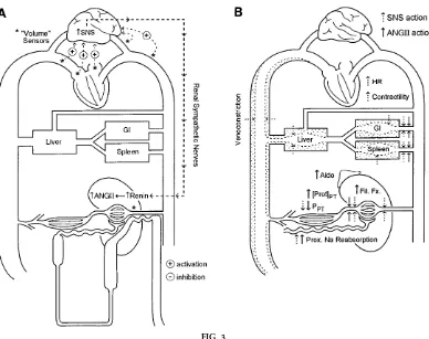
Graphics used to describe homeostatic systems are derived from the premise that incorporating the whole organism into a ‘‘single eyeful’’ helps organize and visually imprint details while preserving a clini-cally meaningful whole body context (Figs. 3 and 4).
Link Conceptual and Clinical Practice Aspects of ECF Volume/ Na Content Evaluation
Along with concepts developed in lectures and small group cases, a practical lecture describing ECF volume assessment by physical examination is immediately followed by a hands-on exercise providing demonstra-tion of physical exam skills and opportunities for practice with immediate feedback.
COURSE CHRONOLOGY AND CONTENT
Lectur e: Review of Physiology—ECF Volume/ Na Content and Water/ Osmolarity Homeostasis
This lecture is based on material covered in the first year.
Overview Lectur e: The Clinical Language of Fluid/ Electr olytes
compart-ment volumes and compositions are reviewed. Distri-bution of ingested water versus NaCl versus saline solutions of varying concentration in normals are presented in a semi-interactive format, providing an introduction to the first small group sessions.
Small Gr oup Case-Oriented Session A: Nor mal Body Fluid Physiology
The purpose of session Ais to address quantitative distribution of ingested salt/water in physiologica l
contexts, and thus to orient student thinking in terms of tracking ingested solutions (salt vs. water vs. NaCl solutions) into specific body fluid compartments in a whole person. (Cases and written summary of key points are available on request.) Students are asked to revisit body fluid compartments, calculate the respec-tive compartment volumes based on body weight, and calculate how each changes with ingestion of water only, salt only, or both in varying proportions. Stu-dents are encouraged to envision ingested milliequiva-FIG. 3.
lents of salt and cubic centimeters of water in terms of volumes of normal saline versus ‘‘free’’ (solute-free) water. This exercise emphasizes the clinical reality that different fluids distribute into distinct compart-ments with dramatic consequences for therapeutic decision making. This session also prepares the way for the subsequent small group sessions addressing clinical disorders of salt/water homeostasis and
requir-ing estimation of body fluid compartment sizes and calculation of water deficits.
Lectur e: Dual Systems of Body Fluid Regulation
Instructional objectives for the three major lectures are presented in Table 2.
This lecture opens with a general description of the two homeostatic systems from a clinical perspective. Their uniqueness, their respective sensors/effectors, and their distinct requirements for clinical detection are carefully outlined (Fig. 2). Three points of interac-tion are then described. First, the presence/normal operation of the osmolarity/water regulating system is what perm its regulation of ECF Na content to also achieve regulation of the size of ECF volume. [Ex-ample used: salt tablet ingestion, with stepwise re-sponses of the osmolarity/water regulatory system, followed by the activation of ECF Na/ECF volume regulating systems (see above and Fig. 1)]. Second, primary abnormalities of the TBF osmolarity/water regulating system, which include the ECF compart-ment in the relative excess or deficit of water, activate ECF volume sensors and initiate ECF volume regula-tion; this protects the ECF volume from uncontrolled osmoregulation. Third, severe (pathological) reduc-tion in ECF volume or in ‘‘effective arterial volume’’ activates ADH secretion nonosmotically, thus recruit-ing a component of the osmolarity/water system to protect ECF volume at the cost of impairing normal water excretion capacity. This interaction underlies the clinical approach to differential diagnosis of body fluid osmolarity/water disorders based on the status of the ECF volume.
The clinical indicators for assessing the volume status of each of the ECF subcompartments are then intro-duced and explained. These include the jugular ve-nous pressure (JVP, an index of the veve-nous compo-nent of vascular volume), arterial blood pressure measured supine and standing (‘‘postural BP,’’ an index of effective arterial volume), and edema (as evidence of excessive interstitial fluid volume and excessive total ECF volume). We establish this clinical connection early and reemphasize the crucial point that clinical diagnosis of ECF volume/Na content disorders depends primarily on the bedside exam. This is in distinct contrast to the clinical diagnosis of FIG. 4.
TBF osmolarity/water abnormalities, which is based solely on a laboratory blood test (TBF osmolarity, measured as serum osmolality or estimated by serum Na concentration as an index of TBF osmolarity) (Fig. 2).
The ‘‘Na concentration5Na content’’ cognitive error and its clinical implications are then described (as outlined inSTRATEGIES). Its universality and power are
acknowledged, and the students are encouraged to actively participate with faculty in detecting and eradicating this sneaky neural pathway that stands between the student and metabolic enlightenment.
An optional demonstration (which can be done using color slides) involves hidden beakers of varying sizes, each filled with fluid and representing an ECF volume of distinct (low, normal, or high) size; each beaker contains a different concentration of solute (blue dye) that varies independent of beaker volume. Test tubes containing equal aliquots from each beaker are pre-sented to the students, who are then asked to predict the volume of the beaker (ECF volume) from the solute concentration of the test tube sample. Most will recognize the fallacy of this effort when the task is thus presented. The similar fallacy of predicting ECF volume or Na content from serum Na concentration can then be emphasized.
Lectur e: Clinical Disor ders of ECF Volume/ Na Content Regulation
Once the duality of the two regulatory systems is presented, the components of the ECF volume/ECF Na content regulating system is specifically outlined using whole body/single-eyeful graphs to permit detail while retaining a whole body context (Fig. 3). The components of the ECF volume regulating system are initially presented in sequential steps as a response to ECF volume/Na content depletion. Thus initial tion of low-pressure stretch receptors and early activa-tion of renal sympathetic nerve system (SNS) traffic leads to the initiation of renin release and both circulating and intrarenal ANG II formation (Fig. 3A). Subsequent ANG II and SNS actions on proximal Na reabsorption reduce distal NaCl delivery and thus activate the macula densa mechanism of renin release, bringing into play two of the three renin-activating pathways (Fig. 3A). It is pointed out that activation of the renal baroreceptor by lowered pressure at the
afferent arteriole, while activated in pathological states of ECF volume depletion with hypotension, is not necessarily activated in physiological states such as dietary Na restriction. Next, hemodynamic effects of ANG II are outlined, including—and specifically under states of circulatory stress—a dominant efferent arterio-lar constriction. This provides rela tive protection of glomerular filtration rate (GFR) via sustaining glomeru-lar capilglomeru-lary pressure and increasing filtration fraction. It is important to stress that GFR is not increased, rather it is decreased to a lesser degree than is renal blood flow (RBF). Increased filtration fraction yields increased protein concentration and lowered hydro-static pressure in the peritubular capillary, thus alter-ing Starlalter-ing forces to promote Na/water reabsorption. Finally, direct actions of ANG II, the SNS, and aldoste-rone on respectively proximal and distal tubular Na reabsorption are reviewed (Fig. 3B).
(We do not emphasize pressure natriuresis as a physi-ological effector of ECF volume/Na content regula-tion, because this homeostatic system is extremely effective in the absence ofclinica lly detecta blechanges in blood pressure. This obviously does not exclude important intrarenal pressure changes. Pressure natri-uresis is instead presented in the context of long-term blood pressure regulation.)
Clinical indicators of ECF volume status are then reviewed for expected changes in true ECF volume depletion: reduced JVP (venous), postural hypoten-sion (arterial), or absent edema (interstitial). (Major focusing on physical exam details is deferred to a dedicated lecture and clinical ECF volume exam exercise on the following day.) A simple differential diagnosis of causes of ECF volume depletion is briefly reviewed.
(min-eralocorticoid excess, renal failure) logically falls here but is deferred to later lectures on secondary hyperten-sion and renal failure, respectively. These categories are thus noted for completeness, but primary atten-tion is directed to the three classic states of general-ized edema formation: congestive heart failure (CHF), hepatic cirrhosis, and nephrotic syndrome. The preced-ing detailed description of the normal response to dietary Na restriction/ECF volume depletion has also provided the pathophysiological information needed to understand the systemic and renal responses to these classic edematous states. Each is presented as a pathophysiological abnormality that leads to func-tional maldistribution of ECF volume and thereby to deficits of effective arterial volume; the latter then activates some or all of the same Na-retaining mecha-nisms involved in true ECF volume/Na content deple-tion. A second and extremely important aspect of these complex clinical states is the typically discor-dant changes in individual ECF subcompartments. Thus effective arterial volume, as assessed by postural BP changes, may be significantly decreased and organ perfusion threatened despite an unequivocal increase in interstitial (and total ECF) volume as manifested by edema or ascites. It is crucial to equip students to clinically evaluate each ECF subcompartment sepa-rately and to acknowledge the clinical priority of restoring the arterial circuit and organ perfusion when it is threatened.
Congestive heart failur e. CHF (Fig. 4A) is taken stepwise from the first subnormal stroke volume with consequent increased filling volume/pressures. In-creased venous pressures distend the veins, sequester-ing excess blood volume in the venous compartment; elevated hydrostatic pressure promotes transfer of ECF volume from vascular to interstitial compart-ments, augmented by increased circulating ANF. The impaired cardiac pump action creates a ‘‘functional dam’’ at the level of the heart, initially producing ‘‘ineffective arterial volume’’ at normal filling pres-sures. Accordingly, ECF volume is maldistributed at two levels: arterial volume shifts to the venous side; and vascular volume is shifted to the interstitial compartment. Inadequate arterial volume activates central arterial and renal components of the ECF volume/Na content regulating system (e.g., arterial high-pressure stretch receptors with SNS response; macula densa and potentially renal baroreceptor
regulation of renin release). In addition to these mechanisms of Na retention, mechanisms of edema formation are outlined (increased venous/capillary hydrostatic pressure, ANF-dependent increase in endo-thelial permeability). If pump dysfunction is not severe, then the increased filling pressure can restore cardiac output, offsetting the dam effect at the expense of increased pressures (i.e., ‘‘compensated’’ CHF).
Hepatic cirrhosis.Hepatic cirrhosis (Fig. 4B) is next outlined as an abnormality that creates a functional dam within the liver, increasing intrahepatic sinusoi-dal and portal venous pressures. The increased pres-sure distends the veins, sequestering excess blood volume in the splanchnic venous circulation, and forms ascites by increasing hydrostatic force for fluid transudation at the liver surface and across capillary beds. In addition (and perhaps as a compensation for high venous pressure), excess nitric oxide-dependent vasodilation localized to the splanchnic circulation expands splanchnic arterial volume and further seques-ters blood volume. These forces yield maldistribution of ECF volume at two levels: within the vascular tree (extrasplanchnic arterial to splanchnic venous1 arte-rial) and between vascular and interstitial/peritoneal compartments (from splanchnic venous to peritoneal space). Both, if uncompensated, would tend to ‘‘steal’’ volume from the arterial tree, contributing to ineffec-tive arterial volume and activating all renal and sys-temic components of ECF volume/Na content regula-tion, inc luding the low-pressure c entral venous elements. Mechanisms of renal Na retention are empha-sized: reduced effective extrasplanchnic vascular volume, intrahepatic activation of a sympathetic hepa-torenal reflex circuit, and the direct intrarenal Na-retaining effects of commonly associated hypoalbumin-emia. Mechanisms of generalized edema formation include compressive effects of ascites on lower extrem-ity venous return and the lowered oncotic pressure of hypoalbuminemia.
the natural charge-based barrier to albumin (an anion) leads not only to proteinuria but also to generalized transudation of albumin, reducing plasma oncotic pressure and redistributing ECF from vascular to interstitial space. Unopposed, this reduction in intra-vascular volume would activate all components of the ECF volume/Na content regulating system and thus causeseconda ryrenal Na conservation and Na reten-tion together with water in isotonic amounts. Evi-dence for a direct intrarenal effects of hypoalbumin-emia to promote prim a ry renal Na retention is also noted, and the balance of these factors may vary, yielding a low (dominance of hypoalbuminemia), normal, or high (dominance of primary renal Na retention) intravascular subcompartment. Recent stud-ies have raised technical questions about methods used to show increased vascular volume in some nephrotics. Nonetheless, expanded tota l ECF vol-ume/Na content, with edema as the hallmark, remains a classic finding in nephrotic syndrome, and the clinician’s challenge is to accurately assess the intravas-cular subcompartment in planning therapeutic inter-vention. (Other aspects of nephrosis are addressed in the renal section of the course.)
Lectur e: Disor ders of TBF Osmolarity/ Water Regulation
Instructional objectives are outlined in Table 2.
The lecture begins with a brief reminder of body fluid spaces and their composition. The normal physiology of osmotic regulation and water balance is reviewed in detail because this information is crucial to the under-standing of the disorders (Fig. 5). Acute versus chronic renal actions of ADH at the level of aquaporin recruit-ment to the apical membrane are briefly introduced. It is emphasized again that these disorders, typically presenting as altered serum Na concentra tion (an index of TBF osmolarity), are in fact abnormalities of TBF osmolarity/water regulation and are not reflec-tions of altered ECF Na content. This point is reiter-ated in many different ways to the students, once again directly confronting the ‘‘Na concentration 5
Na content’’ error.
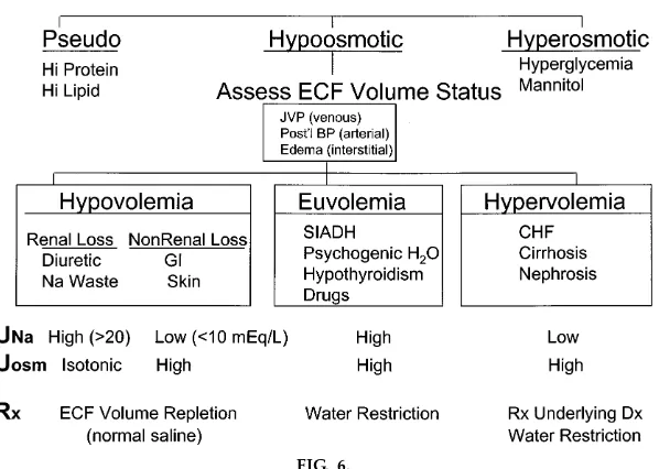
A classic clinical approach to the differential diagnosis ofhypona trem ia is presented in depth, including the requirement for clinical estimation of the ECF volume
FIG. 5.
to categorize hypoosmotic hyponatremias into hypo-, hyper-, and euvolemic subsets (Fig. 6). The general mechanism of hyponatremia in each subset is intro-duced and linked to the therapeutic approach in each case. Thus hypovolem ic hypona trem ia reflects the capacity of severe ECF volume depletion to
nonosmoti-cally activate ADH secretion; this, together with re-duced GFR, impairs free water excretion at the expense of hypoosmolarity. Correction requires ECF volume repletion with NaCl and water.Hypervolem ic hypona trem ia also reflects inadequate effective arte-rial blood volume, causing nonosmotic ADH
stimula-FIG. 6.
tion and impaired free water excretion. However, in this case, total ECF volume/Na content is in excess, indicating discordant directional changes in the ECF subcompartments (i.e., increase in total ECF volume but decrease in the effective arterial subcompartment, and thus ECF maldistribution). These two categories of hyponatremia, hypo- and hypervolemic, are in fact complications of, and thus secondary to, primary disturbances of ECF volume/Na content regulation and are especially apt to occur when the primary ECF disorder is severe. In contrast,euvolem ic hypona tre-m iais a subset of disorders in which TBF osmolarity/ water regulation is the primary body fluid disturbance, induced by inappropriate production of ADH or
ADH-like molecules or by other disturbances that impair renal free water excretion.
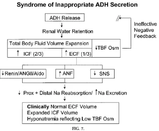
A specific example of a prototypical disorder that presents with hyponatremia is then discussed in depth for each of the three subtypes (Fig. 6). The syndrome of inappropriate antidiuretic hormone secre-tion (SIADH) is detailed as a type of euvolemic hyponatremia. Simple pathophysiological diagrams are used to explain the pathogenesis of the disorder (Fig. 7). The inappropriately retained water distrib-utes evenly throughout the TBF. When the compo-nent retained within the ECF is sufficient to activate stretch receptors, activation of ECF volume/Na
con-FIG. 7.
tent regulation promotes transient Na loss, maintain-ing a steady-state ECF volume within a clinically normal range. Thus, although the ECF compartment shares in the retained water/hypoosmolarity, the de-gree of ECF volume expansion under these circum-stances is homeostatically limited and not clinically detectable. We apply the term ‘‘clinically euvolemic’’ despite the recognition that TBF is increased and subtle ECF expansion is present.
Similarly, specific examples of hypovolemic hyponatmia and hypervolemic hyponatrehyponatmia are each re-viewed stepwise with emphasis on the mechanism of water retention. It is emphasized again to the students that the hypo- and hypervolemic hyponatremias are very common clinically and most often develop as a complication of the primary ECF Volume/Na content disorders. Finally, a case of hypervolemic hyponatre-mia due to CHF is presented and the pathogenesis reviewed to demonstrate the simultaneous presence of a TBF osmolarity/water disorder and an ECF vol-ume/Na content disorder in the same patient (Fig. 4C) and the need to address their respective treatments separately.
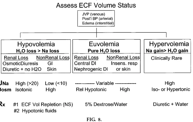
In a parallel manner, hyperna trem ia is discussed in the context of a generalized clinical approach to the patient (Fig. 8) with subcategorization based on clini-cal assessment of the ECF volume status. Euvolemic hypernatremia is discussed along with the types of diabetes insipidus, namely, pituitary and nephrogenic. Because of its uncommon occurrence, hypernatremia in the setting of ECF volume overload is mentioned only briefly, whereas hypernatremia in the setting of ECF volume depletion is discussed in more depth. In these latter disorders, we emphasize that a Na content deficit is present (as shown by physical exam) and is accompanied by a proportionally larger water deficit (as manifested by the increased TBF osmolarity). We also stress that restoring the intravascular compart-ment of the ECF with normal saline, thus protecting vital organ perfusion, is always given priority over therapeutic manipulation of the osmolarity.
As a final synthesis, the distinction between regulation of excess and deficiency of ECF volume/Na content and regulation of excess and deficiency of TBF osmo-larity/water is once again revisited (Fig. 2).
FIG. 8.
Case-Oriented Small Gr oup Session B
Paper cases on ECF volume depletion/excess, hypona-tremia, and hypernatremia are provided using the same format as outlined inSmall Gr oup Session A.
Lectur e: Clinical Evaluation of ECF Volume
This practical session describes the standard physical examination of ECF volume, including procedures for determining and guidelines for interpreting jugular venous pressure, postural BP changes, edema, and ascites.
Small Gr oup Session C: Hands-On Clinical ECF Volume Ex am
This 2-hour session provides, for 50 students at each of 2 sessions, 10–12 stretcher stations, each with a patient volunteer, a stretcher with easily adjustable headpiece, Hg manometer with three BP cuff sizes, a centimeter ruler, and a faculty member familiar with the patient/volunteer’s findings and with course objec-tives. Stations are set up in wide hallways surrounding the dialysis unit; two stations involve a stable dialysis patient undergoing hemodialysis. Students spend 15– 20 min per station; they are shown the findings in the first two stations encountered and at subsequent stops are asked to do their own exam and assess the status
of the ECF volume/Na content. Faculty are careful to acknowledge when jugular veins are not able to be evaluated and why. A faculty timekeeper kept the student groups moving from station to station on time.
Case-Oriented Small Gr oup Session D
Paper cases are provided in which somewhat more complex fluid/electrolyte challenges are presented. Students are encouraged not only to develop their own conceptual analysis of the patient’s problems but also to extend their clinical wings by independently calculating body fluid deficits/excesses and by writing orders specifying types and volumes of fluids to administer and over what time period they should be administered.
The success of these instructional strategies and teaching tools is greatly dependent on a cadre of dedicated faculty with substantial expertise in fluid/ electrolyte issues, commitment to the preparation time essential to achieve consistency, and uncommon enthusiasm. We have been privileged to work with such a group and gratefully acknowledge their many contributions.