BASIC CONCEPTS IN
• • • • • • • • • • • •
A STUDENT'S SURVIVAL GUIDE
BIOCHEMISTRY
Second Edition
HIRAM F. GILBERT,
Ph.D.
Professor of Biochemistry Baylor College of Medicine
Houston, Texas
McGraw-Hill
Health Professions Division
New York St. Louis San Francisco
Auckland Bogotá Caracas Lisbon London Madrid Mexico City Milan Montreal New Delhi San Juan
BASIC CONCEPTS IN BIOCHEMISTRY, 2/E
Copyright © 2000, 1992 by the McGraw-Hill Companies, Inc. All rights reserved. Printed in the United States of America. Except as per-mitted under the United States Copyright Act of 1976, no part of this publication may be reproduced or distributed in any form or by any means, or stored in a data base or retrieval system, without the prior written permission of the publisher.
1234567890 DOCDOC 99
ISBN 0-07-135657-6
This book was set in Times Roman by Better Graphics, Inc. The edi-tors were Steve Zollo and Barbara Holton; the production supervisor was Richard Ruzycka; the index was prepared by Jerry Ralya. R. R. Donnelley and Sons was the printer and binder.
This book is printed on acid-free paper.
Basic Concepts in Biochemistry: A Student’s Survival Guideis not a
con-ventional book: It is not a review book or a textbook or a problem book. It is a book that offers help in two different ways—help in understanding the concepts of biochemistry and help in organizing your attack on the subject and minimizing the subject’s attack on you.
This book presents what are often viewed as the more difficult con-cepts in an introductory biochemistry course and describes them in enough detail and in simple enough language to make them understand-able. We surveyed first- and second-year medical students at a national student meeting asking them to list, in order, the parts of biochemistry they found most difficult to understand. The winner (or loser), by far, was integration of metabolism. Metabolic control, pH, and enzyme kinetics ran closely behind, with notable mention given to molecular biology and proteins.
Biochemistry texts and biochemistry professors are burdened with the task of presenting facts, and the enormity of this task can get in the way of explaining concepts. Since I don’t feel burdened by that necessity, I’ve only outlined most of the facts and concentrated on concepts. My rationale is that concepts are considerably easier to remember than facts and that concepts, if appropriately mastered, can minimize the amount of material that has to be memorized—you can just figure everything out when required. In Basic Concepts in Biochemistry, central concepts are
developed in a stepwise fashion. The simplest concepts provide a review of what might have been forgotten, and the more complex concepts pre-sent what might not have been realized.
•
P R O L O G U E
•xv
Preface xiii
Prologue xv
CHAPTER 1 WHERE TO START 1
Instructions 1
What Do I Need to Know? 2
Instructions for Use 2
Studying and Exams 2
Trivia Sorter 4
CHAPTER 2 PROTEIN STRUCTURE 6
Amino Acid Structure 6
Interactions 8
Water 9
Hydrophobic Interaction 9
van der Waals Interactions and London Dispersion Forces 11
Hydrogen Bonds 11
Secondary Structure 12
Protein Stability 15
Favorable (Good) Interactions 17 Unfavorable (Bad) Interactions 17 Temperature-Sensitive Mutations 19 Ligand-Binding Specificity 20
Global Conclusion 21
CHAPTER 3 MEMBRANES AND
MEMBRANE PROTEINS 22
General Membrane Function 22
Membrane Composition 23
Phospholipid Bilayer 24
Membrane Structure 25
Posttranslational Modification 26
Membrane Fluidity 27
Diffusion in Membranes 28
Movement of Ions and Molecules Across Membranes 28
•
C
O
N
T
E
N
T
S
•Transport Across Membranes 29
The Nernst Equation 31
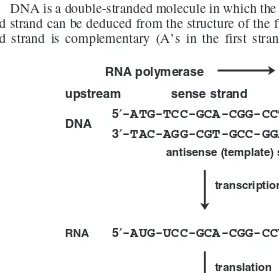
CHAPTER 4 DNA-RNA STRUCTURE 35
DNA Structure 35
DNA Stability 37
RNA Secondary Structure 38
CHAPTER 5 EXPRESSION OF GENETIC
INFORMATION 40
Information Metabolism 40
Directions and Conventions 41
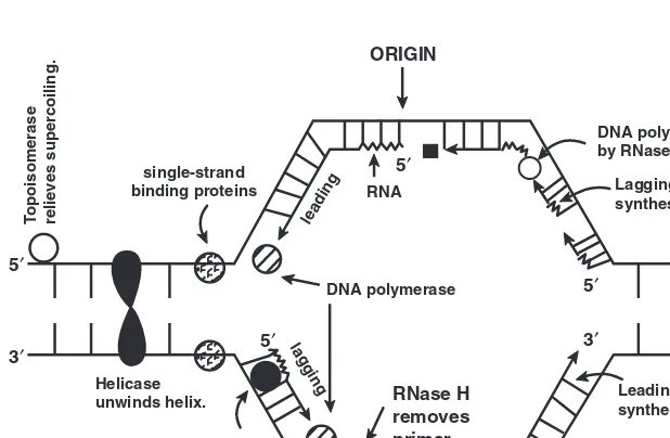
DNA Replication 42
Types of DNA Polymerase 45
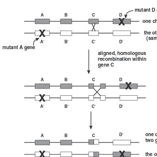
Recombination 47
Regulation of Information Metabolism 49
Transcription 53
Regulation of Transcription 55
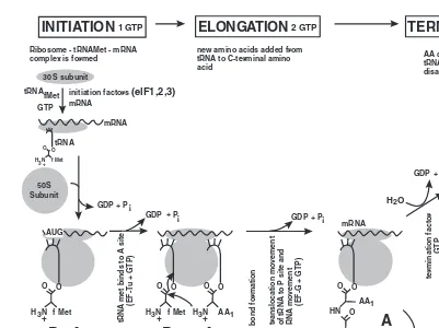
Translation 57
Use of High-Energy Phosphate Bonds During Translation 60
CHAPTER 6 RECOMBINANT-DNA METHODOLOGY 61
Restriction Analysis 61
Gels and Electrophoresis 65
Blotting 67
Restriction Fragment-Length Polymorphism 69
Cloning 70
Sequencing 73
Mutagenesis 75
Polymerase Chain Reaction 76
CHAPTER 7 ENZYME MECHANISM 80
Active Site 81
Transition State 81
Catalysis 83
Lock and Key 83
Induced Fit 83
Nonproductive Binding 85
Entropy 87
Strain and Distortion 88
Transition-State Stabilization 88 Transition-State Analogs 91
Chemical Catalysis 93
CHAPTER 8 ENZYME KINETICS 95
S, P, and E (Substrate, Product, Enzyme) 96 Amounts and Concentrations 96
Active Site 97
Assay 98
Velocity 98
Initial Velocity 100
Mechanism 101
Little k’s 102
Michaelis-Menten Equation 103
Vmax 106
kcat 106
Km 107
Special Points 107
kcat/Km 107
Rate Accelerations 108
Steady-State Approximation 109 Transformations and Graphs 111
Inhibition 112
Allosterism and Cooperativity 117 The Monod-Wyman-Changeaux Model 119
CHAPTER 9 SIGNAL TRANSDUCTION PATHWAYS 123
Signal Transduction Pathways 123
Organization 124
Signals 125
Receptors 126
Soluble Receptors 126
Transmembrane Receptors 128 Enzyme Coupled Receptors 128 G-Protein Coupled Receptors 131 Ion-Channel Coupled Receptors 132
Second Messengers 133
Amplifiers 136
Integrators 137
Inhibitors 138
CHAPTER 10 GLYCOLYSIS
AND GLUCONEOGENESIS 141
Glycolysis Function 143
Glycolysis Location 143
Glycolysis Connections 143
Glycolysis Regulation 143
Glycolysis ATP Yields 144
Glycolysis Equations 144
Effect of Arsenate 144
Lactate or Pyruvate 145
Gluconeogenesis Function 145 Gluconeogenesis Location 145 Gluconeogenesis Connections 145 Gluconeogenesis Regulation 146 Gluconeogenesis ATP Costs 146 Gluconeogenesis Equations 146
CHAPTER 11 GLYCOGEN SYNTHESIS
AND DEGRADATION 147
Function 147
Location 147
Connections 148
Regulation 148
ATP Yield 150
ATP Cost 150
Molecular Features 150
CHAPTER 12 TCA CYCLE 153
TCA Cycle 153
CHAPTER 13 FAT SYNTHESIS
AND DEGRADATION 155
Fatty Acid Synthesis Function 156 Fatty Acid Synthesis Location 156 Fatty Acid Synthesis Connections 157 Fatty Acid Synthesis Regulation 157 Fatty Acid Synthesis ATP Costs (for C16) 157
Fatty Acid Synthesis Equation 159 Elongation and Desaturation 160 Triglyceride and Phospholipid Synthesis 162
-Oxidation Function 164
-Oxidation Location 164
Carnitine Shuttle 164
-Oxidation Connections 164 -Oxidation Regulation 164 -Oxidation ATP Yield 166
-Oxidation Equation 167
-Oxidation of Unsaturated Fatty Acids 168 -Oxidation of Odd-Chain-Length Fatty Acids 172
CHAPTER 14 ELECTRON TRANSPORT
AND OXIDATIVE PHOSPHORYLATION 173
Oxidation and Reduction 173 The Electron Transport Chain 174
Connections 176
Regulation 178
P/O Ratios 178
Uncouplers 179
Inhibitors 180
CHAPTER 15 PENTOSE PHOSPHATE
PATHWAY 183
Pentose Phosphate Pathway 183
CHAPTER 16 AMINO ACID METABOLISM 186
Nonessential Amino Acid Synthesis 186
Essential Amino Acids 187
Amino Acid Degradation 187 Generalities of Amino Acid Catabolism 187 Products of Amino Acid Degradation 188
CHAPTER 17 INTEGRATION OF ENERGY
METABOLISM 190
Integrating Metabolic Pathways 191
ATP 192
Glucose 192
Storage Molecules 193
Metabolic States and Signals 194
Insulin 195
Glucagon 196
Epinephrine 197
Secondary Signals 198
Generalities of Metabolism 199
Phosphorylation 202
Glycogen 204
Metabolic Movements of Glycogen 205
Fat 207
Metabolic Movements of Fat 207
Protein 209
Metabolic Movements of Protein 209
Tissue Cooperation 211
Liver 212
Muscle 212
Adipose 212
Brain 213
Connection of Storage Pools 213
Feeding 214
Fasting 214
Starvation 217
Excitement 219
Interorgan Cycles 221
Cori Cycle 221
Alanine Cycle 222
Ketone Bodies 223
CHAPTER 18 UREA CYCLE 225
Urea Cycle 225
CHAPTER 19 PURINE METABOLISM 227
Purine Synthesis 227
Purine Salvage 228
Deoxynucleotides 228
Purine Degradation 229
CHAPTER 20 PYRIMIDINE METABOLISM 230
Pyrimidine Synthesis 230
Pyrimidine Salvage 231
Pyrimidine Degradation 232
CHAPTER 21 ONE-CARBON METABOLISM 233
One-Carbon Metabolism 233
Oxidation States of Carbon 233
CHAPTER 22 TRACKING CARBONS 236
Glucose to Pyruvate 236
TCA Cycle 238
CHAPTER 23 ph, pKA, pROBLEMS 241
Proton: H⫹or H
3O⫹ 242
Acid 242
Base 242
Not All Acids and Bases Are Created Equal 243
pKa⫽ ⫺log (Ka) 244
Weak Acids Make Strong Bases (and Vice Versa) 244
Who Gets the Proton? 245
Don’t Forget Stoichiometry 245 The Sadistic Little p 246
Taking log10(x) 247
Taking ⫺log10(x) 247
pH ⫽ ⫺log10[H⫹] 248
pKa⫽ ⫺log10(Ka) 248
Buffers 248
Henderson-Hasselbalch Equation 249
Titration Curves 250
pI—Isoelectric Point 254
The Bicarbonate Buffer 255
Imbalance in Blood pH 257
Acidosis and Alkalosis 258
CHAPTER 24 THERMODYNAMICS
AND KINETICS 261
Thermodynamics 261
Free Energy 263
Adding Free-Energy Changes 268 Coupling Free Energies 268
Thermodynamic Cycles 269
⌬G⫽ ⌬H⫺T⌬S 272
Driving Force 273
• xii • Contents
Kinetics 275
Velocity 275
Transition State Theory 276
Rate Constants 277
Rate Constants and Mechanism 283
Appendix 284
Glossary 287
Since the first edition of this series, we have witnessed the birth of “mol-ecular medicine,” using biochemistry, cell biology, and genetics to diag-nose and treat disease. Consequently, the basic sciences are becoming more important to the practice of medicine. This puts a new pressure on the student—to understand the basis of molecular medicine and the mol-ecular sciences. I still think that it’s easier to remember things that you understand, things that make sense. That’s the idea behind the Basic Concepts series and that’s why I have been so pleased with the expansion of the Basic Concepts series beyond Biochemistry.
The revisions in the second edition include two new chapters, “Membranes and Membrane Proteins” and “Signal Transduction Pathways.” These topics are related to the explosion of new information about cell signaling and signal transduction pathways. In addition, I’ve added some tables of information that I think will be helpful in seeing the big picture (and remembering some of the more important details). As before, the major topics and things to remember are set off in boxes so that if you already know everything in the box, you can skip the rest of the sec-tion.
•
P
R
E
F
A
C
E
•• • • • • • • • • • • •
•
C H A P T E R
•
1
•
WHERE TO START
•
Instructions
What Do I Need to Know?
Instructions for Use
Studying and Exams
Trivia Sorter
• • • • • • • • • • • •
The first page of each chapter presents an index. A title-summary box for each section presents a short summary and memory jogger intended to be helpful for review. If you already know what the boxed terms mean and feel comfortable with them, don’t bother to read the text section that follows—proceed until you find a heading you don’t understand, and then read till you understand. The first rule (it may not really be the first rule, but it is a rule) is not to waste time reading things you already know. Keep on not reading the text until you find something you don’t understand—then read the text till you do. The sections are generally arranged in order of increasing complexity and build on previous tions. So if you screwed up and jumped in over your head, back up a sec-tion or two. Another opsec-tion is just to look at the pictures. Pictures and diagrams, if extensively annotated and carefully designed (by you), can be an enormous review aid.
INSTRUCTIONS
Read for understanding. Read only what you don’t know. Organize, organize, organize.
Medicine and biology are becoming increasingly molecular in nature, so one answer to the question is that you need to know things down to the last atom. Everythingis not the right answer. You can’t
pos-sibly learn it all. Therefore, you will have to be selective.
Another answer is that you just need to know the things on the exam.
Later ends at the final. In reality, later may be longer than this. Try to
pick out the major concepts of biochemistry as you go along. Concepts are generally easier to remember than factual details—particularly if the concepts make sense.
General concepts don’t need to be memorized. Once you understand them, they provide a framework to hang the rest of the material on. Since they don’t need to be memorized, they can be learned (or thought about) almost anywhere. To remember something, write it down. Don’t just highlight it with a colored pen or pencil. Highlighting is a great way to forget to read the material.
•
1. ALWAYS REMEMBER THAT IT IS POSSIBLE TO BE AWORTHWHILE HUMAN BEING REGARDLESS OF (OR IN SPITE OF) HOW MUCH BIOCHEMISTRY YOU KNOW. This won’t
nec-essarily help you with biochemistry, but it may help you keep your sanity.
•
2. MINIMIZE THE AMOUNT OF MATERIAL THAT YOU HAVETO MEMORIZE. If you understand a general concept, you can often
figure out the specific details rather than memorize them. For example,
STUDYING AND EXAMS
Organize, understand, condense, memorize.
INSTRUCTIONS FOR USE
Understand the concepts first. Make notes. Never use a colored highlighter.
WHAT DO I NEED TO KNOW?
You need to know only the things you will need later.
does phosphorylation activate or inactivate acetyl-CoA carboxylase? You could just memorize that it inactivates the enzyme. However, this would not help when it came to the phosphorylation of glycogen synthase. Try the following line of reasoning. We store energy after eating and retrieve it between meals. Storage and retrieval of energy do not happen at the same time. Protein phosphorylation generally increases when you’re hun-gry. Since both acetyl-CoA carboxylase and glycogen synthase are involved in energy storage (fat and glucose, respectively), they will both be inactivated by phosphorylation. For just two enzymes, it might be eas-ier to just memorize all the regulatory behaviors—but for several hundred?
•
3.ARRANGE NOTES AND STUDY TIME IN ORDER OFDE-CREASING IMPORTANCE. During the first (or even second and
third) pass, you can’t possibly learn everything biochemistry has to offer. Be selective. Learn the important (and general) things first. If you have enough gray matter and time, then pack in the details. Organize your notes the same way. For each topic (corresponding to about a chapter in most texts) write down a shortsummary of the really important concepts
(no more than one to two pages). Don’t write down the things that you already know, just the things you’re likely to forget. Be really cryptic to save space, and use lots of diagrams. These don’t have to be publication-quality diagrams; they only have to have meaning for you. The idea is to minimize the sheer volume of paper. You can’t find yourself at finals time with a yellow-highlighted 1000-page text to review 2 days before the exam. An enormous amount of information can be crammed onto a diagram, and you learn a significant amount by creating diagrams. Use them extensively.
•
4. SORT OUT THE TRIVIA AND FORGET ABOUT IT. The mostdifficult part may be deciding what the important things actually are. After all, if you’ve never had biochemistry, it all sounds important (or none of it does). Use the following trivia sorter (or one of your own invention) to help with these decisions. To use this sorter, you must first set your trivia level. Your trivia level will depend on whether you just want to pass or want to excel, whether you want to devote a lot of time or a whole lot of time to biochemistry, and your prior experience. Once you set this level, make sure you know almost everythingabove this level
and ignore almost everything below it. Setting your trivia level is not irre-versible; the setting can be moved at any time. You should consider lev-els 7 to 10 as the minimal acceptable trivia level (passing). The trivia sorter shown here is generic. You can make your own depending on the exact demands of the course you’re taking. Levels 21 and 22 might be too trivial for anybody to spend time learning (again, this is opinion).
•
5. DON’T WASTE TIME ON ABSOLUTE TRIVIA UNLESS YOU HAVE THE TIME TO WASTE. It is possible to decide that somethingis just not worth remembering; for example, cleavage specificities of pro-teases or restriction endonucleases, and protein molecular weights, are
TRIVIA SORTER
1. Purpose of a pathway—what’s the overall function?
2. Names of molecules going into and coming out of the
path-way
3. How the pathway fits in with other pathways
4. General metabolic conditions under which the pathway is
stimulated or inhibited
5. Identity (by name) of control points—which steps of the
path-way are regulated?
6. Identity (by name) of general regulatory molecules and the
direction in which they push the metabolic pathway
7. Names of reactants and products for each regulated enzyme
and each enzyme making or using ATP equivalents
8. Names of molecules in the pathway and how they’re
con-nected
9. Structural features that are important for the function of
spe-cific molecules in the pathway (this includes DNA and pro-teins)
10. Techniques in biochemistry, the way they work, and what
they tell you
11. Molecular basis for the interactions between molecules 12. Genetic diseases and/or specific drugs that affect the pathway 13. Essential vitamins and cofactors involved in the pathway 14. pH
15. Enzyme kinetics
16. Specific molecules that inhibit or activate specific enzymes 17. Names of individual reactants and products for nonregulated
steps
18. Chemical structures (ability to recognize, not draw)
19. Structures of individual reactants and products for all
enzymes in pathway
20. Reaction mechanism (chemistry) for a specific enzyme 21. Cleavage specificity for proteases or restriction endonucleases 22. Molecular weights and quaternary structures
obvious choices. You can set the “too trivial to bear” level anywhere you want. You could decide that glycolysis is just not worth knowing. How-ever, if you set your limits totally in the wrong place, you will get another chance to figure this out when you repeat the course. The trivia line is an important line to draw, so think about your specific situation and the requirements of the course before you draw it.
6
•
C H A P T E R
•
2
•
PROTEIN STRUCTURE
•
Amino Acid Structure
Interactions
Water
Hydrophobic Interaction
Van der Waals Interactions and London Dispersion Forces
Hydrogen Bonds
Secondary Structure
Protein Stability
Favorable (Good) Interactions
Unfavorable (Bad) Interactions
Temperature-Sensitive Mutations
Ligand-Binding Specificity
Global Conclusion
• • • • • • • • • • • •
Proteins start out life as a bunch of amino acids linked together in a head-to-tail fashion—the primary sequence. The one-dimensional information contained in the primary amino acid sequence of cellular proteins is enough to guide a protein into its three-dimensional structure, to deter-mine its specificity for interaction with other molecules, to deterdeter-mine its ability to function as an enzyme, and to set its stability and lifetime.
AMINO ACID STRUCTURE
Remembering something about the structures of the amino acids is just one to those basic language things that must be dealt with since it crops up over and over again—not only in protein structure but later in metabolism. You need to get to the point that when you see Asp you don’t think snake but see a negative charge. Don’t memorize the amino acids down to the last atom, and don’t spend too much time worrying about whether glycine is polar or nonpolar. Methylene groups (–CH2–)
may be important, but keeping track of them on an individual basis is just too much to ask. Organize the amino acids based on the functional group of the side chain. Having an idea about functional groups of amino acids will also help when you get to the biosynthesis and catabolism of amino acids. Might as well bite the bullet early.
HYDROPHILIC (POLAR)
•
CHARGED POLAR Acidic (–COO⫺) and basic(–NH⫹3) amino acidside chains have a charge at neutral pH and strongly “prefer” to be on the exterior, exposed to water, rather than in the interior of the protein. The terms acidic and basic for residues may seem a little strange. Asp
and Glu are called acidic amino acids, although at neutral pH in most proteins, Asp and Glu are not present in the acidic form (–COOH) but are present in the basic form (–COO⫺). So the acidic amino acids, Asp and Glu, are really bases (proton acceptors). The reason that Asp and Glu are called acidic residues is that they are such strong acids (proton donors) they have already lost their protons. Lys, Arg, and His are con-sidered basic amino acids, even though they have a proton at neutral pH. The same argument applies: Lys, Arg, and His are such good bases (pro-ton acceptors) that they have already picked up a pro(pro-ton at neutral pH.
FUNCTIONAL GROUP AMINO ACID
Hydrophilic, Polar
Acidic Carboxylates —COO⫺ Asp, Glu
Basic Amines —NH⫹
3 Lys, Arg, His
Neutral Amides —CONH2 Asn, Gln
Alcohols —OH Ser, Thr, Tyr
Thiol —SH Cys
Hydrophobic, Apolar
Aliphatic —CH2— Ala, Val, Leu, Ile, Met
Aromatic C Rings Phe, Trp, Tyr
Whatever Pro, Gly
Charged groups are usually found on the surface of proteins. It is very difficult to remove a charged residue from the surface of a protein and place it in the hydrophobic interior, where the dielectric constant is low. On the surface of the protein, a charged residue can be solvated by water, and it is easy to separate oppositely charged ions because of the high dielectric constant of water.1If a charged group is found in the
inte-rior of the protein, it is usually paired with a residue of the opposite charge. This is termed a salt bridge.
•
NEUTRAL POLAR These side chains are uncharged, but they havegroups (–OH, –SH, NH, C“O) that can hydrogen-bond to water. In an unfolded protein, these residues are hydrogen-bonded to water. They pre-fer to be exposed to water, but if they are found in the protein interior they are hydrogen-bonded to other polar groups.
HYDROPHOBIC (APOLAR)
Hydrocarbons (both aromatic and aliphatic) do not have many (or any)
groups that can participate in the hydrogen-bonding network of water. They’re greasy and prefer to be on the interior of proteins (away from water). Note that a couple of the aromatics, Tyr and Trp, have O and N, and Met has an S, but these amino acids are still pretty hydrophobic. The hydrophobic nature usually dominates; however, the O, N, and S atoms often participate in hydrogen bonds in the interior of the protein.
INTERACTIONS
A few basic interactions are responsible for holding proteins together. The properties of water are intimately involved in these interactions.
• 8 • Basic Concepts in Biochemistry
1The dielectric constant is a fundamental and obscure property of matter that puts a
The properties of water dominate the way we think about the inter-actions of biological molecules. That’s why many texts start with a lengthy, but boring, discussion of water structure, and that’s why you probably do need to read it.
Basically, water is a polar molecule. The H—O bond is polarized— the H end is more positive than the O end. This polarity is reinforced by the other H—O bond. Because of the polarity difference, water is both a hydrogen-bond donor and a hydrogen-bond acceptor. The two hydrogens can each enter into hydrogen bonds with an appropriate acceptor, and the two lone pairs of electrons on oxygen can act as hydrogen-bond accep-tors. Because of the multiple hydrogen-bond donor and acceptor sites, water interacts with itself. Water does two important things: It squeezes out oily stuff because the oily stuff interferes with the interaction of water with itself, and it interacts favorably with anything that can enter into its hydrogen-bonding network.
The driving force for a chemical reaction is what makes it happen.
It’s the interaction that contributes the most to the decrease in free energy. For protein (and DNA) folding, it’s the hydrophobic interaction that provides most of the driving force. As water squeezes out the hydrophobic side chains, distant parts of the protein are brought together into a compact structure. The hydrophobic core of most globular proteins is very compact, and the pieces of the hydrophobic core must fit together rather precisely.
HYDROPHOBIC INTERACTION
Proteins fold in order to put as much of the greasy stuff out of con-tact with water as possible. This provides much of the “driving force” for protein folding, protein–protein interactions, and protein– ligand interactions (Fig. 2-1).
WATER
Water’s important. Polar amino acid chains can participate in hydrogen bonding to water, or hydrophobic side chains can inter-fere with it.
Putting a hydrophobic group into water is difficult to do (unfavor-able). Normally, water forms an extensive hydrogen-bonding network with itself. The water molecules are constantly on the move, breaking and making new hydrogen bonds with neighboring water molecules. Water has two hydrogen bond donors (the two H—O bonds) and two hydrogen bond acceptors (the two lone electron pairs on oxygen), so a given water molecule can make hydrogen bonds with neighboring water molecules in a large number of different ways and in a large number of different directions. When a hydrophobic molecule is dissolved in water, the water molecules next to the hydrophobic molecule can interact with other water molecules only in a direction away from the hydrophobic molecule. The water molecules in contact with the hydrophobic group become more organized. In this case, organization means restricting the number of ways that the water molecules can be arranged in space. The increased organization (restricted freedom) of water that occurs around a hydrophobic molecule represents an unfavorable decrease in the entropy of water.2In the absence of other factors, this increased
organi-zation (decreased entropy) of water causes hydrophobic molecules to be insoluble.
The surface area of a hydrophobic molecule determines how unfa-vorable the interaction between the molecule and water will be. The
big-• 10 • Basic Concepts in Biochemistry
ORGANIZED WATER
ORGANIZED WATER
DISORGANIZED WATER
smaller surface area for total volume larger total surface area
per total volume
+
+
A
B
A-B
Figure 2-1 The Hydrophobic Interaction
As hydrophobic surfaces contact each other, the ordered water molecules that occupied the surfaces are liberated to go about their normal business. The increased entropy (disorder) of the water is favorable and drives (causes) the association of the hydrophobic surfaces.
2As with most desks and notebooks, disorder is the natural state. Order requires the input of
energy. Reactions in which there is an increasing disorder are more favorable. Physical chemists
(and sometimes others) use the word entropyinstead of disorder. There’s a discussion of
ger the surface area, the larger the number of ordered water molecules and the more unfavorable the interaction between water and the hydrophobic molecule. Bringing hydrophobic residues together mini-mizes the surface area directly exposed to water. Surface area depends on the square of the radius of a hydrophobic “droplet,” while volume depends on the cube of the radius. By bringing two droplets together and combining their volume into a single droplet of larger radius, the surface area of the combined, larger droplet is less than that of the original two droplets. When the two droplets are joined together, some of the orga-nized water molecules are freed to become “normal.” This increased dis-order (entropy) of the liberated water molecules tends to force hydrophobic molecules to associate with one another. The hydrophobic interaction provides most of the favorable interactions that hold proteins (and DNA) together. For proteins, the consequence of the hydrophobic interaction is a compact, hydrophobic core where hydrophobic side chains are in contact with each other.
When the hydrophobic effect brings atoms very close together, van der Waals interactions and London dispersion forces, which work only over very short distances, come into play. This brings things even closer together and squeezes out the holes. The bottom line is a very compact, hydrophobic core in a protein with few holes.
HYDROGEN BONDS
Hydrogen bonding means sharing a hydrogen atom between one
atom that has a hydrogen atom (donor) and another atom that has a lone pair of electrons (acceptor):
—C“O$H2O H2O$H—N— —C“O$H—N— H2O$H2O
The secondary structure observed in proteins is there to keep from losing hydrogen bonds.
VAN DER WAALS INTERACTIONS AND LONDON
DISPERSION FORCES
These are very short-range interactions between atoms that occur when atoms are packed very closely to each other.
A hydrogen bond is an interaction between two groups in which a weakly acidic proton is shared (not totally donated) between a group that has a proton (the donor) and a group that can accept a proton (the accep-tor). Water can be both a hydrogen-bond donor and a hydrogen-bond acceptor. In an unfolded protein, the hydrogen-bond donors and accep-tors make hydrogen bonds with water. Remember that the polar amino acids have groups that can form hydrogen bonds with each other and with water. The peptide bond [–C(“O)–NH–] that connects all the amino acids of a protein has a hydrogen-bond donor (NH) and a hydrogen-bond acceptor (“O). The peptide bond will form hydrogen bonds with itself (secondary structure) or with water.
Everything is just great until the hydrophobic interaction takes over. Polar peptide bonds that can form hydrogen bonds connect the amino acid side chains. Consequently, when hydrophobic residues aggregate into the interior core, they must drag the peptide bonds with them. This requires losing the hydrogen bonds that these peptide bonds have made with water. If they are not replaced by equivalent hydrogen bonds in the folded structure, this costs the protein stability. The regular structures (helix, sheet, turn) that have become known as secondary structure
pro-vide a way to preserve hydrogen bonding of the peptide backbone in the hydrophobic environment of the protein core by forming regular, repeat-ing structures.
Secondary structure exists to provide a way to form hydrogen bonds in the interior of a protein. These structures (helix, sheet, turn) provide ways to form regular hydrogen bonds. These hydrogen bonds are just replacing those originally made with water.
As a protein folds, many hydrogen bonds to water must be broken. If these broken hydrogen bonds are replaced by hydrogen bonds within
SECONDARY STRUCTURE
Secondary structure is not just hydrogen bonds.
␣ Helix: Right-handed helix with 3.6 amino acid residues per
turn. Hydrogen bonds are formed parallel to the helix axis.
Sheet: A parallel or antiparallel arrangement of the polypeptide
chain. Hydrogen bonds are formed between the two (or more) polypeptide strands.
 Turn: A structure in which the polypeptide backbone folds
back on itself. Turns are useful for connecting helices and sheets.
the protein, there is no net change in the number of hydrogen bonds (Fig. 2-2). Because the actual number of hydrogen bonds does not change as the secondary structure is formed, it is often argued that hydrogen bonds don’t contribute much to the stability of a protein. However, hydrogen bonds that form after the protein is already organized into the correct structure may form more stable hydrogen bonds than the ones to water. Hydrogen bonding does contribute somewhat to the overall sta-bility of a protein; however, the hydrophobic interaction usually domi-nates the overall stability.
Small peptides generally do not form significant secondary structure in water (there are some that do). For small peptides that do not form sta-ble secondary structure, there are often other favorasta-ble interactions within the peptide that stabilize the formation of the helix or sheet struc-ture.
The stability of secondary structure is also influenced by surround-ing structures (Fig. 2-3). Secondary structure may be stabilized by inter-actions between the side chains and by interinter-actions of the side chains with other structures in the protein. For example, it is possible to arrange the amino acid sequence of a protein or peptide into a helix that has one face that is hydrophobic and one that is hydrophilic. The helix wheel shown in Fig. 2-3 illustrates how this is possible. View the helix as a long cylinder. The peptide backbone spirals up and around the cylinder. The
2 Protein Structure • 13 •
Unfolded Protein 2 H-Bonds
Folded Protein 2 H-Bonds H
O H O=C
NH
C=O H-N
H O H
H O H
H O H
Figure 2-2 Solvation in Protein Folding
side chains of the amino acid residues point out from the helix. Each amino acid residue moves up the helix and around the helix at an angle of 100° (360°/turn ⫽3.6 residues/turn ⫽100°/residue). What you see in Fig. 2-3 is a view looking down the helix axis. The side chains are on the side of the circle (cylinder). One surface of the helix has only hydrophobic side chains, while the other side has hydrophilic side chains. This is termed an amphipathic helix (or amphiphilic, depending on
whether you’re a lover or a hater). With these kinds of helices, the hydrophobic face is buried in the interior while the hydrophilic face is exposed to water on the surface. There are two ways to look at this. The formation of the helix allows it to interact in a very specific way with
• 14 • Basic Concepts in Biochemistry
BOTTOM TOP
1 Gln
3 Ser
5 Lys
7 Arg
9 Thr Gly 8
Val 2
Leu 4
Ala 6
Looking at the side of a -sheet. Every other residue is on the same face of the sheet.
Looking down the axis of an ␣-helix. Residue sequence is numbered. The angle between residues is 3608/3.6 residues or 1008.
HYDROPHOBIC FACE HYDROPHILIC FACE Phe 1 5 Ala 9Trp 2 Leu 6 Gly 10 Lys 3 Asp Gln7 Arg11 4 Ser Met 8 Figure 2-3
SECONDARY-STRUCTURE STABILIZATIONis not provided by just the
hydrogen bonds. On the left, you’re looking at a representation of a  sheet in
which the amino acid side chains alternately stick up and down. If every other side chain is hydrophobic, one side of the sheet will be hydrophobic and the other side will be hydrophilic. Interaction of the hydrophobic side with a hydrophobic region on the protein will add stability to the sheet. On the right
an ␣ helix is shown with a hydrophobic and a hydrophilic face. Again, putting
the rest of the protein. Alternatively, you could suppose that the interac-tion with the rest of the protein allows the helix to form. These are equiv-alent ways to view things, and energetically it doesn’t make any difference (see linked thermodynamic functions in Chap. 24 if you dare)—the result is that the presence of a hydrophobic and a hydrophilic side of a helix and a complementary hydrophobic region in the interior of the protein makes it more favorable to form a helix. Secondary struc-ture can be stabilized by interactions with other parts of the protein.
 Sheets can also have a hydrophobic face and a hydrophilic face. The backbone of the  sheet is arranged so that every other side chain points to the same side of the sheet. If the primary sequence alternates hydrophobic–hydrophilic, one surface of the sheet will be hydrophobic and the other will be hydrophilic.
It’s a miracle that we’re here at all. Most proteins are not very sta-ble even though there are a large number of very favorasta-ble interactions that can be seen in the three-dimensional structure. The reason is that the favorable interactions are almost completely balanced by unfavorable interactions that occur when the protein folds. A reasonably small net protein stability results from a small net difference between two large numbers. There are lots of favorable interactions but also lots of unfa-vorable interactions.
Protein stability is just the difference in free energy between the cor-rectly folded structure of a protein and the unfolded, denatured form. In the denatured form, the protein is unfolded, side chains and the peptide backbone are exposed to water, and the protein is conformationally mobile (moving around between a lot of different, random structures). The more stable the protein, the larger the free energy difference between the unfolded form and the native structure.
You can think about the energy difference in terms of an equilibrium constant if you want. For the folding reaction, the equilibrium constant Keq ⫽[native]/[denatured] is large if the protein is stable. Proteins can
be denatured (unfolded) by increasing the temperature, lowering the pH, or adding detergents, urea, or guanidine hydrochloride. Urea and guani-dine hydrochloride denature proteins by increasing the solubility of the hydrophobic side chains in water. Presumably these compounds, which
PROTEIN STABILITY
Protein stability is proportional to the free-energy difference between an unfolded protein and the native structure (Fig. 2-4).
are polar, alter water structure in some way to make it easier to dissolve hydrophobic molecules.3
Protein structure (and also the interactions between proteins and small molecules) is a compromise. It may be necessary to sacrifice a hydrogen bond or two in order to gain two or three hydrophobic inter-actions. In contrast, it may be necessary to place a hydrophobic residue in contact with water in order to pick up a few more hydrogen bonds in
• 16 • Basic Concepts in Biochemistry
U
N
Unfolded Folded
[N]
[U]
=
K
eqU
∆
G
∆
G
N
N
U
More stable protein
More favorable equilibrium constant More negative ∆G
Figure 2-4
The FREE-ENERGY CHANGEduring a reaction such as the folding of a
pro-tein is related to how big the equilibrium constant is. For reactions that are down-hill and favorable, the free energy of the product is lower than that of the reactant. The change in free energy (products ⫺reactants) is less than zero (neg-ative). Very downhill reactions have very large equilibrium constants.
3You may have figured out from this sentence that it’s not exactly known how urea and
secondary structure. So it’s all a compromise—a constant game of give and take. The game involves getting as many favorable interactions as you can while doing as few of the unfavorable things as possible.
These are the favorable interactions that were discussed above. They work together to provide stabilizing interactions that hold the structure together.
There are numerous bad things (energetically speaking) that can hap-pen when proteins fold into a three-dimensional structure. The worst thing that has to happen is that lots of covalent bonds in the protein must assume relatively fixed angles. They’re no longer free to rotate as they were in the unfolded form. Protein folding requires a large loss in the conformational entropy (disorder) of the molecule. Restriction of the con-formational freedom is probably the biggest unfavorable factor opposing the folding of proteins.
UNFAVORABLE (BAD) INTERACTIONS
Avoid as many of these as possible:1. Organizing anything into a structure (decreasing entropy) 2. Removing a polar group from water without forming a new
hydrogen bond to it
3. Removing a charged group from water without putting an
opposite charge nearby or putting two like charges close together
4. Leaving a hydrophobic residue in contact with water 5. Putting two atoms in the same place (steric exclusion)
FAVORABLE (GOOD) INTERACTIONS
Try to get as many of these as possible:1. Hydrophobic interactions 2. van der Waals interactions 3. London dispersion forces 4. Hydrogen bonds
5. Charge–charge interactions
When a protein folds, most of the hydrophobic side chains pack into the interior. As they move into the interior, they must drag the polar amides of the polypeptide backbone with them. These backbone amides must lose contact with water and break hydrogen bonds to the solvent.4
If these hydrogen bonds that were formed with the solvent aren’t replaced by new hydrogen bonds between the different polar groups that now find themselves in the interior, there will be a net loss in the number of hydro-gen bonds upon folding—this is not good. Secondary structure provides a way to allow much of the polypeptide backbone to participate in hydro-gen bonds that replace the ones made with water. But then there’s the odd residue that just may not be able to find a suitable hydrogen-bond-ing partner in the folded protein. This costs energy and costs the protein stability. The same thing happens with charged residues (although they’re almost always ion-paired). By the same token, it may occasionally be necessary to leave a hydrophobic group exposed to water. It may not be possible to bury all the hydrophobic residues in the interior. If not, this is also unfavorable and destabilizes the protein. All these unfavorable interactions sum up to make the protein less stable.
Don’t get the impression that proteins need to be as stable as possi-ble and that the unfavorapossi-ble interactions are necessarily bad. Proteins shouldn’t live forever. A good bit of metabolism is regulated by increas-ing and decreasincreas-ing the amount of a specific enzyme or protein that is available to catalyze a specific reaction. If a protein were too stable, it might not be possible to get rid of it when necessary.
The net result of all the favorable and unfavorable interactions is that they’re almost balanced. For a 100-residue protein, it is possible to esti-mate roughly that the sum of all the favorable interactions that stabilize the three-dimensional, native structure is on the order of ⫺500 kcal/mol. This comes from all the favorable hydrophobic, van der Waals, hydro-gen-bonding, and electrostatic interactions in the native protein. In con-trast, the sum of all the unfavorable interactions that destabilize the structure is probably near ⫹490 kcal/mol. These come from conforma-tional entropy losses (organization of the protein into a structure) and other unfavorable effects such as leaving a hydrophobic group exposed to water or not forming a hydrogen bond in the interior after having lost one that was made to water in the unfolded state. The net result is that the three-dimensional structure of a typical protein is only about ⫺5 to ⫺15 kcal/mol more stable than the denatured, structureless state.
• 18 • Basic Concepts in Biochemistry
2 Protein Structure • 19 •
A single methylene group (–CH2–) involved in a hydrophobic
inter-action may contribute as much as ⫺1.5 to ⫺2 kcal/mol to the stability of a protein that is only stable by ⫺10 kcal/mol. A single hydrogen bond might contribute as much as ⫺1.5 to ⫺3.5 kcal/mol. If a mutation dis-rupts interactions that stabilize the protein, the protein may be made just unstable enough to denature near body (or culture) temperature. It might strike you as strange that we were talking earlier about how hydrogen bonds didn’t contribute much to the net stability of proteins and now I’m telling you they contribute ⫺1.5 to ⫺3.5 kcal/mol. Both statements are more or less right. In the first case we were considering the folding process in which a hydrogen bond to solvent is replaced by a hydrogen bond in the folded protein—the result is a small contribution of a hydro-gen bond to stability. What we’re talking about now is messing up a pro-tein by changing one amino acid for another by mutation. Here we’re destroying an interaction that’s present in the intact, folded protein. For any hydrogen-bonded group in the folded protein, there must be a com-plementary group. A donor must have an acceptor, and vice versa. Mak-ing a mutation that removes the donor of a hydrogen bond leaves the acceptor high and dry, missing a hydrogen bond. In the unfolded protein, the deserted acceptor can be accommodated by water; however, in the folded protein the loss of the donor by mutation hurts. It costs a hydro-gen bond when the protein folds. The result: a loss in stability for the protein. Loss in stability means that the protein will denature at a lower temperature than before.
Temperature-sensitive mutations usually arise from a single tion’s effect on the stability of the protein. Temperature-sensitive muta-tions make the protein just unstable enough to unfold when the normal temperature is raised a few degrees. At normal temperatures (usually 37°C), the protein folds and is stable and active. However, at a slightly higher temperature (usually 40 to 50°C) the protein denatures (melts) and becomes inactive. The reason proteins unfold over such a narrow tem-perature range is that the folding process is very cooperative—each inter-action depends on other interinter-actions that depend on other interinter-actions.
For a number of temperature-sensitive mutations it is possible to find (or make) a seond mutation in the protein that will suppress the effects
TEMPERATURE-SENSITIVE MUTATIONS
• 20 • Basic Concepts in Biochemistry
of the first mutation. For example, if the first mutation decreased the pro-tein stability by removing a hydrogen-bond donor, a second mutation that changes the acceptor may result in a protein with two mutations that is just as stable as the native protein. The second mutation is called a sup-pressor mutation.
The specificity of the interaction between a protein and a small mol-ecule or another protein is also a compromise. We’ve just said that charge–charge and hydrogen-bond interactions don’t contribute a lot to the stability of a protein because their interaction in the folded protein simply replaces their individual interaction with water. The same may be said of the interaction between an enzyme and its substrate or one pro-tein and another. However, there is a huge amount of specificity to be gained in these kinds of interactions. For tight binding, the protein and its ligand must be complementary in every way—size, shape, charge, and hydrogen-bond donor and acceptor sites.
Both the protein and the ligand are solvated by water when they are separated. As the two surfaces interact, water is excluded, hydrogen bonds are broken and formed, hydrophobic interactions occur, and the protein and ligand stick to each other. As in protein folding and for the same reasons, the hydrophobic interaction provides much of the free energy for the association reaction, but polar groups that are removed
LIGAND-BINDING SPECIFICITY
This is also a compromise (Fig. 2-5).C=O…
H-O H
CH3 CH3
HN
O
O-H –O-H
H
H
H
H
+ – + –
C=O·HN
H
H-O H-O
H
H-O H-O
H
H ·· · · ··
+
Figure 2-5
The ASSOCIATIONof two molecules uses the same interactions that stabilize
2 Protein Structure • 21 •
from water by the interaction must find suitable partners in the associ-ated state.
Consider what happens when a nonoptimal ligand binds to the pro-tein. The binding of this modified ligand is much weaker not because it’s not the right size to fit into the protein-binding site, but because the com-plementary group on the protein loses a favorable interaction with water that is not replaced by an equally favorable interaction with the ligand (Fig. 2-6).
As with the formation of secondary structure, the multiple, cooper-ative hydrogen bonds that can be formed between the ligand and the pro-tein may be stronger and more favorable than hydrogen bonds that the ligand might make to water. Hydrogen bonding may, in fact, make some contribution to the favorable free energy of binding of ligands to pro-teins.
Now that you understand the basis for the interactions between func-tional groups in water, you also understand the basis for most in-teractions: DNA–DNA, DNA–RNA, DNA–protein, RNA–protein, protein–protein, protein–ligand, enzyme–substrate (Get the picture?), antibody–antigen, protein–chromatography column—it’s all the same stuff.
GLOBAL CONCLUSION
C=O…
H-O H
CH3 CH3
–O-H H
+ – + –
C=O ))))
H H H-O H
H-O
H-O O
H
H
·· ·· no H-bond
donor
no H-bond formed
Figure 2-6
SPECIFICITYin the association of two proteins or a protein and a small
22
•
C H A P T E R
•
3
•
MEMBRANES AND
MEMBRANE PROTEINS
•
General Membrane Function
Membrane Composition
Phospholipid Bilayer
Membrane Structure
Posttranslational Modification
Membrane Fluidity
Diffusion in Membranes
Movement of Ions and Molecules Across Membranes
Transport Across Membranes
The Nernst Equation
• • • • • • • • • • • •
Membranes separate one part of the cell from the other. Proteins and other molecules can be localized in the membrane. Membrane local-ization concentrates the molecules and makes it easier for them to find each other (two-dimensional diffusion) than it is for two molecules in solu-tion (three-dimensional diffusion). Because most molecules can’t pass through the membrane by themselves, the cell machinery can create
con-GENERAL MEMBRANE FUNCTION
1. Separates one area of the cell from another 2. Provides a diffusion barrier
centration gradients across membranes by pumping specific molecules out of the cell and/or by allowing specific molecules into the cell. As we’ll see later, these gradients are a source of energy for the cell and can be used for signaling.
Lipids are biological molecules that are soluble in certain organic solvents (whether or not something is a lipid is operationally defined by the solubility). Lipids include a variety of molecules such as triglyc-erides, phospholipids, and cholesterol. The major type of lipid in mem-branes is the phospholipid. They’re called phospholipids because they all contain a phosphate diester.
COMMON PHOSPHOLIPIDS
HEAD ABBRE-GROUP STRUCTURE LIPID NAME VIATION
Neutral Lipids Ethanolamine HOCH2CH2NH3
Phosphatidylethanolamine PE Choline HOCH2CH2N(CH3)3 Phosphatidylcholine PC
also called Lecithin
Acidic Lipids(negatively charged—remember the negative charge on the phosphate group)
Serine HOCH2CH(CO2)NH3 Phosphatidylserine PS
Inositol Phosphatidylinositol PI
Glycerol HOCH2CH(OH)CH2OH Phosphatidylglycerol PG
The other phospholipids that you may encounter are based on sphin-gosine. They are derived from serine instead of glycerol but the concept is the same. They have two long, fatty acid chains, a phosphate diester, and a choline-like charged group. This is a neutral lipid.
MEMBRANE COMPOSITION
This includes negative phospholipids (PG, PS, PI), neutral phos-pholipids (PC, PE, sphingolipids), cholesterol, and asymmetric structure.
3 Membranes and Membrane Proteins • 23 •
OH
OH OH OH OH
HO— —
—
—
—
Glycolipidsare derived from sphingosine, but have a sugar unit, such
as glucose or galactose attached instead of the choline unit. The carbo-hydrate can be extended to form more complex structures, including branches. The sugars point out from the cell surface and are involved in cell-cell recognition.
Cholesterolis an essential component of mammalian membranes. It
is obtained from the diet or can be synthesized from acetyl-CoA.
Phospholipids are detergents; they have a hydrophobic part (the fatty acid tail) and a hydrophilic part (the head) (Fig. 3-1). The phospholipids
PHOSPHOLIPID BILAYER
This consists of two layers: tails inside, heads outside. The hydrophobic part is 30 Å thick.
• 24 • Basic Concepts in Biochemistry
— O—P—O— N— O
— —
H3C— (CH2)12 CH CH CH—
OH — CH NH — CH2 C O —
CH2 CH2— CH3
CH3 CH3 — — — — — — O — —
— phosphoryl choline unit Fatty acid Sphingosine — — + Sphingomyelin Glycerol Phosphate Head group Fatty acid
H2C - O –C
HC - O –C
H2C - O
–P-O-O
O
O
O-==
=
–
Figure 3-1 Structure of Phospholipids
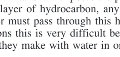
associate with each other through hydrophobic interaction, forming two layers (leaflets) of phospholipid (Fig. 3-2). This buries the hydrophobic fatty acid tails and exposes the polar part (head) to water. Because of the thick layer of hydrocarbon, any molecule that may try to penetrate the bilayer must pass through this hydrophobic region. For polar molecules and ions this is very difficult because they must lose the strong interac-tions they make with water in order to pass through the bilayer.
The membrane establishes in and out. The membrane is asymmetric because the inner and outer leaflets can have a different lipid composi-tion and contain different proteins (Fig. 3-3). Proteins can be associated with either side of the membrane, or they can pass through the membrane using membrane-spanning segments. The functional part of the protein can be on the cytosolic side, the external side, or even in the membrane itself. A common structure for spanning a membrane is an -helix (but
there are examples of sheets spanning a membrane). It takes about 20 amino acid residues arranged in a helix to span to a 30 Å hydrophobic interior of the bilayer.
Proteins that can be removed from membranes by washing them with salt solutions or low pH solutions (disrupts ionic interactions) are called peripheral membrane proteins. Proteins that cannot be removed
without disrupting the membrane with detergents are called integral
MEMBRANE STRUCTURE
Membranes are asymmetric. Integral membrane proteins can’t be washed off. Peripheral membrane proteins can be washed off. Membrane spanning segments and lipid modification (fatty acyla-tion and prenylaacyla-tion), anchor proteins in a fluid bilayer (Singer fluid mosaic model).
3 Membranes and Membrane Proteins • 25 •
[image:38.612.86.280.163.255.2]30 A°
Figure 3-2
PHOSPHOLIPIDSassociate to form a bilayer consisting of a hydrophobic core
membrane proteins. Remember that the distinction between integral and
peripheral membrane proteins is operational rather than structural. All proteins that pass through the membrane one or more times will be inte-gral membrane proteins, but not all inteinte-gral membrane proteins will pass through the membrane—it depends on whether or not the protein can be removed by salt or low pH washes. Peripheral membrane proteins asso-ciate with the membrane or, more usually, with integral membrane proteins.
Some proteins can be posttranslationally modified by the addition of prenyl groups. Prenyl groups are long-chain, unsaturated hydrocarbons that are intermediates in isoprenoid synthesis. The farnesylgroup has 15
carbons, and the geranylgeranylhas 20 carbons. They are attached to a
cysteine residue near the end of the protein as a thiol ether (Protein-S-R). Other proteins can have a long-chain fatty acid (C14“myristoyl, C16“palmitoyl) attached to the amino terminus as an amide. These fatty acid modifications can increase the association of proteins with the mem-brane.
Glycoproteins and glycolipids have complex sugar residues attached. Since they are attached in the ER and Golgi compartments, the sugar
POSTTRANSLATIONAL MODIFICATION
Posttranslational modification can affect membrane association by prenylation (adding C15 or C20 unsaturated hydrocarbons) or fatty acylation (C14 or C16). Glycoproteins and glycolipids on the exte-rior face of the membrane have carbohydrates attached.
• 26 • Basic Concepts in Biochemistry
carbohydrate
phospholipid bilayer
PERIPHERAL PROTEIN
glycolipid
CYTOPLASM
INTEGRAL PROTEIN
Figure 3-3
FLUID-MOSAIC MODELof membrane structure. Proteins and lipids that are
coating will point outward from the cell (will be on the outside surface of the membrane). Membrane proteins as well as phospholipids and gly-colipids are embedded in the lipid bilayer and move around in the plane of the bilayer very rapidly.
The membrane is a dynamic assembly and things are diffusing rapidly in the plane of the bilayer. The middle of the bilayer has been likened to olive oil. As with oil, cooling the lipid bilayer will cause the hydrocarbons to become more ordered (structured). The side chains pack closer to each other, and the fluidity of the membrane is lower. Things that disrupt the ability of the side chains to pack in a regular fashion make the membrane more fluid (Fig. 3-4). These include high temperature, lipids with shorter chains (C16), and lipids with cis-double bonds. The
shorter lipids and the cis-double bonds cause the occurrence of holes
(packing defects).
Cholesterol has a funny effect on membrane fluidity. Because of its shape, cholesterol prevents long-chain fatty acids from packing close to each other. When cholesterol is added to a membrane composed largely
MEMBRANE FLUIDITY
Increasing fluidity makes lateral diffusion faster. Fluidity increases with increased temperature, increased content of short-chain fatty acids, and increased content of cis-fatty acids. Cholesterol increases
the fluidity of membranes that are not very fluid, but decreases the fluidity of membranes that are already fluid.
3 Membranes and Membrane Proteins • 27 •
Cholesterol
OH OH
cis-fatty acid short-chain
fatty acid
Figure 3-4
MEMBRANE FLUIDITY is regulated by altering the chain length of fatty
of saturated, long-chain fatty acids, it will cause the fluidity to increase. However, cholesterol is just the right size to pack into the defects caused by cis-fatty acids. In a membrane (like most mammalian membranes) that
contains significant cis-fatty acids, adding cholesterol will cause the
membrane fluidity to decrease.
To diffuse rapidly in the plane of the membrane (lateral diffusion), a molecule must simply move around in the lipid environment (includ-ing the polar head groups). It need not change how it interacts with phos-pholipids or with water since it is constantly exposed to pretty much the same environment. Lateral diffusion can be slowed (or prevented) by interactions between membrane proteins and the cellular cytoskeleton. This spatially restricts a plasma membrane protein to a localized envi-ronment.
To move through the membrane (change sides or transverse diffu-sion), a molecule must be able to pass through the hydrophobic portion of the lipid bilayer. For ions and proteins, this means that they must lose their interactions with water (desolvation). Because this is extremely dif-ficult, ions and proteins do not move through membranes by themselves. Small molecules such as CO2, NH3 (but not NH4), and water can
dif-fuse through membranes; however, most other small molecules pass through the lipid bilayer very slowly, if at all. This permeability barrier means that cells must develop mechanisms to move molecules from one side of the membrane to the other.
MOVEMENT OF IONS AND MOLECULES
ACROSS MEMBRANES
This requires the participation of a protein transporter. Molecules move spontaneously toward lower concentration (chemical gradi-ent) and opposite charge (electrical gradigradi-ent). Moving in the oppo-site direction requires the input of energy.
DIFFUSION IN MEMBRANES
Lateral diffusion is in the plane of the membrane, and transverse (flip-flop) diffusion is perpendicular to the membrane (through the membrane). Lateral diffusion (in two dimensions) is fast, and trans-verse diffusion is slow (or nonexistent) except for gases (CO2,
NH3) and hydrophobic, uncharged, small molecules (such as
cho-lesterol)
Because the cell membrane is not permeable to ions and most mol-ecules, the cell can regulate the concentrations of things on either side of the membrane. There are two factors that influence the movement of ions and molecules through a membrane. These are the concentration gradi-ent across the membrane (also called the “chemical potgradi-ential”) and the electrical potential of the membrane.
A concentration gradient (chemical potential) exists if the
concen-tration of a given molecule or ion is different on the two sides of the membrane. If you punch a hole in the membrane, the concentration of the molecule will try to equalize itself on the two sides of the membrane (if it is an uncharged molecule).
Normally, cells maintain a slight excess of negative ions inside the cell. This costs energy, but you’ll see it’s worth it. This uneven distribu-tion of charge across the membrane results in a membrane potential(or
electrical potential). The membrane potential is negative, indicating that the inside of the cell is negatively charged as opposed to the outside which is positive. It has a normal value of about 0.06 V (60 mV). As
long as the membrane potential is maintained, it will affect how ions move (the movement of molecules with no charge is not sensitive to the membrane potential). Moving an ion toward the opposite charge (ing a positive ion from outside to inside the cell) will be easier than mov-ing the ion toward the same charge.
The membrane potential and the concentration gradient can reinforce each other or they can be in opposition to each other. The total force tend-ing to move a molecule or ion through a membrane is called the elec-trochemical potential. When the concentration gradient and the electrical
potential work to oppose each other, the stronger effect wins. If someone forces you to get quantitative (this may be a physiologist rather than a biochemist), see the section on the Nernst equation at the end of the chapter.
Because membranes are impermeable to most molecules, you must have a transporter (a protein) in the membrane to help molecules or ions move through it (Fig. 3-5). They are also called channels because they
behave like selective holes in the membrane. Transporters are selective
TRANSPORT ACROSS MEMBRANES
Facilitated diffusion (channel): molecule moves down its
electro-chemical gradient. Active transport (pump): molecule moves up its
electrochemical gradient (requires energy input). Pumps use energy (usually ATP hydrolysis). Na
high outside/K
high inside.
because they usually allow only one type of ion to pass through the mem-brane. For example, the calcium channel that releases calcium from the endoplasmic reticulum, releases only calcium but not other divalent cations. What distinguishes a channel from a pump is that the movement of ions through a channel does notrequire any input of energy.
Channels can be gated. This means that something causes the chan-nel to open. For example, a ligand-gated chanchan-nel opens when a specific ligand binds to a receptor in the membrane. The acetylcholine receptor is a ligand-gated sodium channel that initiates the flow of sodium into the cell (and potassium out) when the receptor binds the neurotransmit-ter, acetylcholine. Voltage-gated channels open and close in response to changes in the membrane potential. The acetylcholine receptor is also voltage-gated. When the membrane potential becomes positive, it opens the channel further, increasing the rate of membrane depolarization.
Pumps move ions and molecules up their electrochemical gradient. Pumps require energy, usually in the form of ATP hydrolysis. Sodium-potassium ATPase is an example of a pump. Cells maintain a higher con-centration of potassium inside the cell than they do outside the cell. Sodium is maintained low inside, high outside. Sodium-potassium ATPase pumps three sodium ions from inside the cell to outside. This is the unfavorable direction—Na
moves from low concentration to a higher one and against the membrane potential. At the same time, it also
• 30 • Basic Concepts in Biochemistry
K+ K+
Channel No energy required
Down gradient
Pump Energy required
Up gradient
ATP ADP+ Pi
K
+
K
+
Figure 3-5
Moving from high to low concentration (CHANNEL)does not require the input
of energy. Moving from low to high concentration (PUMP)does require some
pumps two potassium ions from outside the cell to inside (against the concentration gradient but with the electrical gradient). Both ion move-ments are unfavorable so that the transport process requires energy.
Pumps work by changing their structure and binding characteristics during the cycle of ATP binding, hydrolysis, and release of ADP and Pi. Exactly when and what triggers these changes will vary from pump to pump, but the essential feature is that there must be a cycle that moves a binding site from one side of the membrane to the other while it also changes the affinity for the ligand. When it faces outside, the sodium-binding site must have a low affinity for sodium. This allows sodium to be released where the concentration is high. The potassium-binding site will have a high affinity for potassium when it faces outside. This allows potassium to be taken up outside where the concentration is low. During the movement to the other side of the membrane (this doesn’t happen at the same time for the potassium and sodium sites), the affinity for sub-strate changes. When it faces inside, the sodium-binding site has a high affinity so that it can take up sodium at the low concentration inside. The potassium site that faces inside has a low affinity so that it can release potassium at the high concentration inside the cell.
Ion gradients can also be used to transport other molecules. During digestion, glucose is concentrated in the intestinal epithelium (moves from low concentration outside to a high concentration inside). The energy for this process comes from coupling the transport of glucose to the cotransport of sodium from outside the cell to inside (down its con-centration gradient; Fig. 3-6).
3 Membranes and Membrane Proteins • 31 •
Na
+
GlucoseNa+ Glucose
Figure 3-6
Intestinal epithelial cells concentrate GLUCOSE from the intestinal lumen by
The Nernst equation tells you quantitatively about the energetics of ions moving through membranes. There are two things you need to con-sider to decide which direction an ion will move spontaneously or whether an ion movement will require energy or not. The first