ORIGINAL ARTICLE
ABSTRACT
Address for corespondance :
Email :puji astuti t.k <[email protected]> INTERSTITIAL LuNG DISEASE IN SySTEmIC SCLEROSIS
Puji Astuti Tri K1, Anak Agung Arie1, Cleopas Martin Rumende2, Zulkifli Amin2
1)Internal Medicine Department, Cipto Mangunkusumo National General Hospital-Faculty of Medicine, Universitas Indonesia 2)Division of Respirology and Critical Care, Internal Medicine Department, Cipto Mangunkusumo National General
Hospital-Faculty of Medicine, Universitas Indonesia
Systemic sclerosis (SSc) atau sklerosis sistemik adalah kelainan jaringan ikat yang ditandai dengan disfungsi
sistem imun, kerusakan mikrovaskular dan fibrosis. Systemic sclerosis melibatkan multiorgan, terutama paru, yaitu sebesar 90% pasien dengan SSc. Interstitial lung disease (ILD) atau penyakit paru intersisial merupakan komplikasi tersering pada SSc dan menyebabkan angka kematian yang tinggi. Penanganan kasus ILD pada SSc pada dasarnya sesuai dengan perjalanan skleroderma. Kami melaporkan sebuah kasus dari pasien perempuan 59 tahun, dengan keluhan luka menghitam pada ujung jari manis kirinya yang disertai dengan hilangnya segmen distal jari tersebut, serta batuk berdahak putih dengan sesak nafas yang memberat bila beraktivitas. Pemeriksaan fisik menunjukkan manifestasi skleroderma disertai dengan penyakit paru intersisial. Dalam perjalanannya, keluhan semakin memberat sehingga pasien mendapatkan terapi imunosu -presan dan terapi suportif.
Kata kunci: sklerosis sistemik, penyakit paru intersisial
ABSTRACT
Systemic sclerosis (SSc) is a chronic tissue disorder
char-acterized by immune dysfunction, microvascular injury, and fibrosis. Many organs involved in patients with SSc; especially, pulmonary involvement occurs in up to 90% of patients with SSc. Interstitial lung disease (ILD) is a
major complication in SSc and causing high mortality
rate. The SSc-ILD therapy is basically consistent with the
progress of scleroderma pathophysiology. We reported
a case of 59-years-old female patient with a blackened ulcer on her left hand ring finger with disappearing of her distal finger segment, and also a chronic white phlegm cough followed by dyspnea in exertion. Clinical exami -nation and evaluation showed that she had a scleroderm,
accompanied with ILD. Her complaint did not improve,
so she got an immunosuppresant and supportive therapy to control her worsening disease.
Keywords: systemic sclerosis, interstitial lung disease
INTRODuCTION
Systemic Sclerosis (SSc) is a chronic connective tis-sue disorder involving many organs, with a variety of
manifestations and often leads to disability and mortality. SSc is also characterized by an inflammatory process, followed by functional and structural impairment of the blood vessels to the visceral organ, then continued with fibrosis.1
manifest in SSC are Interstitial Lung Disease (ILD) and
pulmonary hypertension. Both of them cause high
mortal-ity rates in SSC, 33% and 28% of death respectively.2,3
Prevalence of SSc in the US is 50-300 cases per 1 million
inhabitants, with 90% of cases diagnosed with ILD. The 5 years survival rate was 84,1% and 74,9% in 10 years.4 The incidence of SSc-associated ILD (SSc-ILD) mostly
occurs in women aged 30 to 55 years.5
The clinical manifestations of SSc-ILD are difficult to
distinguish from idiopathic pulmonary High Resolution
CT (HRCT) scan fibrosis or other lung diseases.5 The most sensitive investigation to detect ILD is by means of
High Resolution CT (HRCT) scan, with a ground glass
appearance as primary feature. The SSc-ILD therapy is basically consistent with the progress of scleroderma
ABSTRACT
Systemic sclerosis (SSc) is a chronic tissue disorder characterized by immune dysfunction, microvascular injury, and fibrosis. Many organs involved in patients with SSc; especially, pulmonary involvement occurs in up to 90% of patients with SSc. Interstitial lung disease (ILD) is a major complication in SSc and causing high mortality rate. The SSc-ILD therapy is basically consistent with the progress of scleroderma pathophysiology. We reported a case of 59-years-old female patient with a blackened ulcer on her left hand ring finger with disappearing of her distal finger segment, and also a chronic white phlegm cough followed by dyspnea in exertion. Clinical examination and evaluation showed that she had a scleroderm, accompanied with ILD. Her complaint did not improve, so she got an immunosuppresant and supportive therapy to control her worsening disease.
Keywords: systemic sclerosis, interstitial lung disease
INTRODuCTION
Systemic Sclerosis (SSc) is a chronic connective tissue disorder involving many organs, with a variety of manifestations and often leads to disability and mortality. SSc is also characterized by an inflammatory process, followed by functional and structural impairment of the blood vessels to the visceral organ, then continued with fibrosis.1
Lung disease is the most common complication of SSc. About 80% of patients with SSc have lung disease, especially fibrotic lung disease, and is the most common cause of death in SSc patients (33%). Fibrotic lung diseases that manifest in SSC are Interstitial Lung Disease (ILD) and pulmonary hypertension. Both of them cause high mortality rates in SSC, 33% and 28% of death respectively.2,3
Prevalence of SSc in the US is 50-300 cases per 1 million inhabitants, with
90% of cases diagnosed with ILD. The 5 years survival rate was 84,1% and 74,9% in 10 years.4 The incidence of SSc-associated ILD (SSc-ILD) mostly occurs in women aged 30 to 55 years.5
The clinical manifestations of SSc-ILD are difficult to distinguish from idiopathic pulmonary High Resolution CT (HRCT) scan fibrosis or other lung diseases.5 The most sensitive investigation to detect ILD is by means of High Resolution CT (HRCT) scan, with a ground glass appearance as primary feature. The SSc-ILD therapy is basically consistent with the progress of scleroderma pathophysiology.
CASE ILLuSTRATION
weeks before arrival to hospital, accompanied with swelling, redness, pain (visual analog scale: 6), and disappearing of her distal finger segment. Patient also complained a chronic white phlegm cough followed by shortness of breath which was getting more cumbersome. The patient felt nauseated and bloated. There was no fever complained. Such complaints often recur since 3 years ago, accompanied by tightness on the forehead and around the mouth. There were no other diseases histories. She was hemodinamically stable with peripheral oxygen saturation 98% on room air.
Physical examination showed fibrosis in the forehead and around the mouth. From chest auscultation, a basilar
crackle was obtained. There were also atrophy, gangrene, and auto amputation the tip of the thumb of both hands.
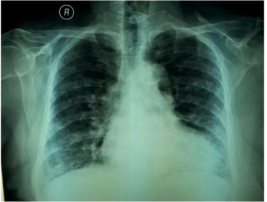
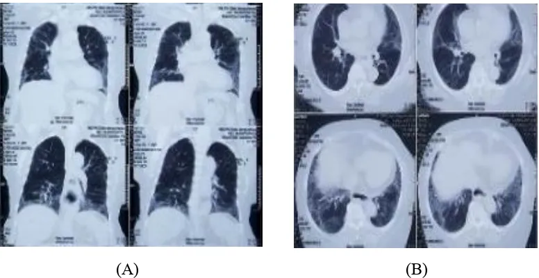
From the laboratories, there was a mild leucocytosis (12.800/mm3). ANA test resulted a positive, 1/1000 homogenous titer with rough speckled pattern. Chest radiography showed a cardiomegaly with aortic calcification and reticular infiltrates in both lungs (Fig.1). Spirometry test showed restrictive results, and failure peripheral airway with FVC 44,1%, FEV1 42,9%. The chest CT scan showed ground glass appearance, with aortic calcification and dilatation along the thoracic esophagus to the esophagogastric junction (Fig.2).
weeks before
by shortness of breath cumbersome ted and bloated
accompanied by
was hemodinamically stable with peripheral oxygen saturation 98% on
Physical examination showed fibrosis
From chest auscultation, a basilar
crackle was obtained. There
the tip of the thumb both hands.
From the laboratories, there was a mild
reticular infiltrates in both lungs
1%, 9%.
in both lungs
(A) (B)
Figure 2. High resolution computed tomography (HRCT) scan of the chest showed graound glass appearance in (A). Coronal view, B). Axial view
During treatment, the complaints of cough and dyspnea worsened. She then treated with targeted dose of unfractionated heparin, antiplatelet, nifedipin, and beraprost for her digital ulcer. For the SSc-ILD, she got mycophenolate mofetil, and morphine sulphate for her pain treatment.
DISCuSSION
SSc is characterized by three mechanisms, such as vasculopathy of small blood vessels, autoantibody formation, and fibroblast dysfunction that increase the deposit of extracellular matrix.6
The initial stage is inflammation, then it involves the blood vessel tissue’s structure and function, and progressively lead to visceral organ dysfunction due to fibrosis.1
Table 1. The American College of Rheumatology / European League Against Rheumatism Collaborative Initiative 2013 Criteria for the classification of systemic sclerosis6
Item Sub-item(s) Weight/score
Skin thickening of the fingers of both hands extending proximal to the metacarpophalangeal
joints (sufficient criterion)
- 9
higher score) Sclerodactyly of the fingers (distal to the
metacarpophalangeal joints but proximal to the proximal interphalangeal joints)
4
Fingertip lesions (only count the higher score) Digital tip ulcers Fingertip pitting scras
2 3
Telangiectasia - 2
Abnormal nailfold capillaries - 2
Pulmonary arterial hypertension and/or interstitial lung disease (max score: 2)
Pulmonary arterial hypertension Interstitial lung disease
2 2
Raynaud’s phenomenon - 3
SSc-related autoantibodies (anticentromere, anti -topoisomerase I (anti Scl-70), anti RNA polymerase III) (maximum score:3)
Anticentomere Anti-topoisimerase I Anti-RNA polymerase III
3
In this case, there was a thickening of the skin of the fingers of both hands up to the metacarpophalangeal joint (9 points), finger swelling (4 points), fingertip ulcers (2 points), nail abnormalities (2 points), interstitial lung disease (2 points) and Raynaud's phenomenon (3 points). With a total of 22 points, the diagnosis of SSc in this patient was upright.
ILD is very common (90% of cases in SSC) and is the leading cause of death in SSc. It occurs interstitial tissue abnormalities of the lung parenchyma. Early ILD enforcement helps to improve the prognosis of SSc.1,3 The pathogenesis of SSc-ILD is not yet fully understood.There are three mechanisms of SSc-ILD; 1. Repeated and persistent endothelial cell damage, 2. Activation of the natural and adaptive immune system, 3. Activation of fibroblasts, resulting from injury and accumulation of extracellular matrix.7
The epithelial or endothelial cell death is known to be a major cause of SSc -ILD. It causes necrosis, apoptosis,
pyroptosis, activation of the coagulation pathway, and inducing the expression of 6 epithelial integrins, then activating TGF, which ultimately activates the response immune to fibrosis. Another mechanism is immune system dysregulation, imbalance and dysfunction of T cell in the circulation and bronchoalveolar fluid. Increased T cells (Th17 and Th22) in the circulation cause pulmonary fibrosis. Activation of myofibroblasts also play a role in pulmonary fibrosis.8,9,10
The SSc-ILD manifestations are characterized by dyspnea on exertion and may be accompanied with a non-productive cough and inspiratory fine crackles at the bilateral basilar area on auscultation, supported by typical SSc symptoms. However, crackles usually can not be detected at the beginning of symptoms. A SSc-ILD patients have positive antinuclear antibody (ANA) results, anti-Scl 70, anti-topoisomerase I (anti-Topo I) and anti-Th / To, and anti-centromere antibodies.11
the
metacarpophalangeal joints but proximal to the proximal interphalangeal joints)
Abnormal nailfold capillaries
titial
related autoantibodies (anticentromere, anti
-70), anti RNA
the skin of the fingers of both ha
to the metacarpophalangeal joint (9
abnormalities (2 points), interstitial
ILD is very common (90% of cases in
abnormalities of
Early ILD enforcement helps to
ILD is not yet
ILD;
of fibroblasts, resulting from injury cumulation of extracellular matrix.7
and bronchoalveolar fluid. Increased T cells (Th17 and Th22) in th circulation cause pulmonary fibrosis.
ofibroblasts also play a role in pulmonary fibrosis8,9,10
ILD
characterized by dyspnea on exertion and may be
rackles at the bilateral basilar area on supported by typical SSc
can not be detected at the beginning of ILD patients positive antinuclear antibody (ANA)
Scl 70
centromere antibodies.
marking, but the sensitivity and
specificity are low in detecting parenchymal abnormalities. High Resolution CT (HRCT) scan was a major radiological examination for this case, with non-specific interstitial pneumonia (NSIP) appearance. At the prognosis. Pulmonary involvement increases more than 20% of mortality rate.12
Pulmonary function tests also very important in determining the severity SSc-ILD. On spirometry test showed restrictive pattern with decreased forced expiratory volume in 1 sec (FEV1) and forced vital capacity (FVC), as well as a normal or slightly increased FEV1/FVC ratio, due to parenchymal damage.13
Inflammatory cells and other inflammation mediators (neutrophil and eusonophil, and may be lymphocytes and mast cells) were present in the brochoalveolar lavage (BAL) fluid of patients with SSc-ILD, reflecting ongoing inflammation in the lungs. In histopathology, a nonspecific interstitial pneumonitis (NSIP) is the most common histological finding. Fewer than 20% of patients with SSc -ILD showed inflammation-dominant lesions at lung biopsy.2,12
Inflammation and fibrosis are the main mechanisms of ILD in SSC. Inflammation is a reversible phase, so the treatment could be given in this
phase. But, in the event of pulmonary fibrosis, which most patients had, there is no specific treatment to overcome it. Therefore, the prevention of disease progression becomes the main goal of therapy.14
Cyclophosphamide is used as an induction regimen. Oral administration of 2 mg/kg/day for 12 months, and 600 mg/m2/month, intravenous for 6 months, coupled with oral prednisolone 20 mg alternate, followed by azathioprine 2,5 mg/kg/day could reduce or prevent the progresivity. Cyclophosphamide is indicated for severe fibrosis for slowing the process.15 During the treatment, the could reduce the risk of side-effects. It is recommended the use of intravenous cyclophosphamide pulse dose (1g / m2 for 6-12 months). The dosage and duration of therapy may be adjusted by age and comorbid factors.14
maintenance therapy after cyclophosphamide.16
The use of azatrioprine is not recommended as an induction therapy, but rather as a maintenance therapy after cyclophosphamide.14 Corticosteroids are not suggested as monotherapy, but in combination with cyclophosphamide (initial dose 500 mg/m2 followed by 750 mg/m2for 1 month later, monthly for 6 months, then every 2 months until 12 months, combined with high doses (prednisolone) 1 mg/kgBB/day for 4 weeks then decreased 5 mg/day, alternate for 2 weeks).17
Rituximab is a monoclonal antibody that works directly against CD20 antigen on the surface of B lymphocytes. Daoussis et al. randomly piloted studies of 14 diffuse type cutaneous scleroderma patients with rituximab plus standard therapy and standard single therapy. Rituximab is given in 2 cycles (as a baseline and repeated 24 weeks later). One cycle consists of administering intravenous every week for 4 weeks (375 mg/m2/week). After one year, patients given rituximab showed improvement in FVC, DLCO, and Rodnan skin scores on scleroderma.18
Tyrosine kinase inhibitor, imatinib mesilate, works by blocking the pro -fibrotic agent c-Abl kinase (as a
TGF- inhibitor). In the few existing studies, patients who received imatinib 200 mg/day provided good results,
while the dose of 600 mg/day did not provide lung function improvement.14
A study by Levy, et al. in 3 cases, obtained one of the three patients had mild restrictive lung disease and then recovered after getting a dose immunoglobulin therapy a total of 56 g. Gived intravenous immunoglobulin (2 g / kg for 5 days, 1 cycle per month) also provide efficacy against percutaneous deployment in case of SSC. However, the benefits of the process of lung fibrosis in ILD until now have not obtain enough data.19 According to a randomized, multicentre study conducted by the
Autologus Stem Cell Transplantation International Scleroderma Trial (ASTIS), which compared hematopoetic stem cell transplantation (HSCT) with cyclophosphamide (monthly administration) in 156 dcSSc patients, showed an increase in FVC and total lung capacity at 1 year of HSCT use.13
but rather as a maintenance therapy
monotherapy, but in combination with
followed by 750 mg/
combined with high doses
17
Rituximab is a monoclonal antibody
rituximab plus standard therapy and py. Rituximab is 2 cycles (as a baseline and
every week for 4 weeks (375
given rituximab showed improvement in FVC, DLCO,
Tyrosine kinase inhibitor, imatinib works by blocking the pro fibrotic agent c Abl k
inhibitor). In the few existing ts who received imatinib
A study by
obtained one of the three patients had
immunoglobulin therapy a total of 56 ntravenous immunoglobulin
SSC. However, the benefits of the process of lung fibrosis in ILD until
not obtain enough data.19
multicentre study conducted by the
Lung transplantation is the last ILD
severe ILD.
numerous comorbidities, such as gastrointestinal reflux, dysmotility,
clearance <50 mL/min/1 73 m
untreated organ failure, extra
spine deformities, neuromuscular degenerative disease, and body mass index less than <15kg/m2
. A one year mortality rate in SSc bilateral lung transplantation patient was 6,6% and 13,6% in patients with lung fibrosis.20
CONCLuSION
Systemic sclerosis (SSc) involves multiple organs and has many clinical manifestations. The most frequent organ involvement in SSc is the lung, manifesting as interterstitial lung disease, which leads to lung fibrosis. The mechanism of SSc-ILD has been known as a result of immune-mediated epithelial and or endothelial cell death.
The enforcement of ILD diagnosis as early as possible provides a better prognosis and could prevent the disease get worsening. It is not only necessary to get an early diagnosis, but also to define the severity of the disease and to predict prognosis. The
risk factors of every patients should be
checked. The earlier we know the
tomography (HRCT) scan. Patients
should be re-evaluated regularly in
order to define if remission has been
reached and therapy should be modified if needed. Cyclophosphamide is the initial choice of therapy for the induction phase in SSc-ILD. However, other therapeutic agents options may be used, although
the effectiveness of each drug remains in the continuous evaluation.
REFERENCES
1. King TE. In: Longo D.L, Kasper D.L, Jameson J.L et al. Harrison’s principles of
internal medicine volume 1. 19th
Ed. New York: The
McGraw-Hill Companies, Inc; 2015. p 1708-715.
2. Scholand MB, Carr E, Frech T,
et al. Interstitial lung disease in
systemic sclerosis: diagnosis
and management: review
article. Rheumatology 2012:
1-5.
3. Steen VD, Medsger TA.
Changes in causes of death in
systemic sclerosis, 1972-2002.
Ann Rheum Dis 2007; 66:940–
944.
4. Walker UA, Tyndall A, Czirják
L, et al. Clinical risk
assessment of organ
manifestations in systemic
sclerosis:a report from the
Ann Rheum Dis 2007; 66: 754–763.
5. Raghu G, Collard HR, Egan JJ,
Martinez FJ, Behr J, Brown
KK,et al, on behalf of the ATS/ERS/JRS/ALAT
Committee onIdiopathic
Pulmonary Fibrosis. An official ATS/ERS/JRS/ALATstatement : idiopathic pulmonary fibrosis:
evidence-based guidelinesfor
diagnosis and management.
Am J RespirCrit Care
Med2011;183:788–824.
6. Hoogen FV, Khanna D,
Fransen J, et. Al. 2013
classification criteria for
systemic sclerosis. Am Col
Rheum 2013: 1-10.
7. Massagué J, Gomis RR. The logic of TGF-β signaling.
FEBS Lett 2006; 508: 2811–
2820.
8. Blackwell TS, Tager AM,
Borok Z, Moore BB, Schwartz
DA,Anstrom KJ, et al. Future
directions in idiopathic
pulmonaryfibrosis research: an NHLBI workshop report. Am J
RespirCritCare Med
2014;189:214–22.
9. Radstake TR, van Bon L,
Broen J, Wenink M, Santegoets
K, DengY, et al. Increased
frequency and compromised
function of Tregulatory cells in
systemic sclerosis (SSc) is
related to a diminishedCD69 and TGF_ expression. PLoS One 2009;4:e5981.
10. Hamaguchi Y. Autoantibody
profiles in systemic sclerosis:
predictive value for clinical
evaluation and prognosis.J Dermatol 2010; 37: 42–53. 11.Goh NS, Desai SR,
Veeraraghavan S, Hansell DM,
Copley SJ, et al. Interstitial
lung disease in systemic
sclerosis: a simple staging
system. Am JRespirCrit Care
Med 2008; 177: 1248-1254. 12. Herzog EL, Mathur A, Tager
AM, et. al. Interstitial lung
disease associated with
systemic sclerosis and
Ann Rheum Dis 2007; 66: 754 763.
KK,et al, on behalf of the ATS/ERS/JRS/ALAT
Pulmonary Fibrosis. An official ATS/ERS/JRS/ALATstatement : idiopathic pulmonary fibrosis:
based guidelinesfor
Med2011;183:788
7.
β signaling.
FEBS Lett 2006; 508: 2811
pulmonaryfibrosis research: an NHLBI workshop report. Am J
2014;189:214
9. Radstake TR, van Bon L,
related to a diminishedCD69 and TGF_ expression. PLoS One 2009;4:e5981.
Hamaguchi Y. Autoantibody
Dermatol 2010; 37: 42
2008; 177: 1248
Herzog EL, Mathur A, Tager
iopathic pulmonary fibrosis. 2014; 66:1967 75.
13.Yasuoka H. Recent Treatments of Interstitial Lung Disease with Systemic Sclerosis.
Clinical Medicine Insights:
Circulatory, Respiratory and
Pulmonary Medicine 2015:9(S1) 97–110.
14.Tashkin DP, Elashoff R,
Clements PJ, et al; Scleroderma Lung Study Research Group. Effects of 1-
year treatment with
cyclophosphamide on
outcomes at 2 years in
scleroderma lung disease. Am J
Respir Crit Care Med. 2007;176:1026–34.
15.Theodore AC, Tseng CH, Li N, Elashoff RM, Tashkin DP.
Correlationof Cough with
Disease Activity and Treatment
with Cyclosphosphamide inScleroderma Interstitial Lung Disease: Findings from the Scleroderma LungStudy. Chest. 2011.
16.Simeón-Aznar CP,
Fonollosa-Plá V, Tolosa-Vilella C,
Selva-O’Callaghan A, Solans-Laqué R, Vilardell-Tarrés M. Effect
of mycophenolate sodium in
scleroderma- related interstitial
lung disease. Clin Rheumatol. 2011;30:1393–8.
17. Campos DP, Del Toro ME, Casanovas AP, et al. Are high
dose of prednisone necessary
for treatment of interstitial lung
disease in systemic sclerosis?
Reumatol Clin. 2012;8:58–62.
18. Daoussis D, Liossis SN,
Tsamandas AC, et al. Experience with rituximab in scleroderma: results from a
1-year,proof-of-principle study. Rheumatology 2010; 49: 271– 280.
19. Levy Y, Sherer Y, Langevitz P, et al. Skin score decrease in
systemic sclerosis patients
treated with intravenous immunoglobulin – a preliminary report. Clin
Rheumatol. 2000;19:207–11. 20.Saggar R, Khanna D, Furst DE,