The in
fl
uence of naturalistic, directionally non-speci
fi
c motion
on the spatial deployment of visual attention in right-hemispheric
stroke
Dario Cazzoli
a,b,1, Simone Hopfner
c,1, Basil Preisig
c,d, Giuseppe Zito
a, Tim Vanbellingen
c,e,
Michael Jäger
a,f, Tobias Nef
a,f, Urs Mosimann
a,d, Stephan Bohlhalter
c,e, René M. Müri
a,c,g,
Thomas Nyffeler
a,c,e,naGerontechnology & Rehabilitation Group, University of Bern, Bern, Switzerland bNuffield Department of Clinical Neurosciences, University of Oxford, United Kingdom
cPerception and Eye Movement Laboratory, Departments of Neurology and Clinical Research, Inselspital, Bern University Hospital and University of Bern,
Freiburgstrasse 10, 3010 Bern, Switzerland
dUniversity Hospital of Old Age Psychiatry and Psychotherapy, University of Bern, Bern, Switzerland eNeurology and Neurorehabilitation Center, Luzerner Kantonsspital, Luzern, Switzerland
fARTORG Center for Biomedical Engineering Research, University of Bern, Bern, Switzerland
gDivision of Cognitive and Restorative Neurology, Department of Neurology, Inselspital, Bern University Hospital and University of Bern, Switzerland
a r t i c l e
i n f o
Article history:
Received 16 October 2015 Received in revised form 4 April 2016
Accepted 17 April 2016
Keywords:
Subacute/chronic stroke Hemispatial neglect Visual attention deployment Motion
Eye-movements Everyday relevant setting
a b s t r a c t
An impairment of the spatial deployment of visual attention during exploration of static (i.e., motionless) stimuli is a commonfinding after an acute, right-hemispheric stroke. However, less is known about how these deficits: (a) are modulated through naturalistic motion (i.e., without directional, specific spatial features); and, (b) evolve in the subacute/chronic post-stroke phase. In the present study, we investigated free visual exploration in three patient groups with subacute/chronic right-hemispheric stroke and in healthy subjects. Thefirst group included patients with left visual neglect and a left visualfield defect (VFD), the second patients with a left VFD but no neglect, and the third patients without neglect or VFD. Eye movements were measured in all participants while they freely explored a traffic scene without (static condition) and with (dynamic condition) naturalistic motion, i.e., cars moving from the right or left. In the static condition, all patient groups showed similar deployment of visual exploration (i.e., as measured by the cumulativefixation duration) as compared to healthy subjects, suggesting that recovery processes took place, with normal spatial allocation of attention. However, the more demanding dynamic condition with moving cars elicited different re-distribution patterns of visual attention, quite similar to those typically observed in acute stroke. Neglect patients with VFD showed a significant decrease of visual exploration in the contralesional space, whereas patients with VFD but no neglect showed a significant increase of visual exploration in the contralesional space. No differences, as compared to healthy subjects, were found in patients without neglect or VFD. These results suggest that naturalistic motion, without directional, specific spatial features, may critically influence the spatial distribution of visual attention in subacute/chronic stroke patients.
&2016 Elsevier Ltd. All rights reserved.
1. Introduction
Eye movements and visual attention move together across the
visual space, at least in situations where the eyes are free to move
(
Hunt and Kingstone, 2003
). This phenomenon, often referred to
as overt attention, is a fundamental behaviour during the activities
of everyday life (
Land, 2006
). Cerebral lesions, in particular to the
right hemisphere, can severely impair visual exploration and thus
the spatial deployment of visual attention. Among the disorders
following a right-hemispheric lesion, spatial neglect is a
particu-larly disabling syndrome. Spatial neglect is commonly de
fi
ned as a
de
fi
cit in orienting towards, responding to, and reporting stimuli
that are presented on the contralesional side of space (
Heilman
et al., 2003
). In patients with acute stroke, this de
fi
cit in the spatial
Contents lists available at
ScienceDirect
journal homepage:
www.elsevier.com/locate/neuropsychologia
Neuropsychologia
http://dx.doi.org/10.1016/j.neuropsychologia.2016.04.017 0028-3932/&2016 Elsevier Ltd. All rights reserved.
n
Corresponding author at: Perception and Eye Movement Laboratory, Depart-ment of Neurology, DepartDepart-ment of Clinical Research, Inselspital, Bern University Hospital and University of Bern, Freiburgstrasse 10, 3010 Bern, Switzerland.
allocation of visual attention towards the contralesional space
results in characteristic patterns during visual exploration. For
instance, patients with neglect present a signi
fi
cant ipsilesional
bias in the spatial distribution of visual
fi
xations, a bias of early
attentional orientation towards the ipsilesional space, frequent
re-fi
xations (i.e., repeated
fi
xations over the same region), and
im-paired saccade metrics (
Karnath et al., 1998
;
Mort and Kennard,
2003
;
Müri et al., 2009
;
Niemeier and Karnath, 2000
;
Sprenger
et al., 2002
). In approximately 75% of neglect patients, patterns of
visual attention allocation in space normalise within six months
after stroke (
Stone et al., 1992
). In these patients, visual
explora-tion is characterized by a re-orientaexplora-tion of visual attenexplora-tion towards
the contralesional side, while only an early attentional bias (i.e.,
fi
rst saccade) towards the ipsilesional side may persist (
Karnath,
1988
;
Mattingley et al., 1994b
;
P
fl
ugshaupt et al., 2004
;
Pizza-miglio et al., 1992
).
Several factors can in
fl
uence the allocation of attention in
space, and these can have a spatial or non-spatial nature. Motion,
which is an omnipresent feature of visual stimuli in everyday
si-tuations, is one of those factors (
Zihl, 1995
). For instance, visual
attention can improve when patients with neglect are instructed
to look at dots on a dark background, all moving coherently
towards the contralesional space (
Kerkhoff et al., 2014
).
Further-more, in acute neglect patients, it could be shown that moving
stimuli in the contralesional space can be a spatial cue and attract
visual attention (
Butter et al., 1990
;
Mattingley et al., 1994a
;
Plummer et al., 2006
). Nevertheless, the direction of the
mod-ulatory effect of motion seems also to depend on the integrity of
the optic radiation, i.e., the presence or absence of an additional
visual
fi
eld defect (VFD). In a recent study, we showed that neglect
patients with an additional VFD may present increased or
un-changed neglect severity when confronted with motion in a
touchscreen-based cancellation task (
Hopfner et al., 2015
).
On the other hand, patients with VFD but no neglect may show
a reversed spatial bias during visual exploration, i.e., they may
produce an increased number of gaze shifts towards the affected
side of space (
Ishiai et al., 1987
;
Pambakian et al., 2000
). This
contralesional spatial bias is thought to re
fl
ect an attempt to
compensate for VFD, and seems also to increase with higher task
demands (
Hardiess et al., 2010
).
However, to date far less is known about the effects on the
spatial attentional allocation in right-hemispheric patients of
motion that has no speci
fi
c, directional spatial features, i.e., of
naturalistic motion in everyday scenes, where elements move in
both the ipsi- and the contralesional space, in heterogeneous
directions.
In the present study, we thus adopted a virtual reality (VR)
approach, and established an innovative setup with large
projec-tion displays. We examined free visual exploraprojec-tion of a virtual
traf
fi
c scene, without (static condition) and with (dynamic
con-dition) naturalistic motion, in which cars could move from the left
to the right or vice versa. This not only allowed the stimulation of
the full
fi
eld of view (i.e., 180
°
as opposed to a small stimulus array,
limited by the computer screen), but also the assessment of visual
exploration in the extrapersonal, far space (as opposed to the
peripersonal, near space, which is commonly assessed in
paper-pencil and computer-based tests). Both aspects contribute to a
more naturalistic and ecologically valid evaluation of the effects of
motion. Furthermore, there is little knowledge about the
mod-ulatory effects of motion in patients with a subacute/chronic
stroke, and about the speci
fi
city of the interactions with VFD. For
this reason, in the present study, we included patients with
sub-acute/chronic left-sided neglect and VFD and, as control groups,
patients with a subacute/chronic left VFD but no neglect, and
patients with right-hemispheric lesions but no neglect or VFD.
Additionally, healthy subjects also performed the task.
Based on previous research, we hypothesized that the in
fl
uence
of naturalistic, directionally non-speci
fi
c motion on the spatial
allocation of visual attention would differ between patient groups
and healthy subjects. In particular, we expected that, in subacute/
chronic neglect patients with VFD, neglect severity would increase
in the dynamic condition.
2. Methods
2.1. Subjects
Twenty-four patients who suffered from afirst ischemic or haemorrhagic right hemispheric stroke (aged between 25 and 76, mean¼54.20, SD¼13.27; 8 women) and 8 healthy subjects (aged between 25 and 78, mean ¼60.67, SD ¼19.43; 3 women) were included in the study after giving written, informed consent. There was no statistically significant difference between patients and healthy subjects with respect to age (t(30)¼ 1.019, p¼.316; 2-tailed) or gender (
χ
2(1)¼.046,p¼.575). The group of twenty-four patients was comprised of 8 patients with left-sided visual neglect and VFD, 8 patients with a left-left-sided VFD but without neglect, and 8 patients without any neglect or VFD. The mean interval between stroke onset and testing was 589 days (range: 28–1737 days, SD¼613). No statistically sig-nificant differences were found between the three patient groups with respect to the mean interval between stroke onset and testing (F(2, 21)¼.932,p¼.409), age (F
(2, 21)¼.231,p¼.796), or gender (
χ
2(2)¼.375;p¼.829). The definition of the be-ginning of the chronic post-stroke period is variable among sources in the litera-ture. Although some sources define the beginning of the chronic stroke period relatively late (i.e., 180 days after stroke), other sources set the beginning of this period already at three weeks after stroke (e.g.,Allen et al., 2012). All patients were included in the present study at the earliest 28 days after stroke, thus overlapping with either the subacute or the chronic post-stroke phase, depending on the ap-plied definition. In order to reflect the non-absolute nature of this definition, we opted to name the post-stroke phase with the more general term“subacute/ chronic”.Since patients were in the subacute/chronic stage after stroke, neglect diag-nosis was based not only on standard neuropsychological testing, but primarily on the observation of neglect manifestations in everyday behaviour. Other studies based neglect diagnosis primarily on the presence of neglect signs in computerized and/or paper-pencil tests (e.g., in at least one or two of these tests). While the present study aimed at a more naturalistic and ecologically valid evaluation of the effects of motion in neglect (and thus stressed the importance of neglect mani-festations in everyday behaviour), it is to note that the definition of different cri-teria may lead to the inclusion/exclusion of different patients, and thus to potential differences in the results. For the assessment of neglect manifestations in everyday behaviour, the Catherine Bergego Scale was applied, which is able to quantify the influence of neglect-related persisting deficits in the activities of daily living (Azouvi, 1996;Azouvi et al., 2003). Two classes of neuropsychological tests were also administered: a cancellation task (either Bells test (Gauthier et al., 1989)or Star Cancellation test (Wilson et al.,1987)), and a line bisection task (Wilson et al., 1987). For the Bells test, we calculated the number of targets omitted on the left side minus the number of targets omitted on the right side, and the cut-off score was set at42 (according toAzouvi et al., 2006). For the Star cancellation test, the cut-off score was set at 15% of omitted left-sided targets (according toFerber and Karnath, 2001), i.e., 4 left-sided target omissions. For the line bisection task, a mean right-ward deviation equal to or larger than 11% from the actual midline was considered as clinically relevant (according toSchenkenberg et al., 1980).
Since an evaluation of whether neglect patients also have an additional VFD is difficult to achieve with clinical confrontation testing or visual perimetry (Kerkhoff and Schindler, 1997;Müller-Oehring et al., 2003), we also assessed whether the right optic radiation was damaged or not by means of a track-wise‘hodological’
lesion-deficit analysis (Thiebaut de Schotten et al., 2014), which is based on a re-cently published DTI atlas (Thiebaut de Schotten et al., 2011;Rojkova et al., 2016). The atlas provides the probability for each voxel in the MNI space to belong to a specific white matter tract. To conduct this analysis, we used the ‘Tractotron’
software (Thiebaut de Schotten et al., 2014). In afirst step, we mapped the in-dividual lesions of the patients in the structural MRI data by means of the MRIcron software (Rorden, Karnath, and Bonilha, 2007). The same procedure as inKarnath, Fruhmann Berger, Küker and Rorden (2004) and Karnath, Himmelbach and Rorden
(2002)was applied: if an MRI was conducted within the 48 h post-stroke, then
behavioural performance, and the accuracy was then checked by a second, in-dependent collaborator. In a second step, we then performed the track-wise‘ ho-dological’ lesion-deficit analysis with the ‘Tractotron’ software (Thiebaut de Schotten et al., 2014). The patients’individual lesion maps were overlapped with the map of the right optic radiation in order to analyse the individual pattern of integrity of the latter. The optic radiation was considered to be disconnected based on a binary measure, i.e., considering the optic radiation to be damaged if the patients’individual lesion map overlapped with a voxel within the optic radiation map with a probability greater than 50% (i.e., above chance level). The analysis revealed that all patients in the group with neglect and VFD, and all patients in the group with VFD only (i.e., no neglect) had a damage to the right optic radiation. Conversely, none of the patients in the group without any VFD or neglect had damage to the right optic radiation. As an illustration,Fig. 1depicts the localisation of the brain lesions and their degree of overlap, transferred to standard atlases and with respect to the right optic radiation, in the three groups of patients.
In patients with VFD and without neglect, the visualfield was assessed by means of perimetry (Octopus Perimetry or Goldman Kinetic Perimetry; Octopus Perimeter 101, Haag-Streit International, Bern-Köniz, Switzerland).
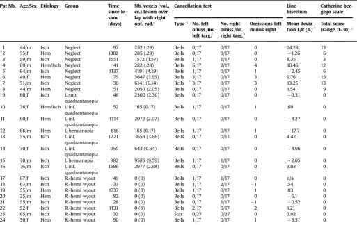
All participants (i.e., patients and healthy subjects) had normal or corrected-to-normal visual acuity. Demographic and clinical details of patients are presented in Table 1.
Ethical approval to conduct the present study was provided by the Ethics Committee of the States of Bern and Lucerne. The study was carried out in ac-cordance with the principles laid down in the latest version of the Declaration of Helsinki.
2.2. Stimulus material and apparatus
The stimulus material consisted of a virtual traffic scene, displaying an inter-section environment, with a two-way road and a pedestrian-crossing, surrounded by buildings and trees. The technical details and a usability evaluation are pub-lished elsewhere (Jäger et al., 2015). The virtual scene was presented under two conditions: a static and a dynamic condition. In the static condition, the virtual scene was presented without any road traffic, i.e., only the motionless environment, without any cars, was displayed. In the dynamic condition, the same virtual scene
was presented as background. However, the dynamic condition also included cars, moving from the left to the right and from the right to the left on the main road. The number of cars driving from the left to the right and vice versa was counter-balanced within each trial, with all cars moving at the same and constant speed. To reduce the potential influence of specific features of the cars (i.e., shape, colour), only cars of the same model and colour were selected (specifically, Ford Mondeo, white). Fig. 2 shows an example of the virtual traffic scene in the dynamic condition.
The virtual scenes were projected by three overhead projectors (Ultra Short Focus LCD projector, Sanyo, Moriguchi Osaka, Japan), with a resolution of 1024768 pixels, onto three canvas projection screens (1.801.39 m each), in-stalled in front of the participant (Fig. 3). Screen 2 and 3 were tilted by 120°with respect to Screen 1, thus creating afield of view of 180°(horizontal) by 40° (ver-tical). Participants were placed in front of Screen 1, with a viewing distance of 155 cm. The simulation was controlled by three computers running Microsoft Windows 7 operating system (Microsoft Corp., Redmond, WA). One of the com-puters calculated and controlled the dynamic aspects of the virtual scene in the dynamic condition, whereas the other two computers refreshed the image at a rate of 35 Hz.
2.3. Eye-movement recording
Eye movements were recorded using an infrared, head-mounted eye-tracking system (SMI iView X HED, SensoMotoric Instruments GmbH, Teltow, Germany), with a sampling rate of 50 Hz, a spatial resolution of typicallyo.1°, and a tracking
Table 1
Demographic and clinical details of the patients.
Pat Nb. Age/Sex Etiology Group Time since le-sion (days)
Nb. voxels (vol., cc.) lesion over-lap with right opt. rad.a
Cancellation test Line bisection
Catherine ber-gego scale
Typeb No. left
omiss./no. left targ.c
No. right omiss./no. right targ.d
Omissions left minus righte
Mean devia-tion L/R (%)f
Total score (range, 0–30)g
1 44/m Isch Neglect 97 292 (.29) Bells 0/17 0/17 0 24.28 13 2 55/f Hem Neglect 1382 285 (.29) Bells 0/17 0/17 0 1.26 6 3 59/m Isch Neglect 1551 1572 (1.57) Bells 1/17 1/17 0 8.35 3 4 69/m Hem/Isch Neglect 41 282 (.28) Bells 6/17 2/17 4 10.46 12 5 64/m Isch Neglect 1137 4191 (4.19) Bells 1/17 0/17 1 2.45 6 6 49/f Hem Neglect 75 3647 (3.65) Bells 3/17 0/17 3 9.76 15 7 51/m Isch Neglect 30 6141 (6.14) Bells 3/17 0/17 3 13.25 13 8 44/m Hem Neglect 51 2050 (2.05) Bells 0/17 0/17 0 1.54 9 9 60/f Isch L sup.
quadrantanopia
46 2300 (2.30) Bells 0/17 0/17 0 0.31 0
10 36/f Hem/Isch L inf. quadrantanopia
52 165 (0.17) Bells 1/17 0/17 1 .69 0
11 60/f Hem L inf. quadrantanopia
1114 2072 (2.07) Bells 0/17 0/17 0 4.27 0
12 66/m Hem L hemianopia 616 165 (0.17) Bells 1/17 0/17 1 17.7 0 13 55/m Isch L inf.
quadrantanopia
1221 3659 (3.66) Bells 0/17 0/17 0 4.42 0
14 30/f Isch L inf. quadrantanopia
959 643 (0.64) Bells 0/17 0/17 0 4.96 0
15 70/m Isch L hemianopia 982 9585 (9.59) Bells 1/17 1/17 0 2.05 0 16 76/m Isch L inf.
quadrantanopia
1599 2977 (2.98) Bells 0/17 0/17 0 3.03 0
17 67/f Isch R.-hemi w/out 49 0 (0) Bells 1/17 1/17 0 n/a 0 18 63/m Isch R.-hemi w/out 33 0 (0) Bells 1/17 2/17 1 .54 0 19 55/m Hem R.-hemi w/out 1737 0 (0) Bells 1/17 0/17 1 .03 0 20 25/m Hem R.-hemi w/out 82 0 (0) Bells 0/17 0/17 0 6.1 0 21 55/m Isch R.-hemi w/out 28 0 (0) Bells 0/17 1/17 1 0.52 0 22 52/f Isch R.-hemi w/out 1131 0 (0) Bells 2/17 0/17 2 1.21 0 23 65/m Isch R.-hemi w/out 32 0 (0) Star 0/27 0/27 0 3.02 0 24 30/f Hem R.-hemi w/out 90 0 (0) Bells 1/17 0/17 1 3.51 0
Note. Hem¼Hemorrhage; inf.¼inferior; Isch¼Ischemia; L¼left; R¼right; R.-hemi w/out¼patients with right-hemispheric lesions without neglect or visualfield defects; sup.¼superior; VFD¼Visualfield defect.
aNumber of voxels (and corresponding volume, in cc., in parentheses) of each individual lesion map overlapping with the right optic radiation, as based on a recently published DTI atlas (Thiebaut de Schotten et al., 2011; probability for voxels to belong to the right optic radiation set at450%).
bBells Test according to:Gauthier et al. (1989). The bells test: A quantitative and qualitative test for visual neglect.International Journal of Clinical Neuropsychology, 11, 49– 54. Star cancellation subtest from the Behavioural Inattention Test (BIT), according to: Wilson, B., Cockburn, J., & Halligan, P. W. (1987). Behavioural Inattention Test. Titchfield, UK: Thames Valley Test Company.
cNumber of omitted targets on the left side/Total number of targets on the left side. For the Start cancellation subtest from the Behavioural Inattention Test (BIT): cut-off set at 15% of omitted left-sided targets (according to:Ferber and Karnath (2001). How to assess spatial neglect–line bisection or cancellation tasks?Journal of Clinical and Experimental Neuropsychology, 23(5), 599–607), i.e., 4 left-sided omissions.
dNumber of omitted targets on the right side/Total number of targets on the right side.
eNumber of omitted targets on the left side minus Number of omitted targets on the right side. For the Bells Test: cut-off set at42 (according to:Azouvi et al. (2006)). A battery of tests for the quantitative assessment of unilateral neglect.Restorative Neurology and Neuroscience, 24, 273–285.
fMean % deviation to right (R,þ) or left (L, ) from the actual midline in the line bisection test. Cut-off deviation set at 11% (according to: Schenkenberg, T., Bradford, D. C., Ajax, E. T. (1980). Line bisection and unilateral visual neglect in patients with neurologic impairment.Neurology, 30, 509–517).
gA total score on the Catherine Bergego Scale was calculated (score range, 0
–30). Mild neglect: score range, 1–10; moderate neglect: score range, 11–20; severe neglect: score range, 21–30 (according to:Azouvi et al. (2003). Behavioural assessment of unilateral neglect: study of the psychometric properties of the Catherine Bergego Scale.
Archives of Physical Medicine and Rehabilitation, 84(1), 51–57).
superimposed to the visual scene recorded by the other camera. The resulting data sets thus consisted of individual video sequences and a datafile, including–for each time frame–a cursor indicating the current point of gaze on the currently explored part of the visual scene. For data pre-processing, the recorded video se-quence and the corresponding datafile were uploaded in BeGaze™analysis soft-ware (SensoMotoric Instruments GmbH, Teltow, Germany). Fixation detection threshold was set at a minimal duration of 100 ms and a maximal dispersion of 100 pixels. Individual gaze position was mapped into the reference space by means of the SMI Semantic Gaze Mapping analysis software tool (SensoMotoric Instruments GmbH, Teltow, Germany). This software enables to map the location of individual gaze positions in the video recordings made by single participants, fixation-by-fixation, on a common reference image. The resulting output is represented by a datafile in which eachfixation of each participant is associated with an exact lo-cation, defined on the x-and y-coordinates, in pixels, on a reference image. In the present study, the reference image was a pngfile, depicting the whole intersection environment (1500368 pixels) used as the virtual scene during the experiment.
2.4. Experimental procedure
Participants were seated in front of the three projection screens, with their mid-sagittal plane aligned with the centre of the middle projection screen (screen 1; seeFig. 3). To ensure the accuracy of gaze position tracking, a 5-point calibration procedure was carried out, in which participants were asked tofixate 5 dots pre-sented at different locations on the central projection screen, in afixed order. During the calibration process, participants were instructed to follow the dots with their eyes only, and to hold their head in a steady position. Once the calibration procedure was completed, participants were allowed and encouraged to freely move their head.
Participants were then asked to freely explore the traffic environment under the two experimental conditions (i.e., the static and the dynamic condition). To familiarize the participants with the experimental setting, a practice sequence from the static and the dynamic condition was presented prior to the experiment proper. The static and the dynamic conditions were administered in the same order for each participant, i.e., the static condition followed by the dynamic condition. The static condition entailed the presentation of the static, virtual traffic scene during 2 min. The dynamic condition was comprised of 15 sequences, where the speed and the direction of the cars were presented in random order, lasting approxi-mately 16 s each, and separated by a black screen during 4 s. Participants were allowed a break of approximately 10 min between the static and the dynamic condition. The overall duration of the experiment was of approximately 16 min.
2.5. Data analysis
In afirst analysis, we aimed to assess early attentional orienting during free visual exploration. For this purpose, we calculated the mean gaze position during early attentional orienting, i.e., the mean coordinates, on the x-axis, of the visual fixation(s) occurring during thefirst second of visual exploration in the static and the dynamic condition, respectively. The data concerning the mean gaze position during early attentional orienting underwent a mixed-model analysis of variance (ANOVA), with the within-subjects factor‘condition’(levels: static; dynamic) and the between-subjects factor‘group’(levels: patients with neglect and VFD; patients with VFD only; patients with right-hemispheric lesions without neglect or VFD; healthy subjects). Moreover, for every participant, we also evaluated whether the mean gaze position during early attentional orienting was located in the left or the right hemispace. For the static and the dynamic condition, we tested whether there was a significant association between the group (patients with neglect and VFD; patients with VFD only; patients with right-hemispheric lesions without neglect or VFD; healthy subjects) and the location of the mean gaze position during early attentional orienting (left hemispace; right hemispace) by means of a Pearson's chi-square test (with Fisher's exact test method).
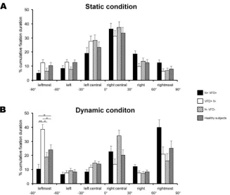
In a second analysis, we assessed the spatial distribution of visual exploration
throughout the task, in both the static and the dynamic conditions. For this pur-pose, the cumulativefixation duration (CFD), i.e., the sum of the duration of all visualfixations, was computed over six vertical columns, covering the wholefield of view: leftmost (from 90°to 60°), left (from 60°to 30°), left central (from 30°to 0°), right central (from 0°to 30°), right (from 30°to 60°), rightmost (from 60°to 90°). The distribution of the CFD over these six columns was calculated with Matlab (7.8.0.347, Mathworks Inc., Natick MA), converting the degrees of visual angle to portions of the reference image (1500 pixels on the horizontal dimension in total): leftmost (0–249 pixels), left (250–499 pixels), left central (500–749 pixels), right central (750–999 pixels), right (1000–1249 pixels), rightmost (1250–1500 pixels). Since the presentation times in the static and the dynamic conditions were not of equal length, a direct comparison of the absolute distribution of the CFD over the six columns was not practicable. We thus decided to convert the absolute CFD values over the six columns into percentage CFD values, in both the static and the dynamic condition, according to the formula: [(absolute CFD in a given column/ sum of the absolute CFD over columns 1–6)*100]. In afirst step, the percentage CFD data underwent an omnibus, mixed-model ANOVA, with the within-subjects fac-tors‘condition’(levels: static; dynamic) and‘column’(levels: leftmost; left; left central; right central; right; rightmost), and the between-subjects factor‘group’
(levels: patients with neglect and VFD; patients with VFD only; patients with right-hemispheric lesions without neglect or VFD; healthy subjects). In a second step, in order to investigate the source of significant interactions, we performedpost-hoc
ANOVAs, adjusting the significance levels according to the Bonferroni procedure (i.e., dividing
α
by the number of performedpost-hocANOVAs).All subsequent post-hoctests were conducted by means of Bonferroni-cor-rectedt-tests.
3. Results
3.1. Early attentional orienting
The analysis of the mean gaze position during early attentional
orienting revealed a signi
fi
cant main effect of the factor
‘
group
’
(F
(3, 28)
¼
4.019,
p
¼
.017), but no signi
fi
cant effect of the factor
‘
condition
’
or of the interaction
‘
condition x group
’
(all
p's
4
.737).
Irrespective of the dynamic or static condition, and as indicated by
Bonferroni-corrected
post-hoc
tests, the mean gaze position during
early attentional orienting was signi
fi
cantly rightward-deviated in
patients with neglect and VFD in comparison to all other groups
(i.e., patients with VFD only, patients with right-hemispheric
le-sions without neglect or VFD, and healthy subjects). The results
concerning the mean gaze position during early attentional
or-ienting and the corresponding
post-hoc
tests are depicted in
Fig. 4
.
Moreover, for every participant, we also evaluated whether the
mean gaze position during early attentional orienting was located
in the left or the right hemispace. For the static condition, the
Fig. 3.Schematic representation of the experimental setup. Schematic top-downview of the experimental setup with the three screens (1-3), and the three pro-jectors (P1–3). Adapted from:Jager et al. (2015). Adapting a Driving Simulator to Study Pedestrians’Street-Crossing Decisions: A Feasibility Study.Assistive Technol-ogy, 27(1), 1–8.
mean gaze position during early attentional orienting was located
in the right hemispace in all patients with neglect and VFD,
whereas this was the case for 3 patients with VFD only (left
hemispace for 5 patients), for 3 patients with right-hemispheric
lesions without neglect or VFD (left hemispace for 5 patients), and
for 2 healthy subjects (left hemispace for 6 healthy subjects). There
was a signi
fi
cant association between the group and whether the
mean gaze position during early attentional orienting was located
in the left or the right hemispace (
χ
2(3)
¼
11.456,
p
¼
.012, Fisher's
exact test). Direct pairwise testing evidenced that this association
was signi
fi
cant when considering the group of patients with
ne-glect and VFD in comparison to all other groups (all
p's
o
.026).
The odds of the mean gaze position during early attentional
or-ienting being located in the right hemispace was thus higher in the
group of patients with neglect and VFD than in the groups of
pa-tients with VFD only, of papa-tients with right-hemispheric lesions
without neglect or VFD, or of healthy subjects. For the dynamic
condition, the mean gaze position during early attentional
or-ienting was located in the right hemispace in 7 patients with
ne-glect and VFD (left hemispace for 1 patient), whereas this was the
case for 2 patients with VFD only (left hemispace for 6 patients),
for 2 patients with right-hemispheric lesions without neglect or
VFD (left hemispace for 6 patients), and for 2 healthy subjects (left
hemispace for 6 healthy subjects). There was a signi
fi
cant
asso-ciation between the group and whether the mean gaze position
during early attentional orienting was located in the left or the
right hemispace (
χ
2(3)
¼
9.080,
p
¼
.033, Fisher's exact test). Direct
pairwise testing evidenced that this association was signi
fi
cant
when considering the group of patients with neglect and VFD in
comparison to all other groups (all
p's
o
.041). The odds of the
mean gaze position during early attentional orienting being
lo-cated in the right hemispace was thus higher in the group of
pa-tients with neglect and VFD than in the groups of papa-tients with
VFD only, of patients with right-hemispheric lesions without
ne-glect or VFD, or of healthy subjects.
3.2. Cumulative
fixation duration (CFD) spatial distribution
The omnibus ANOVA on the percentage CFD data revealed a
signi
fi
cant main effect of factor
‘
column
’
(F(5, 140)
¼
26.731,
p
o
.001), signi
fi
cant two-way interactions between factors
‘
col-umn x group
’
(F(15, 140)
¼
4.393,
p
o
.001) and
‘
condition x
col-umn
’
(F(5, 140)
¼
41.179,
p
o
.001), and a signi
fi
cant three-way
in-teraction between factors
‘
condition x column x group
’
(F(15,
140)
¼
2.364,
p
¼
.005). No other main effects or interactions were
signi
fi
cant (all
p's
4
.999). In order to investigate the source of the
signi
fi
cant three-way interaction in the omnibus ANOVA, we
performed two separate
post-hoc
ANOVAs for the static and the
dynamic conditions (signi
fi
cance level set at
p
¼
.05/2
¼
.025). In
the static condition, there was a signi
fi
cant main effect of the
factor
‘
column
’
(F(5, 140)
¼
47.564,
p
o
.001), but no signi
fi
cant
ef-fect of the factor
‘
group
’
or of the two-way interaction
‘
column x
group
’
(all
p's
4
.151). In contrast, in the dynamic condition, there
was a signi
fi
cant main effect of factor
‘
column
’
(F(5, 140)
¼
20.012,
p
o
.001) and of the two-way interaction
‘
column x group
’
(F(15,
140)
¼
5.382,
p
o
.001), but no signi
fi
cant effect of the factor
‘
group
’
(p
4
.999). This indicates that the distribution of the percentage
CFD over columns was different among groups in the dynamic
condition, but not in the static one. In order to investigate the
source of the signi
fi
cant two-way interaction in the dynamic
condition, we performed separate
post-hoc
one-way ANOVAs for
each of the six columns (signi
fi
cance level set at
p
¼
.05/6
¼
.008).
This analysis revealed a signi
fi
cant effect of the factor
‘
group
’
for
the leftmost column (F(3, 28)
¼
11.859,
p
o
.001), but not for any
other column (all
p's
4
.009). As indicated by Bonferroni-corrected
post-hoc
tests, in the leftmost column, patients with neglect and
VFD showed signi
fi
cantly lower percentage CFD values than
healthy subjects, whereas patients with VFD only showed
sig-ni
fi
cantly higher percentage CFD values than all other groups (i.e.,
patients with neglect and VFD, patients with right-hemispheric
lesions without neglect or VFD, and healthy subjects). The results
concerning the percentage CFD and the corresponding
post-hoc
tests are depicted in
Fig. 5
.
4. Discussion
The present study analysed free visual exploration behaviour in
a static and dynamic, naturalistic virtual traf
fi
c scene in three
groups of subacute/chronic right-hemispheric stroke patients and
in healthy subjects. The
fi
rst group included patients with left
vi-sual neglect and a probable VFD due to an additional lesion to the
optic radiation, the second included patients with a left VFD only,
and the third included patients without neglect or VFD.
In the static condition, the spatial distribution of CFD was
si-milar across all patient groups and the group of healthy subjects.
In the dynamic condition, however, neglect patients with an
ad-ditional VFD showed a signi
fi
cant decrease of CFD in the
con-tralesional, peripheral space, whereas patients with only VFD
showed a signi
fi
cant increase of CFD in the contralesional,
per-ipheral space. No differences compared to healthy subjects were
found in patients without neglect or VFD.
Previous studies of visual exploration with free head and eye
movements found stable patterns in healthy individuals, evidencing
a symmetrical and bell-shaped distribution of visual
fi
xations in
space (
Karnath and Fetter, 1995
;
Karnath et al., 1996
,
1998
). In the
present study, this bell-shaped
fi
xation distribution was also
ob-served in the static condition: longer CFD were found on the central
columns (i.e., left central and right central), and shorter CFD on
more peripheral columns (i.e., leftmost, left, right, rightmost).
Re-markably, all three patient groups showed a similar
fi
xation
dis-tribution compared to healthy subjects in the static condition. This
fi
nding probably re
fl
ects recovery processes, which have been
shown to take place in the subacute/chronic stroke phase (
Nyffeler
et al., 2011
,
2006
;
Werhahn et al., 2003
), restoring a normal spatial
deployment of attention, at least in less demanding, static
situa-tions. However, the analysis of early attentional orienting evidenced
that the mean gaze position was signi
fi
cantly rightward-deviated in
patients with neglect and VFD in comparison to all other groups,
irrespective of the condition, i.e., during both static and dynamic
visual scenes exploration. This
fi
nding is in line with the results of
previous studies, showing that the direction of early attentional
orienting during free visual exploration or search is sensitive to the
presence of neglect, even if residual or partially recovered (in terms
of more ipsilesional than contralesional
fi
rst saccades; e.g.,
Olk et al.,
2002
;
P
fl
ugshaupt et al., 2004
;
Gainotti, De Luca et al., 2009
;
Müri
et al., 2009
;
Cazzoli et al., 2011
; but also see
Niemeier and Karnath,
2000
;
Ro et al., 2001
).
As a new
fi
nding, however, non-homogenous motion, with no
speci
fi
c, directional spatial features, such as in naturalistic scenes,
triggered different patterns in the distribution of CFD in the four
groups. Healthy subjects were able to
fl
exibly shift their spatial
attention allocation towards relevant areas of the stimulus display,
in order to ef
fi
ciently gather information. They showed a
redis-tribution of the CFD from the central columns, as observed in the
static condition, towards the more peripheral columns. Such a
redistribution is congruent and appropriate with respect to task
demands, since, in the dynamic condition, central areas did not
convey important information about the traf
fi
c situation (i.e., cars
were driving from the periphery towards the centre).
shorter CFD in the leftmost column as compared to healthy
sub-jects. Hence, the typical de
fi
cit in directing visual attention
to-wards the contralesional side of space, with a resulting ipsilesional
bias (
Behrmann et al. 1997
;
Chedru et al., 1973
;
Karnath, 1994
;
Müri et al., 2009
), which was absent in the static condition,
re-emerged in the dynamic condition. This suggests that the dynamic
condition with naturalistic, directionally non-speci
fi
c motion
im-posed higher demands on the mechanisms directing attention in
space, which could not be compensated and thus led to the
re-appearance of spatial attentional de
fi
cits. The new
fi
nding that
subacute/chronic neglect patients with an additional VFD
de-monstrated a rightward shift in their attentional allocation in the
far space when naturalistic, directionally non-speci
fi
c moving
stimuli were presented is interesting in the context of results
ob-tained in patients with acute stroke in the near space (
Hopfner
et al., 2015
). Using another approach, with a simple
touchscreen-based cancellation task, the behaviour of neglect patients was
analysed under static and different dynamic conditions. In neglect
patients with an intact optic radiation, the severity of neglect
signi
fi
cantly decreased when dynamic stimuli were presented,
whereas it signi
fi
cantly increased in neglect patients with an
ad-ditional damage to the optic radiation. In line with this
fi
nding, in
the present study all neglect patients presented an additional
damage to the optic radiation, as evidenced by a probabilistic
track-wise lesion analysis. To explain this phenomenon, we
pos-tulate (see also
Hopfner et al., 2015
) that, in neglect patients, an
additional, left-sided visual
fi
eld defect may prevent the cueing
effect exerted by left-sided moving stimuli, thus allowing
right-sided moving stimuli to more readily capture attention, and thus
increasing neglect severity.
In the control group of patients with VFD only (i.e., no neglect),
on the other hand, an opposite visual exploration pattern was
found in the dynamic condition. Patients showed a signi
fi
cant
increase of the CFD in the leftmost column, as compared to all
other groups. Previous research, investigating visual exploration
with different tasks, has consistently evidenced abnormal
scan-ning behaviour in patients with VFD (
Hardiess et al., 2010
;
Mort
and Kennard, 2003
;
Pambakian et al., 2000
;
Tant et al., 2002
;
Zihl,
1995
). In VFD patients, visual exploration is often biased towards
the affected side of space, indicating compensatory strategies
(
Ishiai et al., 1987
;
Pambakian et al., 2000
). Importantly, this
compensatory shift of visual exploration depends on task demands
(
Hardiess et al., 2010
). The higher the task demands, the more the
patients bias their visual exploration towards the affected side of
space. In line with this view, we thus suggest that, in the present
study, the more demanding dynamic condition with naturalistic,
directionally non-speci
fi
c motion triggered a more active
com-pensatory strategy than the static condition in VFD patients, in
order to align a larger amount of critical visual information with
the intact hemi
fi
eld. The results of a recent driving simulation
study, in which the visual exploration behaviour of patients with
VFD was analysed, are also in line with this interpretation (
Papa-georgiou et al., 2012
). In this study, patients were asked to adapt
their driving speed in order to safely cross an intersection in a
virtual car. Successful driving behaviour in more demanding traf
fi
c
situations was characterized by an increased amount of
ex-ploratory eye movements towards the blind hemi
fi
eld.
The visual exploration behaviour observed in patients without
neglect or VFD was similar to the one of healthy subjects, as the
fi
xation distribution in the peripheral columns was more balanced
than in neglect patients with an additional VFD and in patients
with VFD only.
target, thereby giving the participants an incentive to actively
explore the entire scene (i.e., to
fi
nd the target). In the current
study, in contrast, participants were merely asked to freely explore
the traf
fi
c scene. Thus, it is possible that the differences between
conditions in the spatial distributions of
fi
xations are not only
attributable to the presence or absence of motion per se, but also
to strategy differences, i.e., participants would mostly focus on the
central part of the scene in the static condition, and actively
ex-plore the entire scene only when triggered to do so by motion
cues. This would also be in line with the behaviour observed in
neglect patients, as their spatial de
fi
cit is known to depend on the
size of the actively explored area (e.g.,
Karnath and Niemeier,
2002
). Second, some neglect patients seem to be able to direct
their gaze towards left-sided targets in search tasks, but may fail to
acknowledge the presence of these targets (e.g., by a button press;
see, e.g.,
Benson et al., 2012
;
Làdavas et al., 1997
;
Bourgeois et al.,
2015
), suggesting that attention and gaze shifts may not always be
strictly coupled in neglect.
To sum up, the present study shows that the spatial
deploy-ment of visual attention is signi
fi
cantly in
fl
uenced by motion in a
setting simulating an everyday relevant situation. Moreover, the
results suggest that, in subacute/chronic stroke patients with
‘
re-mitted
’
symptoms, biases in attentional allocation might
re-emerge when attentional demands increase with naturalistic,
di-rectionally non-speci
fi
c moving stimuli. Under higher attentional
demands, an ipsilesional bias may re-emerge in neglect patients
with additional VFD, whereas a contralesional bias may emerge in
patients with VFD only. Thus, motion seems to in
fl
uence the
al-location of visual attention differentially.
The present
fi
ndings may also have practical implications. For
instance, it is conceivable that neglect patients with VFD in a
subacute/chronic post-stroke stage may be able to compensate for
their de
fi
cits in static situations, but may present again neglect
symptoms when confronted with naturalistic, moving stimuli. This
is particularly relevant for real-life situations, in which an accurate
response to critical, moving objects is crucial, e.g., when crossing a
busy road as a pedestrian.
Acknowledgements
This work was supported by the Swiss National Science
Foun-dation [Grants no. 320030_140696/1535 16
–
790; PZ00P3_154714/
1 to DC].
References
Allen, L.M., Hasso, A.N., Handwerker, J., Farid, H., 2012. Sequence-specific MR imagingfindings that are useful in dating ischemic stroke. Radiographics 32 (5), 1285–1297.http://dx.doi.org/10.1148/rg.325115760.
Azouvi, P., 1996. Functional consequences and awareness of unilateral neglect: study of an evaluation scale. Neuropsychol. Rehabil. 6 (2), 133–150.http://dx. doi.org/10.1080/713755501.
Azouvi, P., Bartolomeo, P., Beis, J.-M., Perrenou, D., Pradat-Diehl, P., Rousseaux, 2006. A battery of tests for the quantitative assessment of unilateral neglect. Restor. Neurol. Neurosci. 24, 273–285.
Azouvi, P., Olivier, S., de Montety, G., Samuel, C., Louis-Dreyfus, A., Tesio, L., 2003. Behavioral assessment of unilateral neglect: study of the psychometric prop-erties of the Catherine Bergego Scale. Arch. Phys. Med. Rehabil. 84 (1), 51–57. http://dx.doi.org/10.1053/apmr.2003.50062.
Behrmann, M., Watt, S., Black, S.E., Barton, J.J., 1997. Impaired visual search in pa-tients with unilateral neglect: an oculographic analysis. Neuropsychologia 35 (11), 1445–1458.
Benson, V., Ietswaart, M., Milner, D., 2012. Eye movements and verbal report in a single case of visual neglect. PLoS One 7 (8), e43743.http://dx.doi.org/10.1371/ journal.pone.0043743.
Bourgeois, A., Chica, A.B., Migliaccio, R., Bayle, D.J., Duret, C., Pradat-Diehl, P., Bar-tolomeo, P., 2015. Inappropriate rightward saccades after right hemisphere damage: oculomotor analysis and anatomical correlates. Neuropsychologia 73, 1–11.
Butter, C.M., Kirsch, N.L., Reeves, G., 1990. The effect of lateralized dynamic stimuli on unilateral spatial neglect following right-hemisphere lesions. Restor. Neurol. Neurosci. 2 (1), 39–46.
Cazzoli, D., Nyffeler, T., Hess, C.W., Müri, R.M., 2011. Vertical bias in neglect: a question of time? Neuropsychologia 49 (9), 2369–2374.
Chedru, F., Leblanc, M., Lhermitte, F., 1973. Visual searching in normal and brain-damaged subjects (contribution to the study of unilateral inattention). Cortex 9 (1), 94–111.
Ferber, S., Karnath, H.O., 2001. How to assess spatial neglect - Line bisection or cancellation tasks? J. Clin. Exp. Neuropsychol. 23 (5), 599–607.http://dx.doi. org/10.1076/jcen.23.5.599.1243.
Gainotti, G., De Luca, L., Figliozzi, F., Doricchi, F., 2009. The influence of distracters, stimulus duration and hemianopia onfirst saccade in patients with unilateral neglect. Cortex 45 (4), 506–516.
Gauthier, L., Dehaut, F., Joanette, Y., 1989. The Bells test: a quantitative and quali-tative test for visual neglect. Int. J. Clin. Neuropsychol. 11, 49–54.
Hardiess, G., Papageorgiou, E., Schiefer, U., Mallot, H.A., 2010. Functional compen-sation of visualfield deficits in hemianopic patients under the influence of different task demands. Vis. Res. 50 (12), 1158–1172.http://dx.doi.org/10.1016/j. visres.2010.04.004.
Heilman, K.M., Watson, R.T., Valenstein, E., 2003. Neglect and related disorders. In: Heilman, K.M., Valenstein, E. (Eds.), Clinical Neuropsychology, 4 ed. Oxford University Press, London, UK, pp. 296–346.
Hopfner, S., Kesselring, S., Cazzoli, D., Gutbrod, K., Laube-Rosenpflanzer, A., Che-chlacz, M., Nyffeler, T., 2015. Neglect and motion stimuli - insights from a touchscreen-based cancellation task. PLoS One 10 (7), e0132025.http://dx.doi. org/10.1371/journal.pone.0132025.
Hunt, A.R., Kingstone, A., 2003. Covert and overt voluntary attention: linked or independent? Cognit. Brain Res. 18 (1), 102–105.
Ishiai, S., Furukawa, T., Tsukagoshi, H., 1987. Eye-fixation patterns in homonymous hemianopia and unilateral spatial neglect. Neuropsychologia 25 (4), 675–679. Jäger, M., Nyffeler, T., Müri, R.M., Mosimann, U.P., Nef, T., 2015. Adapting a driving
simulator to study pedestrians’street-crossing decisions: a feasibility study. Assist. Technol. 27 (1), 1–8.http://dx.doi.org/10.1080/10400435.2014.929193. Karnath, H.O., 1988. Deficits of attention in acute and recovered visual
hemi-ne-glect. Neuropsychologia 26 (1), 27–43.
Karnath, H.O., 1994. Spatial limitation of eye movements during ocular exploration of simple line drawings in neglect syndrome. Cortex 30 (2), 319–330. Karnath, H.O., Fetter, M., 1995. Ocular space exploration in the dark and its relation
to subjective and objective body orientation in neglect patients with parietal lesions. Neuropsychologia 33 (3), 371–377.
Karnath, H.O., Fetter, M., Dichgans, J., 1996. Ocular exploration of space as a func-tion of neck proprioceptive and vestibular input - observafunc-tions in normal subjects and patients with spatial neglect after parietal lesions. Exp. Brain Res. 109 (2), 333–342.
Karnath, H.O., Fruhmann Berger, M., Küker, W., Rorden, C., 2004. The anatomy of spatial neglect based on voxelwise statistical analysis: a study of 140 patients. Cereb. Cortex 14 (10), 1164–1172.
Karnath, H.O., Himmelbach, M., Rorden, C., 2002. The subcortical anatomy of hu-man spatial neglect: putamen, caudate nucleus and pulvinar. Brain 125 (Pt 2), 350–360.
Karnath, H.O., Niemeier, M., 2002. Task-dependent differences in the exploratory behaviour of patients with spatial neglect. Neuropsychologia 40 (9), 1577–1585. Karnath, H.O., Niemeier, M., Dichgans, J., 1998. Space exploration in neglect. Brain
121, 2357–2367.
Kerkhoff, G., Bucher, L., Brasse, M., Leonhart, E., Holzgraefe, M., Volzke, V., Reinhart, S., 2014. Smooth pursuit“bedside”training reduces disability and unawareness during the activities of daily living in neglect: a randomized controlled trial. Neurorehabil. Neural Repair. http://dx.doi.org/10.1177/1545968313517757. Kerkhoff, G., Schindler, I., 1997. Hemi-neglect versus hemianopia. Differential
di-agnosis. Fortschr. der Neurol.-Psychiatr. 65 (6), 278–289.http://dx.doi.org/ 10.1055/s-2007–996332.
Làdavas, E., Zeloni, G., Zaccara, G., Gangemi, P., 1997. Eye movements and orienting of attention in patients with visual neglect. J. Cognit. Neurosci. 9 (1), 67–74. Land, M.F., 2006. Eye movements and the control of actions in everyday life. Prog.
Retinal Eye Res. 25 (3), 296–324.http://dx.doi.org/10.1016/j. preteyeres.2006.01.002.
Mattingley, J.B., Bradshaw, J.L., Bradshaw, J.A., 1994a. Horizontal visual-motion modulates focal attention in left unilateral spatial neglect. J. Neurol. Neurosurg. Psychiatry 57 (10), 1228–1235.http://dx.doi.org/10.1136/jnnp.57.10.1228. Mattingley, J.B., Bradshaw, J.L., Bradshaw, J.A., Nettleton, N.C., 1994b. Residual
rightward attentional bias after apparent recovery from right hemisphere da-mage: implications for a multicomponent model of neglect. J. Neurol. Neuro-surg. Psychiatry 57 (5), 597–604.
Mort, D.J., Kennard, C., 2003. Visual search and its disorders. Curr. Opin. Neurol. 16 (1), 51–57.http://dx.doi.org/10.1097/01.wco.0000053590.70044.c5.
Müller-Oehring, E.M., Kasten, E., Poggel, D.A., Schulte, T., Strasburger, H., Sabel, B.A., 2003. Neglect and hemianopia superimposed. J. Clin. Exp. Neuropsychol. 25 (8). http://dx.doi.org/10.1076/jcen.25.8.1154.16727.
Müri, R.M., Cazzoli, D., Nyffeler, T., Pflugshaupt, T., 2009. Visual exploration pattern in hemineglect. Psychol. Res. 73 (2), 147–157.http://dx.doi.org/10.1007/ s00426–008–0204–0.
Niemeier, M., Karnath, H.O., 2000. Exploratory saccades show no direction-specific deficit in neglect. Neurology 54 (2), 515–518.
parietal eyefields: a combined longitudinal oculomotor and fMRI study. Clin. Neurophysiol. 122 (6), 1203–1210.http://dx.doi.org/10.1016/j.
clinph.2010.08.026.
Nyffeler, T., Müri, R.M., Pflugshaupt, T., Wartburg, R., Hess, C.W., 2006. Cortical reorganization after brain damage: the oculomotor model. Eur. J. Neurosci. 23 (5), 1397–1402.http://dx.doi.org/10.1111/j.1460–9568.2006.04648.x. Olk, B., Harvey, M., Gilchrist, I.D., 2002. First saccades reveal biases in recovered
neglect. Neurocase 8 (4), 306–313.
Pambakian, A.L., Wooding, D.S., Patel, N., Morland, A.B., Kennard, C., Mannan, S.K., 2000. Scanning the visual world: a study of patients with homonymous hemianopia. J. Neurol. Neurosurg. Psychiatry 69 (6), 751–759.
Papageorgiou, E., Hardiess, G., Ackermann, H., Wiethoelter, H., Dietz, K., Mallot, H. A., Schiefer, U., 2012. Collision avoidance in persons with homonymous visual field defects under virtual reality conditions. Vis. Res. 52 (1), 20–30.http://dx. doi.org/10.1016/j.visres.2011.10.019.
Pflugshaupt, T., Bopp, S.A., Heinemann, D., Mosimann, U.P., von Wartburg, R., Nyf-feler, T., Müri, R.M., 2004. Residual oculomotor and exploratory deficits in pa-tients with recovered hemineglect. Neuropsychologia 42 (9), 1203–1211.http: //dx.doi.org/10.1016/j.neuropsychologia.2004.02.002.
Pizzamiglio, L., Antonucci, G., Judica, A., Montenero, P., Razzano, C., Zoccolotti, P., 1992. Cognitive rehabilitation of the hemineglect disorder in chronic patients with unilateral right brain damage. J. Clin. Exp. Neuropsychol. 14 (6), 901–923. http://dx.doi.org/10.1080/01688639208402543.
Plummer, P., Dunai, J., Morris, M.E., 2006. Understanding the effects of moving visual stimuli on unilateral neglect following stroke. Brain Cogn. 60 (2), 156–165.http://dx.doi.org/10.1016/j.bandc.2005.11.001.
Ro, T., Rorden, C., Driver, J., Rafal, R., 2001. Ipsilesional biases in saccades but not perception after lesions of the human inferior parietal lobule. J. Cognit. Neu-rosci. 13 (7), 920–929.
Rojkova, K., Volle, E., Urbanski, M., Humbert, F., Dell’Acqua, F., Thiebaut de Schotten, M., 2016. Atlasing the frontal lobe connections and their variability due to age and education: a spherical deconvolution tractography study. Brain Struct.
Funct. 221 (3), 1751–1766.
Rorden, C., Brett, M., 2000. Stereotaxic display of brain lesions. Behav. Neurol.12(4), 191–200.
Rorden, C., Karnath, H.O., Bonilha, L., 2007. Improving lesion-symptom mapping. J. Cognit. Neurosci. 19 (7), 1081–1088.
Schenkenberg, T., Bradford, D.C., Ajax, E.T., 1980. Line bisection and unilateral visual neglect in patients with neurologic impairment. Neurology 30 (5), 509–517. Sprenger, A., Kompf, D., Heide, W., 2002. Visual search in patients with left visual
hemineglect. Prog. Brain Res. 140, 395–416.http://dx.doi.org/10.1016/s0079– 6123(02)40065–9.
Stone, S.P., Patel, P., Greenwood, R.J., Halligan, P.W., 1992. Measuring visual neglect in acute stroke and predicting its recovery: the visual neglect recovery index. J. Neurol. Neurosurg. Psychiatry 55 (6), 431–436.
Tant, M.L., Cornelissen, F.W., Kooijman, A.C., Brouwer, W.H., 2002. Hemianopic vi-sualfield defects elicit hemianopic scanning. Vis. Res. 42 (10), 1339–1348. Thiebaut de Schotten, M., Ffytche, D.H., Bizzi, A., Dell’Acqua, F., Allin, M., Walshe, M.,
Catani, M., 2011. Atlasing location, asymmetry and inter-subject variability of white matter tracts in the human brain with MR diffusion tractography. Neu-roimage 54 (1), 49–59.http://dx.doi.org/10.1016/j.neuroimage.2010.07.055. Thiebaut de Schotten, M., Tomaiuolo, F., Aiello, M., Merola, S., Silvetti, M., Lecce, F.,
Doricchi, F., 2014. Damage to white matter pathways in subacute and chronic spatial neglect: a group study and 2 single-case studies with complete virtual
“in vivo”tractography dissection. Cereb. Cortex 24 (3), 691–706.http://dx.doi. org/10.1093/cercor/bhs351.
Werhahn, K.J., Conforto, A.B., Kadom, N., Hallett, M., Cohen, L.G., 2003. Contribution of the ipsilateral motor cortex to recovery after chronic stroke. Ann. Neurol. 54 (4), 464–472.http://dx.doi.org/10.1002/ana.10686.
Wilson, B., Cockburn, J., Halligan, P.W., 1987. The Behavioural Inattention Test. Thames Valley Test Company, Bury St. Edmunds, UK.