Indo. J. Chem., 2007, 7 (3), 346-349
Suyatno,et al.
346
* Corresponding author.
Email address : suyatno_kimunesa@yahoo.com
TWO ALKYLATED FLAVONOID ISOLATED FROM THE STEM OF
THE FERN Chingia sakayensis (Zeiller) Holtt
Suyatno
1,*, Sri Hidayati Syarief
1, Nurul Hidajati
1, and Kuniyoshi Shimizu
21
Natural Products Research Group, Department of Chemistry,Surabaya State University, Jl. Ketintang Surabaya, Indonesia (60231)
2
Interior Design Research Institute, Fukuoka Industrial Technology Center, Department of Forest and Forest Products Sciences, Faculty of Agriculture, Kyushu University, Fukuoka 812-8581, Japan
Received 11 September 2007; Accepted 2 October 2007
ABSTRACT
Two known alkylated flavonoid namely matteucinol dan matteucinol-7-O--D-glucoside was isolated for the first time from the fern Chingia sakayensis (Zeiller) Holtt’s stem. Their structures were elucidated on the basis of spectroscopic evidence and by comparation with those reported data in literature.
Keywords:Fern, Chingia sakayensis, stem, alkylated flavonoid, matteucinol, matteucinol-7-O--D-glucoside INTRODUCTION
Chingia sakayensis was one of the ferns belong the Thelypteridaceae family distributed in Thailand, Malaysia, Serawak, Sumatra, and Java. It usually grew in the forest, often near streams, at altitude 150-1200 m. Because of the difference of environment condition, the specimens from Java and Sumatra were much ticker in texture, with very strongly raised veins and sinus membrane on the lower [1]. The young fronds of the plant can be eaten cooked or raw, an extract of mature fronds in water some times sprinkled on fever, and a decoction was used as tonic after childbirth [2].
Previous work on the chemical constituents of the fernC. sakayensis’s leaves had led to identification three known compounds namely hexacosyl hexadecanoic, -sitosterol, and kaempferol [3,4]. In continuation of our studies, we examined the phenolic constituents of theC. sakayensis’s stem and separated two known alkylated flavonoid namely matteucinol (1) and matteucinol-7-O- -D-glucoside (2). It was isolated for the first time from this species and of the genus Chingia. The structure of the isolated compound was assigned on the basis of their spectroscopic (UV, IR, 1H-NMR, 13C-NMR, HMQC, and HMBC) and mass spectrometric data, as well as by comparison with those reported in the literature. In this paper, we reported the isolation and structure determination of matteucinol and matteucinol-7-O- -D-glucoside isolated fromC. sakayensis’s stem.
EXPERIMENTAL SECTION
General Experimental Procedures
Melting point was measured by Fisher John melting point apparatus and was uncorrected. The UV spectra were recorded on Shimadzu Pharmaspec UV-1700 spectrophotometer. The IR spectrum in KBr film was
determined by JASCO FT/IR-5300 spectrophotometer. The1H and13C NMR spectra were measured by JEOL JNM-AL 400 spectrometer [operating at 400 MHz (1H) and 100.4 MHz (13C)] using tetra methyl silane (TMS) as the internal standard. The Mass spectrum (MS) was recorded on JEOL LX 1000 spectrometer using ion mode FAB+[3-nitro benzyl alcohol (m-NBA) as matrix]. Kieselgel 60 GF-254 (Merck) and silica gel G 60 63-200 m (Merck) were used for vacuum liquid chromatography (VLC) and flash chromatography (FC), respectively. Precoated silica gel 60 F-254 (Merck) 0.25 mm, 20 x 20 cm was used for thin layer chromatography (TLC) and spots were detected by spraying with the sulphuric acid solution 5% (v/v) in ethanol followed by heating.
Plant Material
The stem of C. sakayensis was collected from Kletak forest, Nongkojajar, Pasuruan, East Java, Indonesia in January 2002. A voucher specimen was deposited at the herbarium of the Purwodadi Botanical Garden, Indonesia.
Isolation
Indo. J. Chem., 2007, 7 (3), 346-349
Suyatno,et al.
347
combined fractions of 34-50 (216 mg) were purified by FC with n-hexane-EtOAc (3:2) as eluen yielded compound 1 (30 mg). Recrystalization of the combined fractions of 128-135 (360 mg) in CHCl3-MeOH afforded compound2(58 mg).
Compound 1was obtained as pale yellow crystal (benzene), mp. 167-168oC, []D
20
= -26o(MeOH,c.0.1), gave positive test with FeCl3 (green) and shinoda test (Mg-HCl)(red). It showed a single spot by TLC on silica gel with Rf = 0.27 (n-hexane-EtOAc = 4: 1), 0.44 (CHCl3), and 0.73 (CHCl3-EtOAc = 1 :1). UV (MeOH) maks (log ) : 294 (3.47), 350 (sh) (2.77) nm; (MeOH + NaOH): 339 (3.72)nm; (MeOH+AlCl3): 295 (3.45), 360 (sh)(2.69) nm; (MeOH+AlCl3+HCl): 297 (3.42), 358 (2.69) nm; (MeOH+NaOAc): 340 (3.51) nm; (MeOH+NaOAc+H3BO3): 296 (3.44), 345 (sh)(2.99) nm. IR (KBr)maks : 3453 (OH), 3005 (aromatic C-H), 2922, 2840 (alkyl C-H), 1630 (chelated C=O), 1520 (aromatic C=C), 1454, 1397 cm-1. 1H-NMR (400 MHz,CDCl3) (ppm) : 2.03(3H, s, 8-CH3), 2.05 (3H, s, 6-CH3), 2.78 (1H,dd,J= 17 Hz, 3 Hz, H-3), 3.03 (1H,dd,J= 17 Hz, 13 Hz, H-3), 3.83 (3H,s, 4’-OCH3), 5.32 (1H,dd,J= 13 Hz, 3 Hz, H-2), 6.95 (2H,d,J = 9 Hz, H-3’,5’), 7.39 (2H, d, J = 9 Hz, H-2’,6’), 12.29 (1H,s, chelated 5-OH).13 C-NMR (100.4 MHz,CDCl3) (ppm) : 6.9 (8-CH3), 7.6 (6-CH3), 43.1 (3,3), 55.2 (OCH3), 78.2 (C-1), 102.3 (C-8), 102.7 10), 103.5 6), 114.0 3’,5’), 127.4 2’,6’), 131.0 1’), 157.7 9), 158.8 5), 159.6 (C-4’), 162.1 (C-7), 196.5 (C-4). EIMS, m/z (rel.int.): 314 (M+)(100), 207 (M-C7H7O)
+
(9), 206 (M-C7H7O-H) +
(6), 180 (M-C9H10O)
+
(89), 152 (M-C9H10O-CO) +
(70), 134 (M-C9H8O4)
+
(36), 121 (24), 91 (10), 77 (5), 69 (5), 55 (4). Compound 2 was obtained as pale yellow crystal (MeOH-CHCl3), mp. 135-136
o C, []D
20
= +7o (MeOH, c.0.1), gave positive test with FeCl3(green) and shinoda test (Mg-HCl)(pale red). It showed a single spot by TLC on silica gel with Rf = 0.14 (CHCl3- EtOAc = 1 : 4), 0.28 (CHCl3 -MeOH = 9 :1) and 0.38 (CHCl3-MeOH = 5:1). UV (MeOH) maks(log ) : 282 (3.41), 361 (sh) (2.77) nm; (MeOH + NaOH): 284 (3.33), 372 (sh) (2.88)nm; (MeOH+AlCl3): 281 (3.37), 362 (sh)(2.73) nm; (MeOH+AlCl3+HCl): 283 (3.36), 363 (sh) (2.76) nm; (MeOH+NaOAc): 282 (3.41), 362 (sh) (2.77) nm; (MeOH+NaOAc+H3BO3): 282 (3.41), 363 (sh) (2.74) nm. IR (KBr) maks : 3432 (OH), 2928 (alkyl C-H), 1636 (chelated C=O), 1516 (aromatic C=C), 1456, 1356, 1125, 1069, 835 cm-1. 1H-NMR (400 MHz,DMSO-d6)
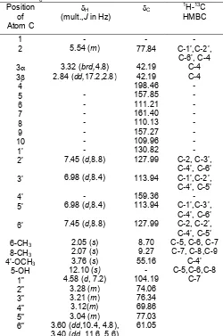
(ppm) : 2.05(3H, s, 8-CH3), 2.07 (3H, s, 6-CH3), 2.84 (1H,dd,J= 17.2 Hz, 2.8 Hz, H-3), 3.04 (m, H-5”), 3.12 (1H, m, H-4”), 3.21 (1H, m, H-3”), 3.28 (1H, m, H-2”), 3.32 (1H,brd,J = 4.8 Hz, H-3), 3.60 (1H, dd, J = 10.4 Hz, 4.8 Hz, H-6”), 3.40 (1H,dd, J = 11,6 Hz, 5.6 Hz, H-6”), 3.76 (3H,s, 4’-OCH3), 4.58 (1H,d, J= 7.2 Hz, H-1”), 5.54 (1H,m, H-2), 6.98 (2H,d,J= 8.4 Hz, H-3’,5’), 7.45 (2H, d, J = 8.8 Hz, H-2’,6’), 12.10 (1H, s, chelated 5-OH).13C-NMR (100.4 MHz, DMSO-d6)(ppm) : 8.70
(6-CH3), 9.27 (8-CH3), 42.19 (3,3), 55.16 (OCH3), 61.05 (C-6”), 69.86 (C-4”), 74.06 (C-2”), 76.34 (C-3”), 77.03 (C-5”), 77.84 (C-2), 104.19 (C-1”), 109.96 (C-10), 110.13 8), 111.21 6), 113.94 3’,5’), 127.99 (C-2’,6’), 130.82 (C-1’), 157.27 (C-9), 157.85 (C-5), 159.36 (C-4’), 161.40 (C-7), 198.46 (C-4). FABMS, m/z (rel.int.): 515 (M+K+)(2), 477 (M+H+) (1), 345 (2 m-NBA+K+)(22), 315 (aglycone+H+), 314 (aglycone) (3), 307 (2 m-NBA+H+)(14), 192 (m-NBA+H+)(100), 136 (m-NBA-OH) (86).
RESULT AND DISCUSSION
Compound 1 was isolated from ethyl acetate soluble fraction of methanol extract of the C. sakayensis’s stem as a pale yellow needles (benzene), mp.167-167 oC, []D
20
Indo. J. Chem., 2007, 7 (3), 346-349
Suyatno,et al.
348
aromatic protons in the B-ring indicated the presence of a methoxy group at C-4’. The other significant correlations of 1 can be seen in Table 1. Further supporting evidence of structure 1 for matteucinol came from comparison of the 1H-NMR, 13C-NMR, and EIMS spectral data with those of reported data in literature [6,7]. From the above results, compound 1 was identified as matteucinol (5,7-dihydroxy-4’-methoxy-6,8-dimethyl flavanone).
Compound 2 was isolated from the ethyl acetate soluble fraction of methanol extract of the C. sakayensis’s stem as a pale yellow powder (CHCl3 -MeOH), mp. 135-136 oC, gave positive test with FeCl3 test (green) and Shinoda-test (Mg-HCl) (pale red). The
O
Indo. J. Chem., 2007, 7 (3), 346-349
Suyatno,et al.
349
signals atH2.84 (dd, H-3), 3.32 (brd, H-3), and 5.54 (m, H-2) in the 1H-NMR spectrum (Table 2) also supported that compound 2had the flavanone skeleton. No batochromic shift of band II on adding NaOH and NaOAc reagent showed that 2 didn’t have a free hydroxyl group at C-7. No batochromic shift on adding NaOAc + H3BO3 reagent supported that 2 didn’t have ortho-dihydroxy group at A-ring. The chelated proton signal at H 12.10 (s) indicated the existence of a hydroxyl group at C-5. The 1H-NMR spectrum of 2 exhibited proton signals due to a 4’-methoxyphenyl group at H 3.76 (3H, s, 4’-OCH3), 6.98 (2H, d, J=8.4 Hz), and 7.45 (2H, d, J=8.8 Hz), two aromatic methyl group at H 2.05 (3H, s) and 2.07 (3H, s) as well as a glycosyl group atH4.58 (1H, d, J = 7.2 Hz, H-1”) andH 3.04-3.60 (6-H glycosyl)(Table 2). The glycosyl group of 2 could be identified as a glucosyl group because its carbon signals resembled those of reported data in literature [5]. In the HMBC spectrum of 2, proton signal of methoxyphenyl group (H 3.76) showed correlation with carbon signal of C-4’ (C 159.36), proton signal of the first aromatic methyl group (H 2.05) correlated with carbon signals of C-5 (C157.85), C-6 (C 111,21), C-7 (C 161.40), and the proton signal of the second aromatic methyl group (H 2.07) correlated with carbon signals of C-7 (C 161.40), C-8 (C 110.13), C-9 (C 157.27) (Table 2). These results suggested the presence of methoxyphenyl group at C-4’ and aromatic methyl group at C-6 and C-8, respectively. The correlation between proton signals of anomeric proton of glucosyl group (H 4.58) with carbon signal of C-7 (C 161.40) in the HMBC spectrum of 2 showed the presence of glucosyl group at C-7. Meanwhile the coupling constant value of the anomeric proton was 7.2 Hz, indicated the presence of a -glycosydic linkage to a aglycone [8]. Further supporting evidence of structure 2 for matteucinol-7-O--D-glucoside came from comparison of the 1H-NMR and 13C-NMR spectral data with those of reported data in literature [6]. From above results compound 2 was suggested to be a matteucinol-7-O- -D-glucoside.
CONCLUSION
Two alkylated flavanone, matteucinol and matteucinol-7-O--D-glucoside had been isolated from the ethyl acetate soluble fraction of the methanol extract of the fernC. sakayensis’s stem. This is the first report of the chemical constituents from this species and the genusChingiaof the fern.
ACKNOWLEDGEMENT
We thank the Directorate General of Higher Education, Ministry of National Education, Indonesia, for financial support through the Fundamental Research Grant 2007 and Mr. Wardaya from the Purwodadi Botanical Garden, Pasuruan, Indonesia, for help in collecting and identifying the plant material. REFFERENCES
1. Steenish, V, and Holttum, R.E., 1982, Flora Malesiana.Junk Publisher, London, 392.
2. Piggott, A.G., 1988, Fern of Malaysia. Kualalumpur, 192.
3. Suyatno, Zaini, N.C., Indrayanto, G., and Tori, M., 2005,Indo. J. Chem., 5 (2) 173-176.
4. Suyatno, Zaini, N.C., Indrayanto, G., and Tori, M., 2006,Indo. J. Chem., 6 (2) 82-84.
5. Markham, K.R., 1982, Techniques of Flavonoid Identification. Academic Press, London, 38-50. 6. Tanaka, N., Murakami, T., Wada, H., Gutierrez,
A.B., Saiki, Y., and Chen, C.M., 1985, Chem. Pharm. Bull.,33(12) 5231-5238.
7. Miraglia, M.D.C., Padua, A.P.D., Mesquita, A.A.L., and Gottlieb, O.R., 1985,Phytochem., 24(5) 1120. 8. Markham KR, and Geiger H., 1994, 1H nuclear