EISSN: 2086-4094 DOI: 10.4308/hjb.21.4.187
_________________
∗Corresponding author. Phone/Fax: +62-251-8622833,
E-mail: [email protected]
SHORT COMMUNICATION
Characterization of an Endophytic Bacterium G062 Isolate
with Beneficial Traits
ALINA AKHDIYA1,2, ARIS TRI WAHYUDI1*, ABDUL MUNIF3, LATIFAH KOSIM DARUSMAN4
1Department of Biology, Faculty of Mathematics and Natural Sciences, Bogor Agricultural University, Darmaga Campus, Bogor 16680, Indonesia
2ICABIOGRRD, Research & Development Institute, Ministry of Agriculture, Jalan Tentara Pelajar 3A, Cimanggu, Bogor 16111, Indonesia
3Department of Plant Protection, Faculty of Agriculture, Bogor Agricultural University, Darmaga Campus, Bogor 16680, Indonesia
4Department of Chemistry, Faculty of Mathematics and Natural Sciences, Bogor Agricultural University, Darmaga Campus, Bogor 16680, Indonesia
Received January 17, 2014/Accepted November 21, 2014
An endophytic bacterium isolate G062 was characterized base on its molecular genetic potents, morphology, physiology, and biochemistry reactions. Analysis of 16S rDNA sequences of G062 showed the highest similarity to
Paracoccus halophilus (98%). Detection of the phlD and prnC genes occurrence indicated that the bacterium had this antibiotic-like genes of Diacethylphloroglucinol (DAPG) and pyrrolnitrin. The cells are rod shaped (0.59-0.89 x 1.85-3.3 µm), aerobic, Gram negative, non motile, non spore forming, positive catalase, positive oxydase, could reduce NO3 to N2, nitrogen fixing, producing siderophore and plant growth hormones-like compounds (IAA, Gibberellin, and zeatin), and solubilizing phosphate. The G062 isolate could grow on media containing 2.5% NaCl. Range of the temperature and pH growth were 15-40 and 5.0-9.5 oC, respectively. The bacterium did not cause red blood cells lysis. There was no hypersensitive response when it was injected into tobacco leaves, and it was not pathogenic against potato plantlets. Moreover, the bacterium promoted the growth of the potato plant and had high colonization ability.
These results suggested that the bacterium had beneficial and good traits as biological agent candidate to promote
potato plant growth.
Key words: characterization, endophytic bacterium, Paracoccus halophilus, potato plant
___________________________________________________________________________
INTRODUCTION
Endophytic bacteria are defined as bacteria that can be isolated from the plant tissue or from tissue that had been surface sterilized and does not harm
to the plant (Hallmann et al. 1997). Today, the
development of microbiology and biotechnology
have prove that the presence of endophytic bacteria is essential for plant growth, plant resistance to biotic and abiotic stresses, and crop productivity. The roles of the endophytic bacteria are related to its abilities in nitrogen fixing, phytohormones producing, biocontrol of pathogens, as well as inducers of plant immunity (Mano & Morisaki 2008; Andreotte et. al. 2010).
Endophytic bacteria have great potential to be applied to seeds or plant seedlings as a way to
increase/promote its growth and resistance; for its early application may protect their host plants from pathogen colonization. Research experiments showed that plantlets of potato, grape, and banana which were grown together with endophytic bacteria grown better and had higher resistancy against pathogens than the non-inoculated plantlets (Frommel et al. 1991; Krechel et al. 2002; Sessitsch et al. 2005; Faltin et al. 2004; Compant et al. 2005; Kavino et al. 2007).
Useful bacteria can be isolated from nature; furthermore its can be developed and used as artificial inocula to improve plant growth and resistance. Physiological and molecular chararacters of the
bacteria are very important information which are
needed as scientific basic to develop a good and safe bacterial inocula. Considering that reason, this
research was conducted and addressed. The aims of
MATERIALS AND METHODS
Bacteria and Preservation of the Culture.
G062 isolate was an endophytic bacterium isolated from the surface of sterilized stem of potato plant (Atlantic) taken from Garut, West Java Indonesia (unpublished data). Working cultures was maintained routinely by growing on TSA (30 g/L TSB, 15 g/L agar) slant at room temperature (29-29 oC) and stored
in refrigerator. The isolate was preserved in 40% glycerol at -20 oC and in lyophilized form for
long-term storage.
Pathogenicity Potent Test of the Isolate.
The isolate was targeted to haemolytic and plant pathogenicity tests. Haemolytic activity of the isolate were evaluated by streaking G062 on blood agar. Plant pathogenicity tests were conducted against non host plant by Hipersensensitive Respon (HR) bioassay on tobacco leaves (N. tobaccum) and host plantlets (S. tuberosum) in 5 replicates. E. coli (as negative control) was grown by streaking in LA (5 g yeast extract, 10 g trypton, 10 g NaCl, and 15 g agar) for 2 days, while R. solanacearum (as positive control) and G062 bacterium isolate were grown by streaking in TSA for 5 days. The bacterial colonies were scrabbed using loop inoculation. The bacterial cells were suspended in 1 mL of physiological saline solution (± 107 CFU/mL). Each suspension was
injected into tobacco leaves (Nicotiana tabaccum) according to the method described by Zou et al. (2006). Tobacco leaves were observed for 7 days to determine the hypersensitive response of the infected tissue around the infected site. A total of 100 µL bacterial suspension (± 107 CFU/mL in distilled
water) were dropped onto the medium around the plantlet root in culture tubes containing the 2-weeks old plantlets. The effect of bacterial inoculation on plantlets was observed for 10 days.
Molecular Characterization. The genomic DNA were extracted using XPrep Stool DNA Mini Kit (Philekorea Technology, INC. Seoul
Korea). Molecular identification of bacteria were
done based on sequence analysis of 16S rRNA
gene. The 16S rRNA gene amplification used 63f
(5’-CAGGCCTAACACATGCAAGTC-3’) and 1387r (5’-GGGCGGWGTGTACAAGGC-3’) primers and PCR condition as described by Marchessi et al. (1998). A pair of IGK (TACGGYAARGCBGGYATCGG) and NDR-1 (TTGGAGCCGGCRTANGCRCA) primers published by Poly et al. (2001) and Valdes
et al. (2005) were used as forward and reverse
primers, respectively, for amplification of the nif
gene. PCR reactions of nif gene were conducted in 30 cycles (2 min initial denaturation at 95 oC,
1 min denaturation at 94 oC, 2 min annealing at
56.5 or 57 oC, 2 min elongation at 72 oC, and 7
min post elongation at 72 oC). Genetic potent of
2,4-diacetylphloroglucinol (DAPG) and pyrrolnitrine production were examined by using pairs of phl2a
(5’-GAGGACGTCGAAGACCACCA-3’) and phl2b
(5’-ACCGCAGCATCGTGTATGAG-3’), phen1 (5’-CCCCTGTTGACAATTAATC-ATCGG-3’) and phen2 (5’-ACCTTGACGTTGTACCATTCCCAA-3’), and
prnCf (5’-CCACAAGCCCGGCCAGGAGC-3’) and prnCr (5’-GAGAAGAGCGGGTCGAT
GAAGCC-3’) primers, respectively. Amplification
condition of the two metabolite genes conducted as published by Raaijmakers et al. (1997) and Mavrodi
et al. (2001), respectively. CLUSTAL W program (Thompson et al. 1994) was used for performing the 16S gene sequences alignment. Construction of the phylogenetic tree was made by using the neighbour-joining method with Maximum Composite Likelihood model in Mega 5.05 (Tamura et al. 2011). To confirm
the reduction activity of NO3 to N2, occurrence of the
nos gene was tested by PCR using a pair of nos661F (5’-CGGCTGGGGGCTGACCAA-3’) and nos1773R primers (5’-ATRTCGATCARCTGBTCGTT-3’) as stated by Scala and Kerkhof (1988). PCR conditions of nos gene were conducted in 30 cycles (2 min initial denaturation at 95 oC, 1 min denaturation at 94 oC,
2 min annealing at 55 oC, 2 min elongation at 72 oC,
and 7 min post elongation at 72 oC).
M o r p h o l o g i c a l O b s e r v a t i o n . C o l o n y morphologies (color, shape, elevation, and consistency of the colony) were examined on TSA and KBA (20 g/L protease pepton, 1.5 g/L K2HPO4.3H
2O, 1.5
g/L MgSO4.7H
2O. 20 ml/L glycerol, 15 g/L agar).
Morphology and size of log phase cells (from 24 h TSB culture) were observed using Scanning Electron Microscope (Jeol JSM-5310 LV) at
10,000 magnification. The specimens were fixed in
cacodylate buffer and glutaraldehyde, dehydrated in alcohol, and then freeze dried. Before observed, the specimen were cut, attached to stub specimens
with double cello-tape, coated with gold 400 A◦ in
Eico I-B2 ion coater (Goldstein 1992). Motility of the cells were examined by observation of hanging
drop preparation slides under bright field microscope
(Olympus CH20BIMF200).
Physiological and Biochemical Characterization.
All of the cultures used for the tests were incubated at room temperature (29-30 oC) unless otherwise stated. Nitrogen fixation ability was tested by growing on
Nitrogen Free Agar (NFA) and LGI Agar which
was modified (0.2 g K2HPO4; 0.6 g KH2PO4; 0.2 g
MgSO4.7H
2O; 0.02 g CaCl2; 0.002 g Na2MoO4
.2H
2O;
acid; and 15 g agar). Acetylene reduction activity test
was conducted toward modified the LGI semisolid
culture in triplicate.
Capability of G062 to produce plant growth hormones-like were determined in TSB. A loop full of colony which were grown on TSA for 48 h were inoculated into the test tube containing 5 mL TSB, then the tube was incubated on incubator by shaking (100 rpm) at room temperature (29-30 oC).
After 24 h, 1 mL of the liquid culture was inoculated
into a flask containing 50 mL new sterile TSB. The flask was incubated at room temperature (29-30 oC). After 7 days, the culture was centrifuged at 4 oC, 10,000 rpm, for 10 min. Quantification of
IAA-like in the supernatant cultures were conducted in 4 replicates by using the Salkowsky methods which has
been modified by Glickmann and Dessaux (1995).
Gibberellin (GA)-like, and zeatin-like contained in the supernatant, were extracted and determined
triplicates by using the methods described by Ergűn
et al. (2002).
TheGram reaction of the cells grown on TSA (24 h) were examined by using non staining (Buck 1982) and staining techniques (Benson 2001) under
bright field microscope (Olympus CH20BIMF200) at 1000x magnification. Chitinolytic, proteolytic,
and amylolytic activities were observed to the culture that has grown for 7 days on 1% (w/v) TSA containing 1% colloidal chitin, 1% skim milk, and 1% soluble starch, respectively. Hydrolysis of CMC (Carboxy methyl cellulose) was observed by streaking of isolate on CMC agar. Capability of the isolate to produce siderophore was tested according to the method described by Hu and Xu (2011). Phosphate solubilization activity was observed on Pikovskaya agar plate (Pikovskaya 1948). Catalase activity was tested by bubbles formation on 3% H2O2 solution that was mixed with a loop of bacterial colony grown for 24 h on TSA. Oxidase activity was determined by using oxidase paper (Difco). NaCl tolerance was tested using LB medium contained appropriate concentration of NaCl. The bacterium were inoculated into TSB or adjusted TSB (by using NaOH and HCl) and then incubated at various temperature (13, 18, 20, 30, 37, 40, and 42 oC) or room temperature (30 ± 1 oC) to determine
its tolerance against temperature and pH of growth. Growth were checked by measuring optical density (600 nm) of the cultures. Other physiological and biochemical reactions were examined by applying API 20NE galleries (bioMerieux SA, Lyon, France).
Evaluation of Colonization Ability. Colonization of G062 isolate on internal stem tissue of potato plantlet was observed by using Scanning Electron
Microscope (Jeol JSM-5310 LV). The specimens were prepared according to Goldstein (1992) as we described above before observed at 750x and 1000x
magnification. Re-isolation of G062 isolate was
also conducted to evaluate the colonization ability of G062 isolate inside the plantlet tissue. The shoot of the plantlets were cut at 1.5 cm above the surface media, weighed, and grinded. The plantlet extract were diluted and plated on 50% TSA.
Observation of Plant Growth Promotion Ability. Potato plantlets (2 weeks after subcultured) were inoculated with G062 suspension as described above. Two weeks after inoculation, the G062 inocula and control plantlets were planted on 3 kg sterile planting media consisted of Chicken’s manure, soil, and risk husk (ratio = 1:1:1). Planting was conducted at Research station of ICABIOGRRD, in Pacet, District of Cianjur, West Java, Indonesia. Three months after planting, the plant biomass (plant shoot and root) were harvested, dried, and weighed; while the tuber were calculated and weighed. Growth parameter and tuber productivity value were calculated from 10 plants.
RESULTS
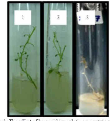
Pathogenicity Test of G062 Isolate. There were no hydrolytic zone showed on Blood agar (data not shown), nor necrotic tissue around the infected sites of tobacco leaves during the observation (7 days after streaking or injection of G062 cells suspension). Pathogenicity test on potato plantlet also showed no disease symptoms or plantlets killed by the bacterium; moreover, all the plantlets looked healthy during observation as shown in Figure 1.
1 2 3
Molecular Characters of G062 Isolate.
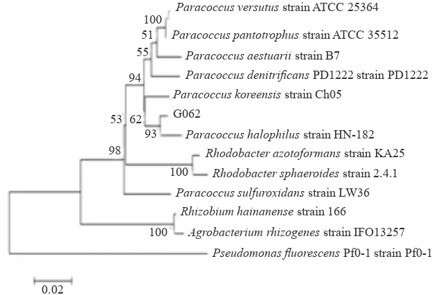
Sequence analysis of 16S rRNA gene showed that G062 isolate was belong to genus Paracoccus, one
of an α proteobacteria group. The closest related
species was Paracoccus halophilus (98%, max. score 1245, query cover 100%, e value 0.0, accession No. NR. 043810.1). P. versutus (98.0%, max. score 1229, query cover 100%), P. pantotrophus (98%, max. score 1223, query cover 100%), P. marinus
(98%, max. score 1221, query cover 98%), and
P. koreensis (98%, max. score 1216, query cover 98%) were the next closely related ones. Sequences alignment of phylogenetic trees were constructed with the neighbour-joining, maximum-parsimony and maximum-likelihood methods. Values of the bootstrap were evaluated by using 1000 replication. The phylogenetic tree placed the isolate in a coherent cluster with P. halophilus and closer relation to P. koreensis (Figure 2). Detection of the nifHD, nos, prnC and phlD gene occurrence by PCR yielded multiband DNA amplicons (Figure 3 & 4). The nif
genes amplicons were approximately 1200, 1400, and 1700 bp in size. Two sharp DNA bands (± 450 and 550 bp) and 4 faint bands (± 850, 1110, 1300, and 1500 kb) were detected on electrophoresis gel of nos
gene amplicons. Two faint bands (both were smaller than 500 bp), and two clear bands (± 720 and 1300 bp) were detected on PCR product of prnC gene; and 5 PCR products (3 clear bands: ± 1200, 500, 300 bp; and 2 faint bands : ± 550 and 265 bp) were obtained by using the phlD primers.
Description of Morphology and Cytological Characters. Colonies of G06 isolate were round, smooth, translucent (on KBA) or cream to faint brown (on TSA), and had soft consistency. The
colonies on modified LGI agar are small (2-5 mm
1 2 3 4 5 6 7
3000
1500
1000
750
500
1500
1000 750
500
250
Figure 3. PCR products of 16SrDNA, nitrogen fixation and
nitrate reduction genes of G062 isolate. The arrowheads indicated the expected DNA amplicon of each genes. 1. 1 kb ladder (Geneaid), 2. 16S rDNA, 3. nif HD gene (56.5 oC), 4. nif HD gene
(57 oC), 5. 1 kb ladder (Thermo), 6. nos gen, 7. 1
kb (Geneaid).
1 2 3 4
3000
1500
1000 750
500
1500
1000 750
500
250
Figure 4. Amplification products of antifungal compounds
genes of G062 isolate. The arrowheads indicated the expected DNA amplicons of each genes. 1. 1 kb ladder (Thermo), 2. prnC gene, 3. 1 kb ladder (Geneaid). 4. phlD gene.
Paracoccus versutus strain ATCC 25364
Paracoccus pantotrophus strain ATCC 35512
Paracoccus aestuarii strain B7
Paracoccus denitrificans PD1222 strain PD1222
Paracoccus koreensis strain Ch05
G062
Paracoccus halophilus strain HN-182 Rhodobacter azotoformans strain KA25
Rhodobacter sphaeroides strain 2.4.1
Paracoccus sulfuroxidans strain LW36
Rhizobium hainanense strain 166
Agrobacterium rhizogenes strain IFO13257
Pseudomonas fluorescens Pf0-1 strain Pf0-1
100 51 55
94
53 62 93 98
100
100
0.02
in 10 days), watery, and translucent or opaque. There were no pellicle on liquid cultures (on TSB
or modified LGI). Shape of the exponential phase
cells grown on TSB were short rod or rod. The cells were non motile and there were no endospore formation observed. Dimension of the exponential phase of single cells grown in TSB were 0.59-0.89
x 1.85-3.3 µm. There were a lot of fibrous material
around and covered the cells as shown in the electron photomicrograph (Figure 5).
Description of Physiological and Biochemical Characters. Cells of G062 isolate are Gram-negative, aerobic, and showed positive of catalase and oxidase activity. Growth observed at 15-40 oC but
not at 13 or 43 oC. Cells growth were detected at pH
5.0-9.5 but not at pH 4.5 or 10.0. Growth of the liquid cultures (on TSB) were observed on TSB contained 2.5% (w/v) NaCl, but there were no increasing optical density at 5% NaCl. This isolate could completely reduced nitrate to dinitrogen. Indol production, urease, arginin dihydrolase, and hydrolysis of aesculin, gelatin, starch, chitin, skim milk, and CMC were negative. It also capable of utilizing and fermenting D-glucose, assimilating L-arabinose,
Figure 5. Scanning electron micrograph of G062 cells (at
10,000 magnification) which were grown on TSB
for 24 hours. Fibrous materials around the cells were produced as indicated by arrowhead.
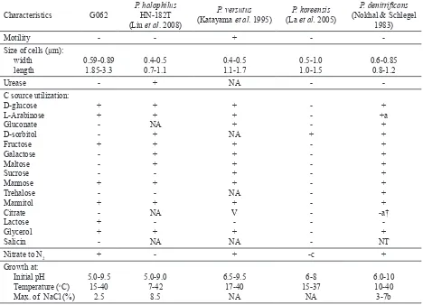
Table 1. Characteristics of closely related Paracoccus species and G062 strain
Characteristics G062
a: most of the strains; a†: most of the strains (except mutant); b: depend on the strains; c: could not reduce NO3, but could reduce NO2; V: various reaction were reported; NA: data not available.
production on simple-double layer Chromo azurol agar.
The result of ARA bioassay showed that G062
isolate could fixed nitrogen from the air. Beside
its ability to fix nitrogen, G062 isolate could produce plant growth hormone-like substances (IAA, Giberellin, and Zeatin-like), even though it was grown in media which was not supplemented with tryptophan. The bioassay result of ARA and measurement of plant growth hormone produced by G062 isolated are show in Table 2.
Micrographs of scanning electron microscope showed occurence of G062 colonies inside stem tissue of the potato plantlet (Figure 7). The microscopic observation were in line with the re-isolation result.
Four weeks after inoculation, the average of G062 cell density on plantlets tissue was 2.8 x 10⁴ ± 4.9 x
103 cfu/g. In comparison to control plants, the G062
inoculated plants produced higher plant biomass. Moreover, G062 plants produced in average 6.8 tuber/plants, while control plants produced only 5.1 tuber/ plants.
DISCUSSION
G062 isolate is not potent as mammalian pathogen, because it did not formed greenish or clear zone under and around the bacterial colonies which mean it did not lyse the red blood cells contained in the blood agar medium. Moreover, hypersensitive response (HR) bioassay result indicated that the isolate is not a phytopathogenic bacterium, because there were no nectrotic tissue observed at and around the infected sites of the tobacco leaves. This bioassay is a common tool to screen phytopathogenic bacteria. HR is a plant mechanism to prevent the recognized phytopathogenic bacteria growth and spreading from its entry sites. Heath (2000) stated that the mechanism involves a complex form of programmed cell death (PCD) which is consistently associated with the induction of local and systemic defence responses, and it can be the result of multiple signaling pathways. All of potato plantlets which were inoculated with the bacterium were also showed healthy condition and there were no disease symptoms appeared during observation time. According to the pathogenicity test results, it will be safe to be developed as bacterial inocula.
Base on partial 16S rDNA sequences, G062 isolate is a member of the metabolically versatile genus Paracoccus. Since the genus was created in 1969, at least 30 species were published as a part of it (Kelly et al. 2006; Roh et al. 2009; Deng et al. 2011; Lee et al. 2011; Zheng et al. 2011). Most
A B
Figure 7. Colonies of G053 inside plantlet stem at 750x (A) and 10000x magnification (B).
Figure 6. Phosphate solubilizing activivity of G062 isolate on Pikovskaya agar incubated at 29-30 oC for 72 hours.
Table 2. Nitrogen fixation activity on modified semisolid LGI
and phytohormones-like compounds produced by G062 isolate in TSB medium
Assay Results
ARA (µmol/mL/h) IAA-like (ppm) Gibberellin-like (ppm) Zeatin-like (ppm)
1.190 ± 0.011 17.4 ± 0.03
of its were isolated from soil, marine, sediment or sludges. Recently, the well characterized (molecular, physiological, and biochemical) of Paracoccus
species that has been reported as endophytic isolate was only P. sphaerophysae Zy-3(T) (Deng et al.
2011). However, the finding that the G062 isolate
was a Paracoccus was not surprising, because some publication on plant bacterial diversity or community studies indicated the occurrence of genus Paracoccus
in plant such as banana cv. Grand Naine shoot tips (Thomas & Soly 2009), P. vulagaris seedlings (Lopez
et al. 2010), eucalyptus seedlings (Ferreira et al. 2008), and mangrove roots (Flores-Mireles et al. 2007). Surprisingly, G062 isolate showed highest similarity to Paracoccus halophilus strain HN-182 which was isolated from marine sediment of South China sea (Liu et al. 2008). The next closest related species were P. versutus and P. pantotrophus. The BLASTN alignment result showed slight different with the phylogenetic tree that the isolate was in a coherent cluster with P. halophilus and closer relation to P. koreensis than P. panthotrophus and P. versutus
(Figure 1).
Micrograph of scanning electron microscope
showed fibrous material around and covered the
cells (Figure 5). Nokhal and Schlegel (1983) also
have reported the finding of fibrous material which
covered P. denitificans cells. They stated that it has indication as extracellular polyanions and acidic mucosubtances, for it has reacted with Ruthenium red.
Compared with published closely related species (Table 2), P. halophilus G062 could tolerate higher pH but lower NaCl concentration (2.5%) than P. halophilus HN-182T. Its lower NaCl tolerance may
correspond to the condition of the internal part of plant as its natural ecological niche. Moreover, G062 was like P. panthotrophus, P. versutus, and P.
denitrificans because they could reduced nitrate to
dinitrogen, while P. halophilus HN-182T could not.
The activity of Nitrate reduction to N2 and ARA bioassay were in line with the result of PCR product of nos and nifHD genes, respectively. The nos gene encodes nitrous oxide reductase which is needed to convert N2O to N2 at the final step of denitrification pathway (Jones et al. 2008), while nifHD encodes
Fe-protein and α subunit of Mo-Fe protein of
dinitrogenase reductase. We have expected that the size of the PCR product will be ± 1112 and 1200 bp for nos and nifHD genes, respectively. But, both of the PCR products showed multiple DNA bands
(Figure 3) that indicated unspecific amplification which may caused by less specificity of the primers
or non optimum PCR condition. The primer set to
amplify nos gene (nos661F and nos1773R) were designed base on nos gene sequences of P. stutzeri
Zobell (Scala & Kerkhof 1988). NDR-1 and IGK were universal primer for nifHD gene (Poly et al. 2001; Valdez et al. 2005), and often use at the first
step of nested PCR of nifHD genes. Moreover,
Paracoccus is a member of α-proteobacteria, which
among it’s group member shared conserved inserts and deletions (indels) in their molecular sequences
(Gupta 2005) that may contributed to this unspesific
in vitro amplification.
A diverse type of secondary compounds have been discovered as the product of endophytic microorganism. PCR of secondary metabolites encoding genes were conducted by using primers,
prnCf/prnCr and phl2a/phl2b designed base on the
Pseudomonas fluorescen genes that we expected to amplify a 719 and 745 bp DNA region of prnC and
phlD, respectively. The prnCfand prnCr primers
were amplified the prnC, one of four genes which involved in pyrrolnitrin biosynthesis (Kirner et al. 1998). While, the phl2a and phl2b primers were
amplified the phlD, one of six genes which involved in the biosynthesis of a secondary metabolite 2,4-diacetylphloroglucinol (Raaijmakers et al. 1997). These substances has been known as broad spectrum antifungal antibiotic. Although PCR product prnC and phlD genes also showed multibands DNA, its indicated that P. halophilus G062 may has potency to produce pyrrolnitrin and 2,4-diacetylphloroglucinol (DAPG) compound. Beside, its may contribute to the
non specific amplifications as we have stated before;
occurrence of indels in region of the target gene may also contribute to the difference of DNA amplicons size of phlD. Insertion or deletion will change the gene length, and may affect its protein function. In addition to these antibiotic-like genes, P. halophilus
G062 could produced siderophore which is also act as antifungal.
Phosphorus place is in the second position as most commonly limiting mineral factor for growth of terrestrial plants. Ironically, large of total P may reserve in soils, but it availability for plants is usually in a small proportion. Its low availability is because majority of its form found in insoluble forms (Verma
Production of single phytohormones such by microorganisms has been reviewed by various scientists over the last 20 years (Verma et al. 2010). IAA, GA, and zeatin was detected on liquid cultures of P. halophilus G062 (Table 2). The ability of P. halophilus G062 to produce the
phytohormone-like compounds is a beneficial character, because
phytohormones analogous produced by endophytic bacteria can promote plant growth. Auxin are responsible for division, extension, and differentiation of plant cells and tissue. Stimulation of seed and tuber germination; increasing of xylem and root formation rate; controlling of vegetative growth,
tropism, florescence, and fructification processes;
affecting of photosynthesis, pigmen formation, metabolites biosynthesis, and plant resistance against stress are correspond with auxin (Tsavkelova
et al. 2006). GA are a class of phytohormones which most commonly associated with modifying plant morphology by the extension of plant tissue, particularly stem tissue (Verma et al. 2010). Zeatin is one of adenine type cytokinin which regulates cell growth, cell differentiation, apical dominance, and leaf senescence (Xu et al. 2012).
Naturally, endophytes and some phytopathogens live in same ecological niche, so colonizing and dominating capacity of the endophythes are important to its host plants. Hallmann and Bergh (2006) had reviewed that average density of natural endophytic
bacteria in various plants root or stem were 104-106
cfu/g. This experiment showed that 2 weeks after inoculation of Paracoccus halophilus G062, cell density of the bacterium on the plantlets was 2.8 x 10⁴ ± 4.9 x 10³ cfu/g. Microscopic observation
results also showed that the bacterium was found at internal tissue (sponge parenchyme) of the plantlet (Figure 7). The result indicated that the bacterium has high colonization ability. High colonization ability and persistency are important characters of a good biological agent candidate. The results also supported and prooved that the bacterium is an endophyte, because beside it was isolated from surface sterilized plant as stated in the materials and methods, the bacterium also showed to live or have life cycle inside the deep area of the plant tissue without harming the host plant, as well as endophytic
defined previously by Wilson (1995).
Plant growth (dry weight of the potato shoot and root) and tuber productivity of G062 inoculated plant were higher than control plants (Figure 8). G062 plants looked vigour and dark green, while the control plants looked dwarf and light green to yellowish. This result obtained from 3 planting were consistent (data from preliminary G0 and next G1 plants parameter observation were not shown). It was suggested that interaction between the bacterium
with the host plant significantly promoted the plant
growth. Capability of Paracoccus halophilus G062
0 10000 20000 30000 40000 50000 60000
T
otal weigh of tuber (mg/plant)
D 23405
30562
0 1 2 3 4 5 6 7 8 9
Number of tuber
C 5.1
6.8
0 50 100 150 200 250 300 350 400 450 500
Dry weigh of root (mg/plant)
256
382
B
0 500 1000 1500 2000 2500
Dry weigh of shoot (mg/plant)
1113
1339
A
to fix Nitrogen, to produce plant growth hormones,
and to solubilize the insoluble phosphate are beneficial characters that contribute to increase the nutrient availability to the plant. Nitrogen is a important element for plant growth. This element is a component of protein, nucleotides, and chlorophyls, three important molecules of plant cells. Compare to the control plants, it seem that nitrogen requirement
of G062 plants were supplied sufficiently than control
plants. Its strongly indicated that P. halophilus G062 contributes to supply the nitrogen requirement of the host. In addition, siderophore production was valuable weapon that may enhance its capacity to combat and compete with phytopathogenic fungi. Therefore, it may dominate inner part of plant tissue and control the pathogens. Beside its antifungal activity, siderophore increase plant capability to absorb Fe (Robin et al. 2006). For all of its beneficial
characters, the bacterium is safe and has great
potency to be developed as a component of artificial
plant growth promoting (PGP) inocula.
REFFERENCES
Andreote FD, UN da Rocha, Araujo WL, Azevedo JL, Overbeek LSV. 2010. Effect of bacterial inoculation, plant genotype and developmental stage on root-associated and endophytic bacterial communities in potato (Solanum tuberosum). Antonie van Leeuwenhoek 97:389-399. http://dx.doi. org/10.1007/s10482-010-9421-9
Benson HJ. 2001. Microbiological application laboratory manual. In: Brown AE (ed). General Microbiology. New York: Mc Graw Hill.
Buck JD. 1982. Nonstaining (KOH) method for determination of Gram reaction of marine bacteria. Appl Environ Microbiol 44:992-993.
Compant S, Reiter B, Sessitsch A, Nowak J, Clement C, Barka EA. 2005. Endophytic colonization of Vitis vinifera L. by Plant growth-promoting bacterium Burkholderia sp. strain PsJN. Appl Environ Microbiol 71:1685-1693. http://dx.doi. org/10.1128/AEM.71.4.1685-1693.2005
Deng ZS, Zhao LF, Xu L, Kong ZY, Zhao P, Qin W, Chang JL, Wei GH. 2011. Paracoccus sphaerophysae sp. nov., a siderophore-producing, endophytic bacterium isolated from root nodules of Sphaerophysa salsula. Intl J Syst Evol Microbiol 61:665-669. http://dx.doi.org/10.1099/ ijs.0.021071-0
Ergűn N, Topcuoģlu SF, Yildiz A. 2002. Auxin (Indole-3-acetic
acid), Gibberellic acid (GA3), Abscisic Acid (ABA), and Cytokinin (Zeatin) production by some species of mosses and lichens. Turk J Bot 26:13-18.
Faltin F, Lottmann J, Grosch R, Berg G. 2004. Srategy to select and assess antagonistic bacteria for biological control of Rhizoctonia solani Kühn. Can J Microbiol 50:811-820. http:// dx.doi.org/10.1139/w04-063
Ferreira A, Quecine MC, Lacava PT, Oda S, Azevedo JL, Araujo WL. 2008. Diversity of endophytic bacteria from Eucalyptus species seeds and colonization of seedlings by Pantoea agglomerans. FEMS Microbiol Lett 287:8-14. http://dx.doi. org/10.1111/j.1574-6968.2008.01258.x
Flores-Mireles AL, Winans SC, Holguin G. 2007. Molecular characterization of diazotrophic and denitrifying bacteria associated with mangrove roots. Appl Environ Microbiol 73:7308-7321. http://dx.doi.org/10.1128/AEM.01892-06 Frommel MI, Nowak J, Lazarovita G. 1991. Growth enhancement
and developmental modifications of in vitro grown potato (Solanum tuberosom ssp. tuberosum) as affected by a
nonfluorescent Pseudomonas sp. Plant physiol 96:928-936. http://dx.doi.org/10.1104/pp.96.3.928
Glickmann E, Dessaux Y. 1995. A critical examination of the
specificity of the Salkowski reagent for indolic compounds
produced by phytopathogenic bacteria. Appl Environ Microbiol 61:793-796.
Gupta RS. 2005. Protein signatures distinctive of alpha proteobacteria and its subgroups and a model for alpha-proteobacterial evolution. Crit Rev Microbiol 31:101-135. http://dx.doi.org/10.1080/10408410590922393
Goldstein JI, Newbury DE, Echlin P, Joy DC, Romig AD Jr, Lyman CE, Fiori C, Lifshin E. 1992. Scanning electron microscopy and X-ray microanalysis: a text for biologist, materials scientist, and cytologists 2nd edition. New York:
Plemun Pr. http://dx.doi.org/10.1007/978-1-4613-0491-3 Hallmann J, Berg G. 2006. Spectrum and population dynamics of
bacteria root endophytes. In: Schulz BJC, Boyle CJC, Sieber TN (eds). Microbial Root Endophytes. Berlin Heidelberg, Germany. Springer. http://dx.doi.org/10.1007/3-540-33526-9_2
Hallmann J, Hallmann A, Mahaffee WF, Kloepper JW. 1997. Bacterial endophytes in agricultural crops. Can J Microbiol 43:895-914. http://dx.doi.org/10.1139/m97-131
H e a t h M C . 2 0 0 0 . H y p e r s e n s i t i v e r e s p o n s e - r e l a t e d death. Plant Molec Biol 44:321-334. http://dx.doi. org/10.1023/A:1026592509060
Hu QP, Xu JG. 2011. A simple double-layered chrome azurol S agar (SD-CASA) plate assay to optimize the production of siderophores by a potential biocontrol agent Bacillus. Afr J Microbiol Res 5:4321-4327.
Jones CM, Stres B, Rosenquist M, Hallin S. 2008. Phylogenetic nalysis of Nitrite, Nitric Oxide, and Nitrous Oxide respiratory enzymes reveal a complex evolutionary history for
denitrification. Mol Biol Evol 25:1955-1966. http://dx.doi. org/10.1093/molbev/msn146
Katayama Y, Hiraishi A, Kuraishi H. 1995. Paracoccus thiocyanatus sp. nov., a new species of thiocyanate-utilizing facultative chemolithotroph, and transfer of Thiobacillus versutus to the genus Paracoccus as Paracoccus versutus comb. nov. with emendation of the genus. Microbiology 141:1469-1477. http://dx.doi.org/10.1099/13500872-141-6-1469
Kavino M, Harish S, Kumar N, Saravanakumar D, Damodaran T, Soorianathasundaram K, Samiyappan R. 2007. Rhizosphere and endophytic bacteria for induction of systemic resistance of banana plantlets against bunchy top virus. Soil Biol Biochem 39:1087-1098. http://dx.doi.org/10.1016/j. soilbio.2006.11.020
Kelly DP, Euzeby JP, Goodhew CF, Wood AP. 2006. Redefining
Paracoccus denitrificans and Paracoccus pantotrophus and
the case for a reassessment of the strains held by international culture collections. Intl J Syst Evol Microbiol 56:2495-2500. http://dx.doi.org/10.1099/ijs.0.64401-0
Kirner S, Hammer PE, Hill DS, Altmann A, Fischer I, Weislo LJ, Lanahan M, Pée KHV, Ligon JM. 1998. Function encoded by pyrrolnitrin biosynthesic genes from Pseudomonas
Krechel A, Faupel A, Hallmann J, Ulrich A, Berg G. 2002. Potato-associated bacteria and their antagonistic potential towards plant-pathogenic fungi and the plant-parasitic nematode Meloidogyne incognita (Kofoid & White) Chitwood. Can J Microbiol 48:772-786. http://dx.doi.org/10.1139/w02-071 La HJ, Im WT, Ten LN, Kang MS, Shin DY, Lee ST. 2005.
Paracoccus koreensis sp. nov., isolated from anaerobic
granules in an upflow anaerobic sludge blanket (UASB)
reactor. Intl J Syst Evol Microbiol 55:1657-1660. http:// dx.doi.org/10.1099/ijs.0.63494-0
Lee M, Woo SG, Park G, Kim MK. 2011. Paracoccus caeni sp. nov., isolated from sludge. Intl J Syst Evol Microbiol 61:1968-1972. http://dx.doi.org/10.1099/ijs.0.017897-0 Liu ZP, Wang BJ, Liu XY, Dai X, Liu YH, Liu SJ. 2008.
Paracoccus halophillus sp. nov., isolated from marine sediment of the South China Sea, China, and emended description of genus Paracoccus Davis 1969. Intl J Syst Evol Microbiol 58:257-261. http://dx.doi.org/10.1099/ ijs.0.65237-0
Lopez AL, Rogel MA, Orrillo EO, Romero JM, Romero EM. 2010. Phaseolus vulgaris seed-borne endophytic community with novel bacterial species such as Rhizobium endophyticum sp. nov. Syst Appl Microbiol 33:322-327. http://dx.doi. org/10.1016/j.syapm.2010.07.005
Mano H, Morisaki H. 2008. Endophytic bacteria in rice plant. Microbes Environ 23:109-117. http://dx.doi.org/10.1264/ jsme2.23.109
Marchesi JR, Sato T, Weighteman AJ, Martin TA, Fry JC, Hiom SJ, Wade WG. 1998. Design and evaluation of useful
bacterium-specific PCR primer that amplify genes coding for
bacterial 16S rRNA. Appl Environ Microbiol 64:795-799. Mavrodi DV, Gardener BBMS, Mavrodi DM, Bonsall RF,
Weller DM, Thomashow LS. 2001. Genetic diversity of
phlD from 2,4-diacetylphloroglucinol-producing fluorescent
Pseudomonas spp. Phytopathology 91:35-43. http://dx.doi. org/10.1094/PHYTO.2001.91.1.35
Nokhal TH, Schlegel HG. 1983. Taxonomic study of Paracoccus
denitrificans. Intl J Syst Bacteriol 33:26-37. http://dx.doi.
org/10.1099/00207713-33-1-26
Pikovskaya RI. 1948. Mobilization of phosphorus in soil connection with the vital activity of some microbial species. Microbiologiya 17:362-370.
Poly F, Monrozier LJ, Bally R. 2001. Improvement in the RFLP procedure for studying the diversity of nifH genes in
communities of nitrogen fixers in soil. Res Microbiol 152:95-103. http://dx.doi.org/10.1016/S0923-2508(00)01172-4 Raaijmakers JM, Weller DM, Thomashow LS. 1997. Frequency
of antibiotic-producing Pseudomonas spp. in natural environments. Appl Environ Microbiol 63:881-887. Robin A, Mougel C, Siblot S, Vansuyt G, Mazurier S, Lemanceau
P. 2006. Effect of ferritin overexpression in tobacco on the structure of bacterial and pseudomonad communities associated with the roots. FEMS Microbiol Ecol 58:492-502. http://dx.doi.org/10.1111/j.1574-6941.2006.00174.x Roh SW, Nam YD, Chang HW, Kim KH, Kim MS, Shin KS, Yoon
JH, Oh HM, Bae JW. 2009. Paracoccus aestuarii sp. nov.,
isolated from tidal flat sediment. Intl J Syst Evol Microbiol 59:790-794. http://dx.doi.org/10.1099/ijs.0.65759-0
Scala DJ, Kerkhof LJ. 1988. Niotrous oxide reductase (nosZ)
gene-specific PCR primers for detection of denitrifiers and
the three nosZ genes from marine sedimens. FEMS Microbiol Lett 162:61-68. http://dx.doi.org/10.1111/j.1574-6968.1998. tb12979.x
Sessitsch A, Coenye T, Sturz AV, Vandamme P, Barka EA, Salles JF, Elsas JDV, Faure D, Reiter B, Glick BR, Pruski GW, Nowak J. 2005. Burkholderia phytoformans sp. nov.,
a novel plant-associated bacterium with plant-beneficial
properties. Intl J Syst Evol Microbiol 55:1187-1192. http:// dx.doi.org/10.1099/ijs.0.63149-0
Tamura K, Peterson D, Peterson N, Stecher G, Nei M, Kumar S. 2011. MEGA5: molecular evolutionary genetics analysis using maximum likelihood, evolutionary distance, and maximum parsimony methods. Mol Biol Evol 28:2731-2739. http://dx.doi.org/10.1093/molbev/msr121
Thomas P, Soly TA. 2009. Endophytic bacteria associated with growing shoot tips of banana (Musa sp.) cv. Grand Naine
and the affinity of endophytes to the host. Microb Ecol 58:952-964. http://dx.doi.org/10.1007/s00248-009-9559-z Thompson JD, Higgins DG, Gibson TJ. 1994. CLUSTAL W:
improving the sensitivity of progressive multiple sequence
alignment through sequence weighing, position-specific
gap penalties and weight matrix choice. Nucleic Acids Res 22:4673-4680. http://dx.doi.org/10.1093/nar/22.22.4673 Tsavkelova EA, Klimova SY, Cherdyntseva TA, Netrusov AI.
2006. Microbial producers of plant growth stimulators and their practical use: A review. Appl Biochem Microbiol 42:117-126. http://dx.doi.org/10.1134/S0003683806020013 Valdes M, Perez NO, Santos PEDL, Mellado JC, Cabriales JJP,
Normand P, Hirsch AM. 2005. Non-frankia actinomycetes isolated from surface-sterilized roots of Casuarina equisetifolia fix nitrogen. Appl Environ Microbiol 71:460-466. http://dx.doi.org/10.1128/AEM.71.1.460-71:460-466.2005 Verma JP, Yadav J, Tiwari KN, Lavakush, Singh V. 2010. Impact
of plant growth promoting Rhizobacteria on crop production. Intl J Agric Res 5:954-983. http://dx.doi.org/10.3923/ ijar.2010.954.983
Wilson D. 1995. Endophyte–the evolution of a term, and
clarification of its use and definition. Oikos 73:274-276. http://dx.doi.org/10.2307/3545919
Xu J, Lin X, Luo L. 2012. Effect of engineered Sinorhizobium meliloti on cytokinin synthesis and tolerance of Alfalfa to extreme drought stress. Appl Environ Microbiol 78:8056-8061. http://dx.doi.org/10.1128/AEM.01276-12
Zheng Q, Wang Y, Chen C, Wang Y, Xia X, Fu Y, Zhang R, Jiao N. 2011. Paracoccus beibuensis sp. nov., isolated from the South China Sea. Curr Microbiol 62:710-714. http://dx.doi. org/10.1007/s00284-010-9768-1