SANGKURIANG CATFISH (CLARIAS GARIEPINUS VAR) SKIN EXTRACT
ACTIVITY ON FIBROBLAST AND COLLAGEN SYNTHESIS FOR SKIN BURN
HEALING
Ary Andini
1, Retno Handajani
2, Soetjipto
2 1Faculty of Health, University of Nahdlatul Ulama Surabaya
2Departement of Biochemistry, Faculty of Medicine, University of Airlangga, Surabaya
Email: [email protected]
Abstract
Background:
Burn healing of IIb degree requires a relatively longer time for healing
followed by hypertrophic scar formation. Sangkuriang catfish (Clarias gariepinus var) skin
extract topical contains amino acids of collagen. Collagen is the main component of skin
wound healing. The aim of this research was to determine the influence of Sangkuriang
catfish skin extract topical treatment on fibroblast and collagen synthesis for burn healing
Methods:
Sangkuriang catfish skin was extracted using HCl 2% for 48 hours and neutralized
by NaOH 1 M. Twenty-eight male Rattus novergicus strain Wistar were used for this
researchwere, divided into K3, P3, K10, and P10 groups. Each member of group was
subjected with IIb degree burn injury on the back, then K3 and K10 groups were treated by
using aquades topically twice a day whereas P3 and P10 groups tpicaly treated with
Sangkuriang catfish skin extract twice a day.
Results:
Sangkuriang catfish skin extract topical treatment increase fibroblast number and
significant extensive collagen percentage on 10
thday
Conclusion:
Topical treatment of Sangkuriang catfish skin extract could accelerate burn
healing by increasing number of fibroblast and percentage of extensive collagen.
Keywords:
Sangkuriang Catfish, Collagen, Fibroblast, Burn Healing
INTRODUCTION
In Indonesia, burn injury cases were higher than other cases of injury. Burn Injury of IIb degree happens when skin tissue damage reaches almost entire of dermis, however low damage in the rest of epithelial
tissue [1]. Based on “Riset Kesehatan Dasar
(Riskesdas)” the prevalence of burn injury in
Indonesia was 70% in 2013[2]. Burn healing
requires a relatively longer time and followed by hypertrophic scar formation. Wound healing itself consist of four phases: haemostasis, inflammation, proliferation and
remodelling[1].
Collagen is the main component in connective tissue that induce wound healing
by generating fibroblast proliferation,
stimulate new granulation and epithel tissue
formation around burn injury[1][3].Fish skin
collagen is better than livestock and poultry
collagen because of its superior
bioavalability (absorbed up to 1.5 times more efficiently) and because of severe infections
(zoonosis), including bovine spongiform encephalopathy, avian and swine inluenza, and tooth-and-mouth disease in bovines,
pigs, and bufalo, occur worldwide[4]. Fish
skin collagen is also halal to be consumed
and it has lower immunoreactivity risk
on wound healing[4][5][6]. It
has relatively high glycine to keep the moist-state of skin and low hydroxyproline causing fish skin collagen tends to be more elastic
than livestock and poultry collagen[6]. The
aim of this research is todetermine if topical
treatment with Sangkuriang catfish skin could accelerate burn healing by increasing synthesis of fibroblast and collagen on wound area.
METHODS
Tanggulangin district, Sidoarjo city. Catfish skin extract was extracted using HCl 2% for 48 hours, neutralized by NaOH 1 M until
collagen fiber appears[4][5][6][7][8].
Twenty-eight male Rattus novergicus strain Wistar
were used for this research, divided into 4 groups, control group for 3 days (K3) and 10 days (K10), treatment group for 3 days (P3) and 10 days (P10). Each member of group was subjected to IIb degree burn injury on its back, K3 and K10 groups were treated with aquades topical, whereas P3 and P10 groups
were topically treated with Sangkuriang
catfish skin extract twice a day.
RESULTS
Topical treatment with Sangkuriang catfish skin extract on groups treatment showed insignificant increase of fibroblast number of fibroblast (p>0,05) on 3 days treatment (K3 and P3), and significant
increased (p>0,05) on 10th day treatment
(K10 and P10). Results on fibroblast increase was presented on table 1. Table 2 showed p-value of differences mean for each group on table 1.
Table 1. Mean of fibroblast number along burn heling fibroblast number along burn healing
Kelompo
Sangkuriang catfish skin extract
topical treatment on treatments groups show insignificant percentage of extensive collagen increase (p>0,05) on 3 days treatment (K3
and P3), and significant(p>0,05) on 10th day
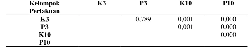
treatment (K10 and P10). The satistic result of extensive collagen mean percentage was presented on table 3. Table 4 showed p-value of post hoc test presenting differences on mean percentage of extensive collagen for each group on table 3. .
Table 3. Mean of percentage of collagen wide-spaced along burn healing
Group N Mean ± Std. Deviation (%)
Table 2. p-value of post hoc test on percentage of extensive collagen on burn injury
(a) (b)
(c) (d)
Figures 1. The histopatology of fibroblast number colored with HE on 10 µm scale, magnitude 400x. a) K3; b) P3; c) K10; d) P10. showed fibroblast number of P3 higher than K3 and P10 higher than K10.
(a) (b)
(c) (d)
DISCUSSIONS
Burn injury induces high morbidity and mortality that could change local and systemic body function, hence patient with burn injury should have special treatment since the early phase until advanced phase of wound care. The burn injury degree is based on wound depth, causative source, agent and
duration contact with calor[1]. Normal wound
healing is characterized by fibroblast
deposition of collagen type I and III[9].
Based on this study, Sangkuriang catfish skin extract topical treatment showed the number of fibroblast was enhanced in treated group, because it contains proline and
glycine. Glycine was used as natural
moisturizing factor on skin and induced collagen synthesi to accelerating burn
healing[3]. The results showed group P had
in inflammation phase, but 10th day of wound
healing in proliferation phase. Hence, Sangkuriang catfish skin extract could increase the number of fibroblast in extracellular matrix synthesis to accelerate wound healing. Proliferation phase happened
along 3-12 days post-injury[1]. Figure 1
showed that P3 has higher fibroblast number than K3 and P10 than K10.
Collagen is biomedically used to promote proliferation and migration of
fibroblast cell into wound area[7].
Collagen plays a key role in each phase of w
ound healing[9][10][11]. The collagen cleavage
products stimulate vascular
endothelial cell proliferation. These cells sec
rete a variety of GFs (VEGF, βFGF, PDGF),
which promote
angiogenesis[9]. Granulation is achieved by
vascularizing ECM. Collagen cleavage
products stimulate also keratinocyte
migration and proliferation[10].
Type I collagen from Clarias
batrachus has significantly enhancing adhesion of fibroblast on culture cell from
mice skin[7], it was used as
chemoattracttant[7][10]. Sangkuriang catfish
skin extract topical treatment on burn injury result showed the percentage of collagen on
P3 is wider than K3 but insignificant and P10 was wider than K10 statistically
significant. It was caused 3rd day of wound
healing during inflammation phase, but 10th
day of wound healing during proliferation phase. Glycine and proline in Sangkuriang catfish skin extract was enhancing deposition and increase of extensive collagen percentage in extracellular matrix. Therefore collagen
extract from Clarias batrachus skin could
generate decomposition of collagen and extracellular matrix around wound area, it could be developed as wound dressing or scaffold for tissue engineering, because it could accelerate burn healing by increasing
fibroblast number and percentage of
extensive collagen.
CONCLUSION
Topical treatment of Sangkuriang catfish skin extract could accelerate burn injury by increasing fibroblast number and
percentage of extensive collagen
significantly on 10th day of wound healing.
Acknowledgment
Integrated Laboratory University of Nahdlatul Ulama Surabaya, Pharmacology Laboratory and Biochemistry Laboratory Faculty Medicine, University of Airlangga. .
REFERENCES
1. Hidayat, Taufiq Sakti Noer. 2013. Peran
Topikal Ekstrak Gel Aloe Vera pada Penyembuhan Luka Bakar Derajat Dalam pada Tikus. Karya Akhir, Departemen/SMF Ilmu Bedah Plastik Rekonstruksi dan Estetik, Fakultas Kedokteran/RSUD Dr. Soetomo, Surabaya
2. Kementrian Kesehatan, 2013. Riset
Kesehatan Dasar: Riskesdas 2013. Badan Penelitian dan Pengembangan Kesehatan Kementrian Kesehatan Republik Indonesia.
3. Gaspar A, Moldovan L, Constantin D,
Stanciuc AM ,. Sarbu PMB, Efrimescu IC.
Collagen-Based Scaffolds for Skin Tissue Engineering, Journal of Medicine And Life
Vol. 4, No.2, 2011, April‐June, pp172‐177.
4. Yamamoto K, Igawa K, Sugimoto K,
Fish (Tilapia) Collagen. BioMed Research International, Volume 2014, 9 pages.
5. Zhang M, Liu W, Li G, 2009. Isolation and
characterization of collagen from the skin of
Largerfin Longbarbel Catfish
(Mystusmacropterus). Food Chemistry 115: 826-831.
6. Singh P, Benjakul S, Maqsood S,
Kishimura H. Isolation and Characterisation of Collagen Extracted from the Skin of
Striped Catfish (Pangasionodon
hipopthalmus). Food Chemistry; 2010: 124:
97105.
7. Leong LM, Sahalan AZ, Tan LH. Mustafa
NH. Rajab, NF. Clarias batrachus collagen
extract increases fibroblast cell adhesion, migration and proliferation. Journal of Applied Pharmaceutical Science ol. 5 (03), 2015, March, pp. 019-023
8. Puspitasari, D.A.P. Bintoro, V.P. Setiani ,
B.E. 2013. The Soaking Effect on Different Hydrocloride Acid Level and Soaking Time
on pH, Swelling Percentage and Collagen
Yield of Chicken Shank Bone. Faculty of
Animal and Agricultural Sciences,
Diponegoro University
9. Sinno H, Malhotra M, Lutfy J, Jardin B,
Winocour S, Brimo F, Beckman L, Watters K, Philip A, Williams B, Prakash S. The effects of topical collagen treatment on wound breaking strength and scar cosmesis in rats. Can J Plast surg 2012;20(3):181-185.
10. Brett D. A Review of Collagen and
Collagen-based Wound Dressings. Wound Management Division, Smith & Nephew Inc. 2008,December, Volume 20- Issue 12
11. Rao H, Pai A, Hussein I, Arun A, Ram HSS,
Pai A, Pai SR, Pain SG. A comparative study between collagen dressings and conventional dressings in wound healing.
International Journal of Collaborative