Redni broj ~lanka: 812 ISSN 1331-2820 (Tisak) ISSN 1848-7769 (Online)
Central nervous system tuberculosis with miliary
tuberculomas in a child: a case report
Tuberkuloza sredi{njeg `iv~anog sustava s milijarnim tuberkulomima u
djeteta: prikaz bolesnika
Maja VRDOLJAK1)
Tihana KNIEWALD1)
Ivona VLADU[I] LUCI]1)
Goran TE[OVI]1,2)
1)University Hospital for Infectious Diseases
"Dr. Fran Mihaljevi}", Pediatric Infectious Diseases Department, Zagreb, Croatia
2)University of Zagreb, School of Medicine,
Zagreb, Croatia Key words
tuberculosis
central nervous system intracranial tuberculomas
Klju~ne rije~i
tuberkuloza
sredi{nji `iv~ani sustav intrakranijalna tuberkuloza Primljeno:2017–02–20 Received:2017–02–20 Prihva}eno:2017–03–29 Accepted:2017–03–29 Case report Central nervous system tuberculosis is a rare form of disease that carries the high mortality and the risk of permanent neurologic sequelae if not recognized early. Clinical, laboratory and radiologic manifestations are usually nonspecif-ic, thus postponing establishment of correct diagnosis and initiation of treat-ment. We report a child with central nervous system tuberculosis, which mani-fested with nontypical laboratory findings, as well as with the rare form of mil-iary intracranial tuberculomas. Signs of active tuberculosis outside the central nervous system in combination with insidious course of disease raised a suspi-cion on tuberculosis. Therefore, antituberculous therapy was initiated promptly, and the patient recovered completely, without permanent neurologic sequelae.
Prikaz bolesnika Tuberkuloza sredi{njeg `iv~anog sustava je rijedak oblik bolesti s visokom smrt-nosti i rizikom trajnih neurolo{kih posljedica, ako se ne prepozna na vrijeme. Klini~ke, laboratorijske i radiolo{ke manifestacije su obi~no nespecifi~ne, {to odga|a dono{enje ispravne dijagnoze i po~etak lije~enja. U radu je prikazano dijete s tuberkulozom sredi{njeg `iv~anog sustava koja se manifestirala neti-pi~nim laboratorijskim nalazima i rijetkim milijarnim intrakranijskim tuberku-lomima. Znakovi aktivne tuberkuloze izvan sredi{njeg `iv~anog sustava u kom-binaciji s prikrivenim tijekom bolesti izazvali su sumnju na tuberkulozu. Anti-tuberkulozno lije~enje je odmah zapo~eto, a bolesnica se oporavila u potpuno-sti, bez trajnih neurolo{kih posljedica.
Introduction
Central nervous system (CNS) tuberculosis (TB) is an uncommon form of disease, which develops in around 4 % of children with TB, but remains the most devastating form of disease, carrying high mortality and a distressing level of neurological morbidity [1]. Since clinical, labora-tory and radiologic manifestations of CNS TB are nonspe-cific, early recognition of the disease is a challenge. However, it is of paramount importance because the clini-cal outcome depends greatly upon the stage at which ther-apy is initiated [2].
Here we report a case of CNS TB with rare form of miliary intracranial tuberculomas and discuss the possible clues for early diagnosis.
Case presentation
A 10-year-old girl presented with a 1-month history of dry cough, occasional low-grade fever, diarrhea, abdomi-nal pain and weight loss of 4 kg. She was examined few times at emergency department when no abnormalities in physical examination were noticed. Laboratory findings showed elevated erythrocyte sedimentation rate (65 – 85 mm/h), normal level of C-reactive protein, unremarkable complete blood count and mildly elevated transaminases (aspartate transaminase 85 IU/L and alanine transaminase 70 IU/L). In the fourth week of disease high temperature appeared, with drowsiness, headache and photophobia, so the girl was admitted to hospital.
She was previously healthy, regularly vaccinated as provided by the Croatian National Immunization Program.
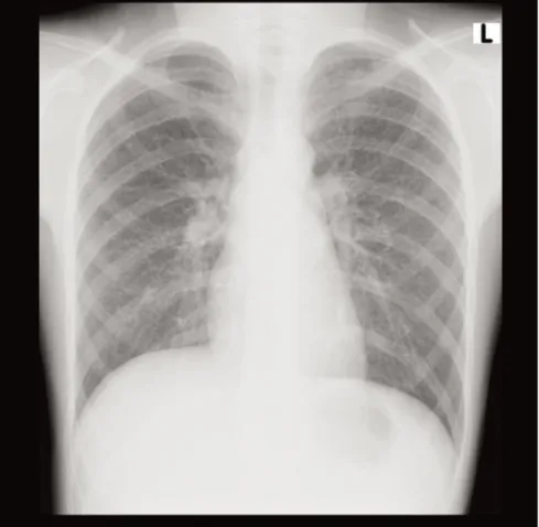
Figure 1.Initial chest x-ray shows diffuse interstitial nodular lesions
Slika 1. Inicijalni radiogram prsnog ko{a pokazuje difuzne intersticijske nodularne lezije
Figure 2.Brain MRI performed on hospital day 4 shows multiple tiny lesions most prominent in cerebellum, but also present in the cere-brum and brainstem
Slika 2. MR mozga u~injen 4. dan hospitalizacije pokazuje multiple sitne lezije najizra`enije u malom mozgu, koje su prisutne i u ve-likom mozgu i mo`danom deblu
Central nervous system tuberculosis with miliary tuberculomas in a child: ... M. VRDOLJAK et al.
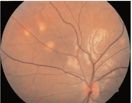
Figure 3.Initial funduscopic examination reveals disseminated chorioretinitis of both eyes Slika 3. Inicijalni pregled fundusa oka pokazuje diseminirani korioretinitis oba oka
Figure 4.Control chest x-ray 3 weeks after initiation of antituberculous therapy shows almost complete resolution of infiltrates Slika 4. Kontrolni radiogram prsnog ko{a nakon po~etka primjene antituberkulotske terapije pokazuje gotovo potpuni nestanak infiltrata
According to her mother's statement, the contacts were healthy, although we later found out the grandmother was in observation because of lung infiltrate with hilar lym-phadenopathy.
On admission neurologic examination revealed nuchal rigidity, but no focal neurologic deficits were detected. La-boratory tests showed similar findings as previously men-tioned. Because of the dry cough in anamnesis, chest x-ray was performed on admission and revealed bilateral nodu-lar infiltrates, highly suggestive of miliary TB (Figure 1).
Lumbar puncture demonstrated moderate pleocytosis of 270 white cells/μL (neutrophils 55 %), mildly elevated protein level (0.83 g/L) and cerebrospinal fluid (CSF)/ serum glucose ratio of 0.35. The patient was immediately started on antituberculous treatment with isoniazid, ri-fampicin, ethambutol and pyrazinamide. Dexamethasone was also added as adjunctive therapy. Lumbar puncture was repeated three days later, when antituberculous thera-py was already initiated. Acid-fast staining, culture and polymerase chain reaction (PCR) for Mycobacterium tu-berculosisremained negative from both CSF samples. The magnetic resonance imaging (MRI) of brain demonstrated multiple, contrast enhanced tiny nodules in cerebellum, cerebrum and brain stem, without signs of basal meningi-tis. This finding highly suggested the miliary pattern of brain tuberculomas (Figure 2).
Active searching for other signs of active TB outside the CNS revealed disseminated chorioretinitis of both eyes (Figure 3).
Our struggle to detect mycobacteria from other sam-ples beside the CSF (i.e. bronchoalveolar lavage fluid, urine, stool) unfortunately wasn't successful. Only blood IFN-γreleasing assay (IGRA) came positive. Although eti-ologic diagnosis wasn't confirmed, there was significant clinical and radiologic improvement on treatment (Figures 4, 5). Repeated lumbar puncture also showed improve-ment of CSF analysis.
HIV test, as well as immunologic screening for possi-ble immunodeficiencies were negative.
The patient was treated with 4-drug regimen men-tioned above for two months, followed by ten months of isoniazid and rifampicin. Dexamethasone was added to therapy for ten weeks. No toxic side effects of therapy were noticed.
Chest x-ray showed complete resolution of nodules two months after initiation of therapy, and brain MRI per-formed six months after admission to hospital demonstrat-ed no pathology. On follow-up one year later, the patient was in good clinical condition and no neurological seque-lae were reported.
Figure 5.Brain MRI reveals significant reduction of the miliary tuberculomas 1 month after initiation of antituberculous therapy Slika 5. MR mozga pokazuje zna~ajnu redukciju milijarnih tuberkuloma mjesec dana nakon po~etka antituberkulotskog lije~enja
Central nervous system tuberculosis with miliary tuberculomas in a child: ...
Discussion
With an overall mortality rate of almost 15 % and per-manent neurological sequelae occurring in around half of patients, CNS TB remains a devastating infection in child-hood [3].
Early recognition of CNS TB is difficult because clini-cal and laboratory manifestations can mimic many infec-tious and noninfecinfec-tious diseases. Our patient presented with 1-month history of occasional low-grade fever with abdominal pain, diarrhea, cough and weight loss. Labora-tory findings of elevated erythrocyte sedimentation rate and slightly elevated transaminases were also nonspecific. Children with CNS TB often initially present with ab-dominal symptoms such as nausea and vomiting. The dis-ease is typically subacute and progresses through three phases: the prodromal, meningitic and paralytic phase. The prodromal phase, lasting two to three weeks, is char-acterized by nonspecific symptoms such as malaise, lassi-tude, headache or low-grade fever. The meningitic phase follows with more pronounced neurologic features, such as meningismus, protracted headache, vomiting, lethargy, confusion, and cranial nerve and long-tract signs. The par-alytic phase is characterized by confusion which progress-es to stupor and coma, seizurprogress-es, and often hemiparprogress-esis [3, 4].
The diagnosis of CNS TB was suspected in our patient in meningitic phase, having in mind one-month history of nonspecific symptoms and finding of small nodules on chest x-ray performed on admission.
It's important to emphasize that abnormalities on chest radiograph may be seen only in half of CNS TB cases, ranging from focal lesions to a subtle miliary pattern [5]. Other sighs of active TB outside the CNS, such as choroid-al tubercles, as seen in our patient, are of diagnostic import if present.
Although the typical analysis of CSF from patients with CNS TB demonstrates lymphocytic pleocytosis, low glucose level and elevated protein level, there are many atypical presentations with normal protein or glucose level or normal white blood cell count. There even may be a pre-dominance of polymorphonuclear cells rather than lym-phocytes in the CSF [6]. Similarly, CSF analysis of our pa-tient wasn't typical for TB: mild pleocytosis with polymor-phonuclear cell predominance (55 %), only slightly elevat-ed protein level (0.83 g/L) and relative hypoglycorachia (1.7 mmol/L or 35 % of blood glucose level).
Definitive diagnosis of CNS TB is based on the detec-tion of the tubercle bacilli in the CSF, either by smear ex-amination or by bacterial culture. However, the demon-stration of mycobacteria in the CNS is not always possible. It is known that the sensitivity of acid-fast staining and TB
culture of CSF is directly dependent on the number and volume of CSF samples examined. That's why repeat lum-bar puncture, meticulous microscopy and culture of a large volume (> 6 mL) of CSF are recommended [7]. Unfortuna-tely, mycobacteria weren't confirmed in CSF samples of our patient, probably because one sample was taken on ad-mission, and second three days later, when therapy was al-ready initiated.
Because of the limitations of conventional tests, the commercial nucleic acid amplification test and other PCR-based assays have emerged as an alternative diagnostic tool. The Xpert MTB/RIF used in this case is reported to have sensitivity of almost 60 % in the diagnosis of CNS TB [8].
We used blood IGRA as a supportive method for diag-nosing CNS TB, although it is reported to have limited di-agnostic value and negative results cannot entirely exclude the disease. As with the tuberculin skin test, IGRAs cannot distinguish latent tuberculosis infection from active dis-ease. The sensitivity of IGRA assays in patients with mil-iary tuberculosis is reported to be 68 – 93 % [9, 10].
CNS TB has various imaging appearances, including meningitis, tuberculoma, miliary tuberculosis, abscess, cerebritis and encephalopathy. Although meningitis is the most common radiologic manifestation of CNS TB, seen most frequently in the children and adolescents [11], our patient's brain MRI didn't show signs of meningeal en-hancement or other radiologic manifestations of possible TB meningitis complications (progressive hydrocephalus, vasculitis, infarction or cranial neuropathies) [12]. On the contrary, MRI showed contrast-enhanced tiny lesions in cerebrum, cerebellum and brainstem. This imaging sug-gested the miliary pattern of intracranial tuberculomas, rare parenchymal form of CNS TB, seen mostly in severe-ly immunocompromised patients [13]. These lesions are usually located at the corticomedullary junctions, are tiny (2 – 3 mm in diameter) and may be invisible on noncon-trast MR sequenced. Postconnoncon-trast, T1-weighted MR im-ages reveal numerous, round, small, homogeneous, en-hancing (usually ring enhancement) lesions [14].
Since our patient raised a high index of suspicion on CNS TB, empiric antituberculous therapy with 4-drug reg-imen was promptly initiated, with significant resolution of symptoms and improvement of chest x-ray and brain MRI. Dexamethasone was also added to therapy regimen as ad-junctive therapy. Because of the relative rarity of TBM and the difficulty of early diagnosis no randomized controlled trial has established an optimal regimen and course for CNS TB. Nevertheless, our patient was treated with an ini-tial 2-month induction therapy regimen including isoni-azid, rifampicin, pyrazinamide and ethambutol, followed by 10 additional months of isoniazid and rifampicin as
maintenance therapy, as suggested by most guidelines [15]. The use of steroid therapy as an adjunctive therapy is also approved by guidelines, and the mechanism for sur-vival benefit may come from reducing hydrocephalus and preventing infarction [16].
Our patient recovered completely, without neurologic sequelae. One of the reasons is early recognition and prompt initiation of treatment. Namely, the overall mortal-ity from CNS TB may be greater than 50 % in those pa-tients recognized late in the course of disease. A large pro-portion of these patients also suffer from severe sequelae [3]. The other reason for good outcome is probably Bacillus Calmette-Guérin (BCG) vaccine, which is a part of national immunization program in Croatia and is given to all newborns. BCG is thought to be 52 – 86 % protective against developing the severe complications of TB such as miliary TB and CNS TB [17], but in cases of CNS TB it protects against a fatal outcome and severe sequelae [3].
In conclusion, we reported a case of CNS TB with rare form of miliary intracranial tuberculomas. Although the etiology wasn't confirmed since the CSF cultures and acid-fast stain remained negative, clinical presentation, epi-demiological history, signs of TB outside the CNS and good clinical and radiologic response to antituberculous treatment indicated the diagnosis.
CNS TB continues to be a condition which carries sig-nificant morbidity and mortality. Early diagnosis and prompt initiation of treatment are essential to improve the poor outcome.
References
[1] Rock RB, Olin M, Baker CA, Molitor TW, Peterson PK. Central Nervous System Tuberculosis: Pathogenesis and Clinical Aspects. Clin Microbiol Rev 2008;21:243–261.
[2] Thwaites GE, Schoeman JF. Update on tuberculosis of the central nervous system: pathogenesis, diagnosis, and treatment. Clin Chest Med 2009;30:745–754.
[3] Farinha NJ, Razali KA, Holzel H, Morgan G, Novelli VM. Tuberculosis of the central nervous system in children: a 20-year survey. J Infect 2000;41:61–68.
[4] Kennedy DH, Fallon RJ. Tuberculous meningitis. JAMA 1979; 241:264–268.
[5] SütlaºPN, Unal A, Forta H, Senol S, KirbaºD. Tuberculous meningitis in adults: review of 61 cases. Infection 2003;31: 387–391.
[6] Klein NC, Damsker B, Hirschman SZ. Mycobacterial meningitis. Retrospective analysis from 1970 to 1983. Am J Med 1985;79:29– 34.
[7] Thwaites GE, Chau TT, Farrar JJ. Improving the bacteriological di-agnosis of tuberculous meningitis. J Clin Microbiol 2004;42: 378–379.
[8] Nhu NT, Heemskerk D, Thu do DA, Chau TT, Mai NT, Nghia HD, et al. Evaluation of GeneXpert MTB/RIF for diagnosis of tubercu-lous meningitis. J Clin Microbiol 2014;52:226–233.
[9] Lee YM, Park KH, Kim SM, Park SJ, Lee SO, Choi SH, et al. Diagnostic usefulness of a T-cell-based assay in patients with mil-iary tuberculosis compared with those with lymph node tuberculo-sis. Clin Infect Dis 2013;56:e26–29.
[10] Kim CH, Lim JK, Yoo SS, Lee SY, Cha SI, Park JY, Lee J. Diagnostic performance of the QuantiFERON-TB Gold In-Tube assay and factors associated with nonpositive results in patients with miliary tuberculosis. Clin Infect Dis. 2014;58:986–989.
[11] Sanei Taheri M, Karimi MA, Haghighatkhah H, Pourghorban R, Samadian M, Delavar Kasmaei H. Central nervous system tuber-culosis: an imaging-focused review of a reemerging disease. Radiol Res Pract 2015;2015:202806.
[12] Chan KH, Cheung RT, Fong CY, Tsang KL, Mak W, Ho SL. Clinical relevance of hydrocephalus as a presenting feature of tu-berculous meningitis. QJM 2003;96:643–648.
[13] Krishnan N, Robertson BD, Thwaites G. The mechanisms and con-sequences of the extra-pulmonary dissemination of Mycobacteri-um tuberculosis. Tuberculosis (Edinb) 2010;90:361–366.
[14] Trivedi R, Saksena S, Gupta RK. Magnetic resonance imaging in central nervous system tuberculosis. Indian J Radiol Imaging 2009;19:256–265.
[15] Thwaites G, Fisher M, Hemingway C, Scott G, Solomon T, Innes J; British Infection Society. British Infection Society guidelines for the diagnosis and treatment of tuberculosis of the central nervous system in adults and children. J Infect 2009;59:167–187.
[16] Thwaites GE, Macmullen-Price J, Tran TH, Pham PM, Nguyen TD, Simmons CP, et al. Serial MRI to determine the effect of dex-amethasone on the cerebral pathology of tuberculous meningitis: an observational study. Lancet Neurol. 2007;6:230–236.
[17] Rodrigues LC, Diwan VK, Wheeler JG. Protective effect of BCG against tuberculous meningitis and miliary tuberculosis: a meta-analysis. Int J Epidemiol. 1993;22:1154–1158.