Color sensitive retina based on bacteriorhodopsin
Michael Frydrych
a, Pertti Silfsten
b, Sinikka Parkkinen
c, Jussi Parkkinen
d,*,
Timo Jaaskelainen
eaDepartment of Information Technology,Lappeenranta Uni6ersity of Technology,Lappeenranta,Finland bDepartment of Electrical Engineering,Lappeenranta Uni6ersity of Technology,Lappeenranta,Finland
cDepartment of Biology,Uni6ersity of Joensuu,Joensuu,Finland
dDepartment of Computer Science,Uni6ersity of Joensuu,PO Box111,80101Joensuu,Finland eVa¨isa¨a¨la¨ Laboratory,Uni6ersity of Joensuu,Joensuu,Finland
Received 5 January 1999; received in revised form 8 October 1999; accepted 15 October 1999
Abstract
Bacteriorhodopsin (BR), a membrane protein of a microorganismHalobacterium salinariumhas been studied since the 80’s as a potential material for information technology. The information processing applications of BR employ either photochromic or photoelectric properties of the protein. In this study we discuss about design principles and describe our study of the use of bacteriorhodopsin as a sensor material for a color sensitive artificial retina. This retina includes low-level processing of input information. The design of a color sensitive matrix element, the self-organizing color adaptation algorithm and a system model for the retina are presented. © 2000 Elsevier Science Ireland Ltd. All rights reserved.
Keywords:Artificial retina; Bacteriorhodopsin; Color space; Color sensor; Vision system
www.elsevier.com/locate/biosystems
1. Introduction
Although all parts of the eye are important for perceiving a good image, the most vital one is the retina. The retina is a part of a brain tissue, which directly interacts with the light and images of the outside. It is the only part of the brain that is visible from outside the skull. The retina contains photoreceptors and five layered neural structure, which is claimed to have some capability of
sim-ple processing of visual information (Dowling, 1987). Even a simple two-layer neural network is capable of low level image processing (Mantere et al., 1992).
The whole human visual system includes also the lateral geniculate nucleus, ‘a relay station’ in the thalamus and the visual cortex. Actually, the image is processed not only on one cortical re-gion, but on a network of over 20 cortical areas (Kosslyn, 1996). The exact meaning of each com-ponent for visual processing is not known, but certain low level processing is done in the retina and a high level conceptual processing in the cortical areas (Zeki, 1993). Like other neural or-* Corresponding author. Tel.: +358-13-2513106; fax: +
358-13-2513290.
E-mail address:[email protected] (J. Parkkinen)
ganism, the visual system is modified through learning, mainly during early years of infancy (Teller, 1997).
An important attribute of human visual system is color. The sense of color originates from re-sponses of three types of color sensitive photore-ceptors in the retina, cones, to a spectrum of electromagnetic radiation originated from an ob-ject observed (Wandell, 1995). Several distinct codings of color exist in the visual system. The triplets of cone responses can be regarded as the initial one. The receptor responses are trans-formed into achromatic and color-opponent sig-nals (Buchsbaum and Gottschalk, 1983). This transformation occurs in the retina and is retained until the early cortical areas. These broadband signals are afterwards transformed to narrow-band responses of cells at the higher cortical areas, namely area V4 (De Valois and De Valois, 1993; Kosslyn, 1996).
The human visual system has been a common source of inspiration in machine vision research (Marr, 1982; Jain, 1989). For more than the last 10 years, many sensors, which include both the photosensors and parallel processing elements have been reported. Mainly these are silicon based special circuits, often called vision chips, smart sensors or artificial retinas (Moini, 1997). There are also some constructions of bacteriorhodopsin-based detectors (Miyasaka et al., 1992; Chen and Birge, 1993; Martin et al., 1997). These detectors use a photoelectric property of bacteriorhodopsin (BR). Miyasaka et al. (1992) constructed BR-based detector in a matrix form. They have immo-bilized BR on a silicon circuit by the Langmuir – Blodgett method, forming a matrix of size 8×8 pixels. They used S-9 strain ofHalobac
-terium salinariumwith the maximum photocurrent
at the wavelength 560 nm, with the width at half maximum being about 100 nm. Therefore, it can be used as a monochromatic detector.
We have developed a model for a color sensi-tive artificial retina, in which photosensisensi-tive ele-ments are based on BR. This retina includes low-level processing of input information. The processing consist in color space transformation. In order for the retina to operate in similar fash-ion as the natural system does, we employ an
artificial neural network learning algorithm, to tune color space transformation parameters for the environment in which the retina operates. In this report, we present the design of a color sensitive matrix element, the learning algorithm and the system model for the color sensitive, protein based, artificial retina.
2. Bacteriorhodopsin
BR is produced by a microorganismH.salinar -ium as a membrane protein. BR is composed of the protein part bound to the chromophore (reti-nal) with Schiff base linkage. Natural function of BR is to act as a light-driven proton pump, converting sunlight into chemical or electric en-ergy (Brauchle et al., 1991; Hampp et al., 1992). The proton pumping is coupled with photochemi-cal conversions, occurring during a photocycle of BR (Lozier et al., 1975) (Fig. 1).
The photocycle has intermediates with different lifetimes and absorption spectra. The transition times vary from ps to ms. In the dark, an equimo-lar mixture of the D and B-state is found which is called dark-adapted BR. When illuminated, a pure population of the B-state occurs, called light-adapted BR. The B-state has a broad absorption band with maximum near 570 nm. Absorption of a photon, causes protein to move from the B-state, through a number of intermediates into the M-state. The M-state has the absorption maxi-mum at 412 nm and it has the longest lifetime of all the excited states. The transition from the B to the M-state takes :50ms, during which a proton
is released from the Schiff base. From the M-state protein it thermally decays in :10 ms back to the
initial B-state. During the transition the Schiff base is reprotonated. The transition from the M to the B-state can be also triggered directly by illumination with blue light (Hampp et al., 1992). The proton release and acceptance, during switching to and from the M-state, causes charge dislocation within bacteria membrane, or an ori-ented protein film (Birge, 1990; Oesterhelt et al., 1991). This photoelectric property is a base for construction of optoelectric devices.
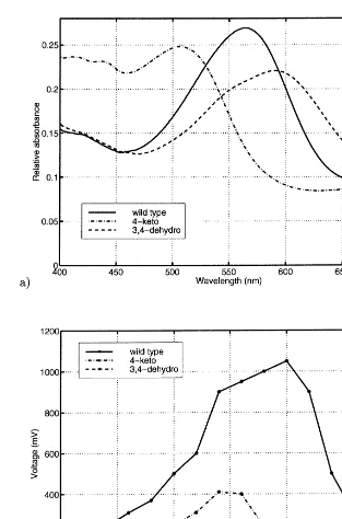
If we use only wild type BR, the sensor has certain wavelength dependency and only different levels of luminosity can be sensed, but not color. The wavelength response can be changed by mod-ifying the BR biotechnically. In our earlier work (Silfsten et al., 1996) we have studied two vari-ants: 4-keto and 3,4-dehydro. As it has been shown also by the others (Drachev et al., 1989; Beischel et al., 1991), both variants retained op-toelectric activity. The measurements have also shown that the optoelectric response with respect to wavelength was different among the protein variants. The differences allow color recognition with BR. Absorption spectra and optoelectric re-sponses of natural form of BR and the two vari-ants are drawn in Fig. 2.
3. The model of the system
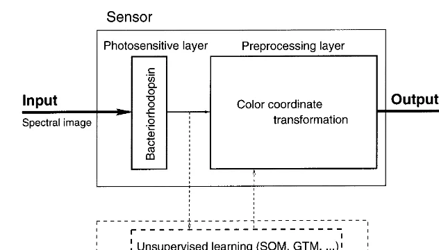
The model of the color sensitive artificial retina is outlined in Fig. 3. The retina consists of two
layers, a photosensitive layer and a preprocessing layer. The input to the retina is an image. The image first interacts with the photosensitive layer, which converts the image into electrical signals. The signals are passed for further transformations to the preprocessing layer. The output of the retina is communicated to the processor.
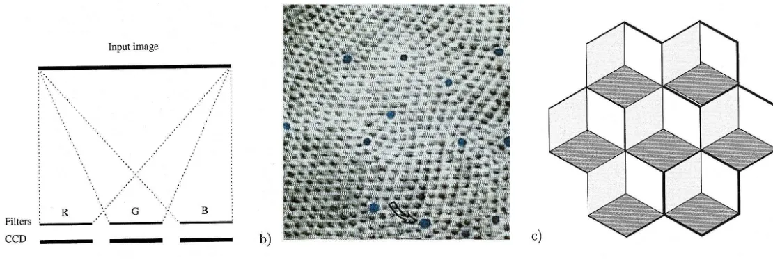
The photosensitive layer consists of one or more arrays of photosensitive elements. The ele-ments can be ordinary CCD eleele-ments, cones in the retina, patches of BR film, or any other suitable material. There is no restriction on a geometrical arrangement of the elements either. Fig. 4 shows some examples of photosensitive layers the model accommodates. An output of the preprocessing layer is an array of n-tuples. The value ofn depends both on physical arrange-ment of elearrange-ments within the layer and on number of those layers (RGB camera is a sensor with n=3).
The preprocessing may include, e.g. simple fea-ture extraction, as edge detection, simple image processing or color coordinate transforma-tions. The preprocessing layer can also do just identity mapping. That in practice, corre-sponds to the case, when the preprocessing layer is missing.
The preprocessing layer can operate in two modes: off and on-line mode. The purpose of off-line or learning mode is to adjust in-ternal parameters of the preprocessing layer, ac-cording to the information about the environ-ment. In on-line mode, the preprocessing layer recodes the output of photosensors according to the parameters adjusted during the learning phase.
4. The constructed system
Fig. 2. Absorption spectra (a) and optoelectric responses; (b) of natural form of BR and 4-keto and 3,4-dehydro variants.
4.1. Photosensiti6e layer
BR in the photosensitive layer was used in a form of purple membrane isolated from H.
salinarium wild type (S9). The membrane was
isolated as described by Oesterhelt and
Fig. 3. The model of the artificial retina based on bacteriorhodopsin.
Polyvinylalcohol (PVA) films were prepared by mixing 750 ml of 15% PVA with 200 ml of BR solution and spread onto a conductive glass substrate. After drying for 24 h, a gold layer of about 40 nm thickness was sputtered on the PVA film to form counter electrode for the conductive glass. Thin wire was connected to the corner of the gold layer by silver paint, to form an electric connection from the gold layer (Fig. 5). The element system containing altogether six such elements was made (Fig. 6). There are three pairs of elements in the system, each pair containing one of the three proteins. All elements have the same size, about 18×17 mm.
Color is represented and communicated using color space. Most of color research and standard color representation is based on some three di-mensional color space (Wyszecki and Stiles, 1982), as CIEXYZ and its linear transformations. This is due to the fact that the human eye have three different classes of color sensitive cells in the retina.
Color representations based on CIEXYZ color space are closely related to the trichromatic hu-man visual system and their use is limited to applications related to human vision. Other living species may have much different color vision. For example, vision of most mammals is dichromatic. Old world primates (Catarrhini), including man,
has evolved trichromatic color vision, but the spectral distribution of the cone-pigment sensitiv-ity peaks differs among subspecies (Osorio and Bossomaier, 1992). Visual pigments can also cover a part of spectrum, which is not visible to hu-mans, as for example in the honey bee (Backhaus, 1993). Another disadvantage of standard CIE color spaces is that they define a fixed representa-tion of color.
The preprocessing layer in the constructed retina, has been designed to learn the color space which makes the retina adaptable to an environ-ment. The learning is done by training the self-or-ganizing map (SOM) with the output of photosensitive layer. Hence, we have a model of an independent color vision, which has protein sensors and which forms its ‘visible’ color space by a self-organization process from shown color samples.
M
.
Frydrych
et
al
.
/
BioSystems
54
(2000)
131
–
140
Fig. 5. The scheme of the BR element.
5. Results
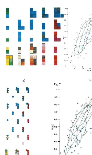
The constructed system has been tested by training it using a collection of 84 plastic filters having different colors. These filters were ‘shown’ to the constructed retina by setting them between the retina and a flash light. A triplet of responses, one for each type of BR, was recorded for each filter (Frydrych et al., 1998). This set of triplets was used for training the SOM. Two SOM were trained, one with rectangular grid of reference vectors and the other one with hexagonal grid, respectively. Both grids consisted of 5×5 vectors. The maps were trained using 10 000 iterations.
Figs. 7 and 8 show the color spaces formed by training the SOM with responses of photosensi-tive layer. Fig. 7a and Fig. 8a show the learned color spaces. Each cluster of color patches are the colors assigned to one reference vector in the map after the training has finished. Fig. 7b and Fig. 8b show in 3-D data space the training data vectors (responses of photosensitive layer) and the self-or-ganizing MAP embedded into the data space. The data vectors are denoted by stars. The reference vectors are the vertices of the grid, lines connect-ing them symbolize the grid connections. It can be seen from Fig. 7a and Fig. 8a, that the colors have been sensibly organized. The similar colors have been clustered to occupy separate regions of the color space.
6. Conclusions
We have built a simple artificial retina using BR protein and its two artificial variants. The particu-lar set of color lights may be regarded as the representation of the environment in which the retina operates. We have given an example of color spaces learned for one such an environment, the artificial environment created using a collec-tion of plastic filters. The color spaces learned with SOM will differ for other environments (Frydrych, 1998).
The number of different proteins in the system is, of course, not limited to three. One can build a retina containing four or more proteins, their actual number will depend on an application and availability of particular proteins.
training data. During training the reference vectors tend to become ordered so that they span the data space. The mapping of the data vectors onto the reference vectors induce a new coordinate system into the data space given by the grid arrangement of the reference vectors.
In the constructed retina, the original signal space is a space of spectral transmittance func-tions of colored lights. Actually, this is a spaceL1
of continuous, Lebesgue integrable functions. The signal space is transformed in the BR-array of the retina into a three-dimensional space of triplets of responses of BR to the transmittance functions. This triplets constitute the data space for SOM. The grid of reference vectors in SOM, represents the learned color space, which the color sensor is adapted into.
The training of the SOM is the off-line mode of operation of the preprocessing layer. After the training has finished the learned reference vectors are stored for use in the on-line mode. While in the on-line mode, the retina is presented with arbitrary colors and it responds with their color coordinates in the learned color space.
The spatial resolution of our system shall be improved, but in this study we have showed that it is possible to produce an artificial color sensi-tive retina based on protein sensors. Compared to the previous study (Miyasaka et al., 1992) our retina is much easier to produce and it is sensitive to color.
The artificial retina based on BR combined with a color space defined using an artificial neu-ral network demonstrates the system that learns colors and adapts to an environment. Because of an ability to learn and use of biological material the system can be thought as an intelligent color sensitive artificial retina.
Acknowledgements
The authors thank to Prof. D. Oesterhelt for providing bacterial strains of H. salinarium wild type S9 and to Dr A. Khodonov for providing retinal analogs. The study was financially sup-ported by the technology development center of Finland — TEKES and the Academy of Finland.
References
Backhaus, W., 1993. Color vision and color choice behavior of the honey bee. Apidologie 24, 309 – 331.
Beischel, C.J., Mani V., Govinjee R., Ebrey T.G., Knapp D.R., Crouch R.K., 1991. Ring oxidized retinals form unusual bacteriorhodopsin analogue pigments. Photochem. Photobiol. 977 – 983.
Birge, R.R., 1990. Photophysics and molecular electronic ap-plications of the rhodopsins. Annul. Rev. Phys. Chem. 41, 683 – 733.
Brauchle, C., Hampp, N., Oesterhelt, D., 1991. Optical appli-cations of bacteriorhodopsin and its mutated variants. Adv. Mater. 3, 420 – 428.
Buchsbaum, G., Gottschalk, A., 1983. Trichromacy, opponent colours coding and optimum colour information transmis-sion in the retina. Proc. Roy. Soc. Lond. B. Bio. 220, 89 – 113.
Chen, Z., Birge, R.R., 1993. Protein-based artificial retinas. Trends. Biotechnol. 11, 292 – 323.
De Valois, R.L., De Valois, K.K., 1993. A multi-stage color model. Vision Res. 33 (8), 1053 – 1065.
Dowling, J.E., 1987. The Retina: An Approachable Part Of The Brain. The Belknap Press, Harvard University Press,
Cambridge, Massachusetts.
Drachev, L.A., Drachev, A.L., Chekulaeva, L.N., 1989. An investigation of the electrochemical cycle of bacteri-orhodopsin analogs with the modified ring. Archiv. Biochem. Biophys. 184 – 197.
Frydrych, N.I. 1998. Color spaces for artificial sensors. In: Proc. Finnish Conference on Artificial Intelligence-STeP ’98, Jyvaskyla, Finland, 144 – 151.
Frydrych, N.I., Parkkinen, J., Parkkinen, S., Silfsten, P., Jaaskelainen, T., 1998. Color recognition with bacteri-orhodopsin. In: Proceedings of the Pacific Symposium on Biocomputing ‘98, Maui, Hawaii, USA, 523 – 534. Hampp, N., Thoma, R., Brauchle, C., et al., 1992.
Bacteri-orhodopsin variants for optical information processing: A new approach in material science. In AIP Conference Proceedings, pp. 181 – 190.
Jain, A.K., 1989. Fundamentals of digital image processing. Prentice Hall, Englewood Cliffs, USA.
Kohonen, T., 1982. Self-organizing formation of topologically correct feature maps. Biol. Cybern. 43 (1), 59 – 69. Kohonen, T., 1995. Self-Organizing Maps. Springer,
Heidelberg.
Kosslyn, S.M., 1996. Image and Brain: The Resolation of the Imagery Debate. A Bradford Book, first paperback edi-tion. The MIT Press, Cambridge, Massachusetts; London, England.
Lozier, R.H., Bogomolni, R.A., Stoeckenius, W., 1975. Bacte-riorhodopsin: A light-driven proton pump in Halobac
-terium halobium. Biophys. J. 15, 955 – 962.
Mantere, K., Parkkinen, J., Jaaskelainen, T., Gupta, M.M., 1992. Wilson – Cowan neural network model in image pro-cessing. J. Math. Imag. Vision 2, 251 – 259.
Marr, D., 1982. Vision: A Computational Investigation into the Human Representation and Processing of Visual Infor-mation. H. Freeman and Company, San Francisco. Martin, C.H., Chen, Z.P., Birge, R.R., 1997. Towards a
bacteriorhodopsin-silicon neuromorphic photosensor. In: Proceedings of the First Pacific Symposiumn, Biocomputing.
Miyasaka, T., Koyama, K., Itoh, I., 1992. Quantum conver-sion and image detection by a bacteriorhodpsin-based ar-tificial photoreceptor. Science 255, 342 – 344.
Moini, A., 1997.Vision chips or seeing silicon. Technical re-port, Centre for High Performance Integrated Technolo-gies and Systems, The University of Adelaide. Available also from WWW atBhttp://www.eleceng.adelaide.edu.au/
Groups/GAAS/Bugeye/visionchips/\.
Oesterhelt, D., Brauchle, C., Hampp, N., 1991. Bacteri-orhodopsin: a biological material for information process-ing. Q. Rev. Biophys. 24 (4), 425 – 478.
Oesterhelt, D., Stoeckenius, W., 1974. Isolation of the cell membrane ofHalobacterinm halobiumand its fractionation into red and purple membrane. Methods Enzymol. 667 – 686.
Silfsten, P., Parkkinen, S., Luostarinen, J., et al., 1996. Color sensitive biosensors for imaging. In: Proc. 11th International Conference on Pattern Recognition, ICPR’96 Vol. III, pp. 331 – 335.
Teller, D.Y., 1997. The development of colour vision in infants. In: Dickinson, C., Murray, I., Carden, D. (Eds.), John Dalton’s Colour Vision Legacy. UMIST, Department of
Optometry and Vision Sciences, Taylor and Francis, Manchester, UK, pp. 217 – 224.
Wandell, B.A., 1995. Foundations of Vision. Sinauer Associates Inc, Sunderland, Massachusetts.
Wyszecki, G., Stiles, W.S., 1982. Color Science, 2nd edition. Wiley, London.
Zeki, S., 1993. A Vision of the Brain. Blackwell Scientific Publications, Osney Mead, Oxford.