Indo. J. Chem., 2007, 7 (3), 342-345
A XANTHONE FROM THE STEM BARK OF MANGGIS HUTAN (
Garcinia bancana
)
Muharni
1,*, Dachriyanus
2, Husein H. Bahti
3and Supriyatna
41
Department of Chemistry Faculty of Mathematics and Natural Sciences, Sriwijaya University, Palembang, Indonesia
2Department of Pharmacy, Faculty of Mathematics and Natural sciences,
Andalas University, Padang, Indonesia
3Department of Chemistry Faculty of Mathematics and Natural Sciences,
Padjadjaran University, Bandung, Indonesia 4
Faculty of Pharmacy, Padjadjaran University, Bandung, Indonesia
Received 13 August 2007; Accepted 14 September 2007
ABSTRACT
A xanthone, 1,5-dihidroxy-3,6-dimethoxy-2,7-bis-(3-methylbutenyl)xanthone had been isolated from the stem bark ofGarcinia bancana Miq. The structure of this compound was elucidated by analysis of spectroscopic data, especially using 1D and 2D NMR spectroscopic data.
Keywords : Xanthone, 1,5-dihidroxy-3,6-dimethoxy-2,7-bis-(3-methylbutenyl)xanthone, G. bancana
INTRODUCTION
Garcinia is the most important genus of the Guttiferae family, widely distributed in tropical Asia, Africa, and Polynesia [1]. The genus is known to be rich in oxygenated xanthone [2], prenylated xanthone [3], and polyisoprenylated benzophenones [4,5]. Some of them exhibit various biological activities such as antimicrobacterial, antioxidant, cytotoxic and anti malaria activities [6-9]. Garcinia bancana Miq. is distributed throughout Southern Thailand, Malaysia and Indonesia. In our continuing phytochemical investigation of Garcinia plants found in Indonesia, we have examined the stem bark of G. bancana. This plant is locally named manggis hutan [1,10]. In this paper we described the isolation and structure elucidation of compound ( 1,5-dihidroxy-3,6-dimethoxy-2,7-bis-(3-methyl-butenyl)xanthone, from the methanol extract of the stem bark of G. bancana. This is the first report on the isolation of compound from this plant. The structure of this compound was determined based on their UV, IR, and NMR 1D and 2D spectroscopic data.
EXPERIMENTAL SECTION
General Experimental Procedure
UV and IR spectra were measured with spectrophotometers Beckman DU-700 and Shimadzu FTIR 8400. 1H and 13C NMR spectra was recorded performed on precoated Si Gel plates (Merck Kiesel gel 60 GF254, 0.25 mm 20 x 20 cm.
Plant material
Sample of the stem bark of G. bancana was collected on April 2006 from the Sarasah Bonta Payah kumbuh, Sumatera Barat. The plant was identified by the staff at the Herbarium Anda, Andalas University, Padang and a voucher specimen had been deposited at the herbarium.
Extraction and isolation
The powdered of the stem bark G .bancana (3 Kg) was extracted by maceration technique three times with hexane, dichloromethane and methanol respectively for 5 days at room temperature. Evaporation of each extract (n-hexane, dichloro-methane and methanol) to dryness in vacuo, afford
Fig 1. Structure of
Indo. J. Chem., 2007, 7 (3), 342-345 vacum liquid chromatography eluted with a gradient system (n-hexana, n-hexana: EtOAc = 9:1; 8:2; 7:3; 6:4 and EtOAc) to afford 5 fractions A1 – A5. Fraction 2 was further purified column chromatography (eluted with gradient system n-hexana, n-hexana : EtOAc = 9:1; 8:2; 7:3 and EtOAc) to afford 4 subfraction. Subfraction 2 after purification with recrystalization gave a pure compound (7 mg).
This compound obtained as a yellow crystal, UV: λmax absorption at 261, 312 and 368 nm. IR (KBr) Vmaks
cm-1 3444 (OH), 2962, 2920 (C-H alifatic), 1651( carbonil chelating), 1600, 1569 dan 1465 cm-1 (benzena
HMBC dan H-H COSY spectrum see Table 2.
RESULT AND DISCUSSION
The methanol extract of the stem bark of G. bancana was subjected to chromatographic purification to afford one xanthone compound. The structure was elucidating using 1D and 2D NMR spectroscopic data. The 13C NMR signals were assigned from DEPT,
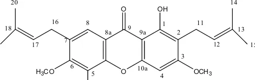
Table 2. NMR correlations data for
Indo. J. Chem., 2007, 7 (3), 342-345
Muharni, et al.
344
2
11
12
13 14
15
6 16 17 18
19 20
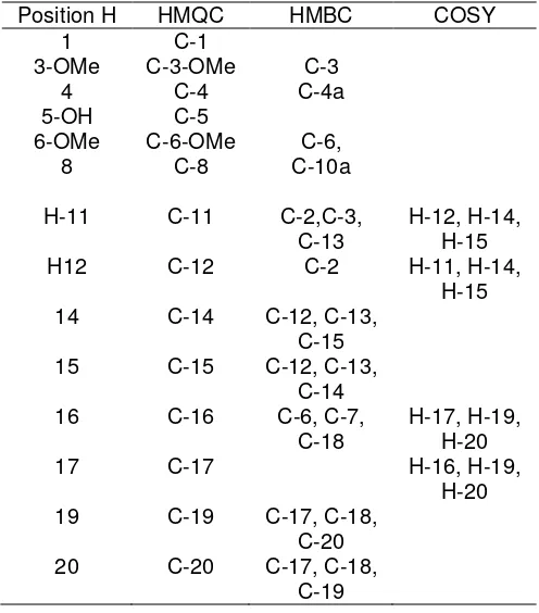
Fig 2. 1H NMR spectrum
Fig 3. HMQC spectrum
Fig 4. HMBC spectrum
Fig 5. COSY spectrum
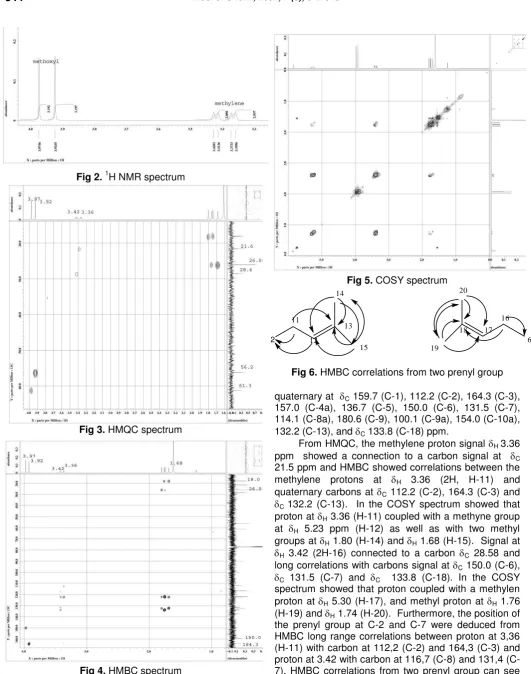
Fig 6. HMBC correlations from two prenyl group
quaternary at C 159.7 (C-1), 112.2 (C-2), 164.3 (C-3),
157.0 (C-4a), 136.7 (C-5), 150.0 (C-6), 131.5 (C-7), 114.1 (C-8a), 180.6 (C-9), 100.1 (C-9a), 154.0 (C-10a), 132.2 (C-13), and C 133.8 (C-18) ppm.
From HMQC, the methylene proton signal H 3.36
ppm showed a connection to a carbon signal at C
21.5 ppm and HMBC showed correlations between the methylene protons at H 3.36 (2H, H-11) and
quaternary carbons at C 112.2 (C-2), 164.3 (C-3) and
C 132.2 (C-13). In the COSY spectrum showed that
proton at H 3.36 (H-11) coupled with a methyne group
at H 5.23 ppm (H-12) as well as with two methyl
groups at H 1.80 (H-14) and H 1.68 (H-15). Signal at
H 3.42 (2H-16) connected to a carbon C 28.58 and
long correlations with carbons signal at C 150.0 (C-6),
C 131.5 (C-7) and C 133.8 (C-18). In the COSY
spectrum showed that proton coupled with a methylen proton at H 5.30 (H-17), and methyl proton at H 1.76
(H-19) and H 1.74 (H-20). Furthermore, the position of
Indo. J. Chem., 2007, 7 (3), 342-345
Muharni, et al.
345
O
O OH
H3CO OCH3
OH
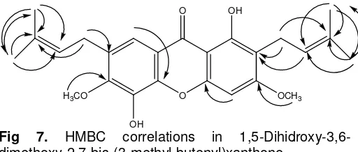
Fig 7. HMBC correlations in
1,5-Dihidroxy-3,6-dimethoxy-2,7-bis-(3-methyl-butenyl)xanthone.
In the HMQC spectrum also showed the proton methyl from methoxyl group at H 3.92 connected to
carbon signal at C 56.2, proton at H 3.97 connected to
carbon at C 61.9. In HMBC spectrum proton at H 3.92
showed long correlation with carbon signal at 164,50 (C-3), and proton at H 3,97 long range correlation to
carbon at C 150 (C-6) The methoxyl group were
placed at C-3 and C-6, adjacent to the prenyl group due to its HMBC correlation with C-3 and C-6. In HMQC spectrum showed proton at H 7.62 connected to carbon
at C 116.7 (C-8) and long correlation to carbon at C
154 (C-10a), proton at H 6.53 connected to carbon at C
89.9 (C-4) and long correlation to carbon at C 157
(C-4a). The position of the hydroxyl group were determined to be at C 159,7 (C-1) and C 136,7 (C-5). Correlation at
HMBC spectrum can see at Fig 7.
Therefore was determined as 1,5-Dihidroxy-3,6-dimethoxy-2,7-bis-(3-methyl-butenyl)xanthone. This is the first report on the isolation of compound from this plant.
CONCLUSION
Xanthone, prenylated xanthone type: 1,5- Dihidroxy-3,6-dimetoxy-2,7-bis-(3-methyl-butenyl)-xanthone, had been isolated from methanol extract of stem bark G. bancana. The structure of this compound was determined based on NMR data.
ACKNOWLEDGEMENTS
The authors thank the Herbarium Anda, Padang,
Indonesia for identification of the plant specimen and
LIPI Research Center for Chemistry, PUSPITEK, Serpong, Indonesia for spectrum measurement.
REFERENCES
1. Whitmore, M.A., 1973, Tree Flora of Malaya: Forest Department, Ministry of Primary Industries, Longman, 218.
2. Gopalakrishnan, G., and Banumathy, B., 2000, Fitoterapia, 71, 607-609
3. Cao, S. G., Valerie, H. L., Wu, X .H., Sim, K.Y., Tan, B. H. K., Pereira, J. T., and Goh, S.H., 1988, Tetrahedron, 54, 10915-10924
4. Baggett, S., Protiva, P., Mazzola, E. P.,Yang, H., Ressler, E.T., Basile, M.J., Weinstein, I.B., and Kennelly, E. J. 2005, J. Nat. Prod., 68, 354-360. 5. Lannang, A. M., Komguem, J., Ngninzeko, F. N.,
Tangmoua, J. G., Lonsti, D., Ajaz, A., Choudhary, M. I., Ranjit, R., Devkota, K. P., and Sondengam, B. L. 2005, Phytochem., 66, 2351-2355
6. Suksamrarn, S., Suwannapoch, N., Phakhodee, W., Thanuhiranlert, J., Ratanahukul, P., Chimnoi, N., and Suksamrarn, A. 2003, Chem. Pharm. Bull, 51(7), 857-859.
7. Hay, A. E., Aumond, M. C., Mallet, S., Dumonted, V., Litaudon, M., Rondeau, D. and Richomne P. 2004, J. Nat.Prod, 67, 707-709.
8. Mackeen, M. M.’ Ali, A. M., Lajis, N. H., Kawazu, K., Hassan, Z., Amran, M., Hasbah, M., Mooi, L.Y., and Mohamed, S. M. 2000, J. Ethnopharmacol., 72, 394-402.
9. Ignatushchenko, M. V., Winter, R. W., Bachinger, H. P., Hinrichs, D. J., and Riscoe, M. K. 1997, FEBS Lett., 409, 67-73.
10. Heyne, K. 1987, Tumbuhan Berguna Indonesia. Jilid III, Yayasan SaranaWana Jaya. Jakarta, 1387.
11. Rukachaisirikul, V., Ritthiwigrom, T., Pinsa, A., Sawanghote, P., and Taylor, W.C. X., 2003, Phytochem., 64, 1149-1156.