Subcellular localisation of cherry leaf roll virus coat protein and
genomic RNAs in tobacco leaves
Paloma Ma´s, M. Amelia Sa´nchez-Pina, Josefa M. Balsalobre, Vicente Palla´s *
Departamento de Mejora y Patologı´a Vegetal,CEBAS(CSIC),Apartado de Correos4195,30080Murcia,Spain Received 28 July 1999; received in revised form 24 November 1999; accepted 24 November 1999
Abstract
The in vivo subcellular location of the coat protein and RNAs of cherry leaf roll nepovirus (CLRV) was studied in infected tobacco plants by two different approaches and it was correlated with the cytopathic structures induced by the virus. Subcellular fractions were obtained by differential centrifugation, visualised by electron microscopy and analysed for their viral RNA and coat protein content by Northern blot and Western blot analysis, respectively. Results indicate that viral RNAs accumulated preferentially at the microsomal fraction. Immunocytochemical studies revealed a clear association of the coat protein of CLRV with the virus-induced cytopathological structures. In situ hybridisation studies confirmed the cytoplasmic location of the virus and allowed one to elucidate the distribution of the CLRV genomic RNAs in the different cell types of infected tissue. © 2000 Elsevier Science Ireland Ltd. All rights reserved.
Keywords:Plant virus; Subcellular fractionation; Immunogold labelling; In situ hybridisation; Nepovirus; CLRV
www.elsevier.com/locate/plantsci
1. Introduction
In their interactions with plants, most viruses induce important changes on plant metabolism that are usually reflected in a variety of character-istic cytological alterations. Some of these changes have been reported to be, directly or indirectly, associated with replication and/or accumulation of the virus. Electron microscopy studies of infected tissues have shown that the synthesis and assembly of virus particles occurred in cytopathological structures classically denominated ‘amorphous in-clusion bodies or viroplasms’ [1]. The size and ultrastructural properties of these inclusion bodies differ widely between distinct groups of viruses but it seems that in all cases, viral-encoded proteins as well as host components participate in the forma-tion of these structures. There are evidences of cytoplasmic inclusion bodies in a large number of
virus-host combinations [see for a review [1,2]]. Cytological alterations have also been described to be associated with a particular organelle such as chloroplasts in tymo- [3], bromo- [4] and po-tyviruses [5]; proplastids in hordeiviruses [6]; mito-chondria in tobraviruses [7] and nuclei in gemini-[8,9]; poty- [10,11], rhabdoviruses [12] and Pea enation mosaic virus [13].
Cherry leaf roll nepovirus (CLRV) belongs to the Comoviridae family of plant viruses [14,15]. Its genome is composed of two positive-sense polyadenylated ssRNAs with a genome-linked protein (VPg) at their 5% ends [16]. The virus is transmitted vertically in pollen to infect seed and seedlings of woody perennial trees [17]. Cyto-pathological alterations induced by CLRV infec-tion in deciduous trees have been described as hyperplasia and severe alterations of the chloro-plasts (e.g. starch accumulation, dilation of thy-lakoids and cytoplasmic invaginations of the chloroplast envelope) [18]. In Nicotiana cle6elandii plants, CLRV has been shown to induce inclusion bodies in palisade leaf cells, forming an apparent * Corresponding author. Tel.: +34-968-215717; fax: +
34-968-266613.
E-mail address:[email protected] (V. Palla´s)
amassing of membranous structures, endoplasmic reticulum and ribosomes [19]. In Nicotiana tabacum cv. ‘Xanthi nc’, CLRV interacts in a compatible manner inducing necrotic ringspots distributed throughout the leaves and often associ-ated with the vascular system [15,20]. Using tissue printing and northern hybridisation techniques, the macroscopic distribution of CLRV RNAs and CP in infected tobacco plants has been previously observed, showing a correlation between macro-scopic symptoms and virus distribution in infected tobacco plants [20 – 22].
To further investigate the subcellular location of the CP and RNAs of CLRV and its relationship with the cytopathic structures induced by the virus, two different experimental approaches have been used. First, biochemical fractionation of cell components from infected tissues revealed a spe-cific association of viral RNAs and CP with mi-crosomal fractions. Secondly, immunocyto-chemical studies have allowed one to visualise the subcellular localisation of the CLRV CP and its correlation with the cytopathological alterations induced by CLRV infection. Furthermore, non-isotopic in situ hybridisation provides a powerful tool to determine the subcellular distribution of CLRV genomic RNAs and its accumulation in specific cell types of infected tissues.
2. Materials and methods
2.1. Virus and plant material
The walnut isolate of cherry leaf roll virus (w-CLRV) was purified from frozen leaf tissue as previously described [23]. Plants of N.tabacum cv. ‘Xanthi nc’ were grown in a growth chamber with a day length of 16 h, day and night temperatures
Fig. 1.
of 24 and 20°C, respectively, and relative humidity of 80%. Six-week-old plants were inoculated with 10 ng/ml of the w-CLRV by rubbing the leaves with carborundum as abrasive.
2.2. Preparation of plant subcellular fractions
The subcellular fractionation procedure was performed basically as described previously [24,25]. Five grams of CLRV-infected and mock-inoculated tobacco plants were homogenised at 4°C in a mortar with 2 vol. of a grinding buffer containing 0.4 M sucrose, 20 mM KCl, 1 mM MgCl2, 0.1% (w/v) bovine serum albumin (BSA),
20 mM Tris – HCl pH 7.5 and 10 mM mercap-toethanol. The homogenate was filtered through four layers of cheesecloth and the filtrate was sequentially centrifuged at 250, 3000 and 10 000×
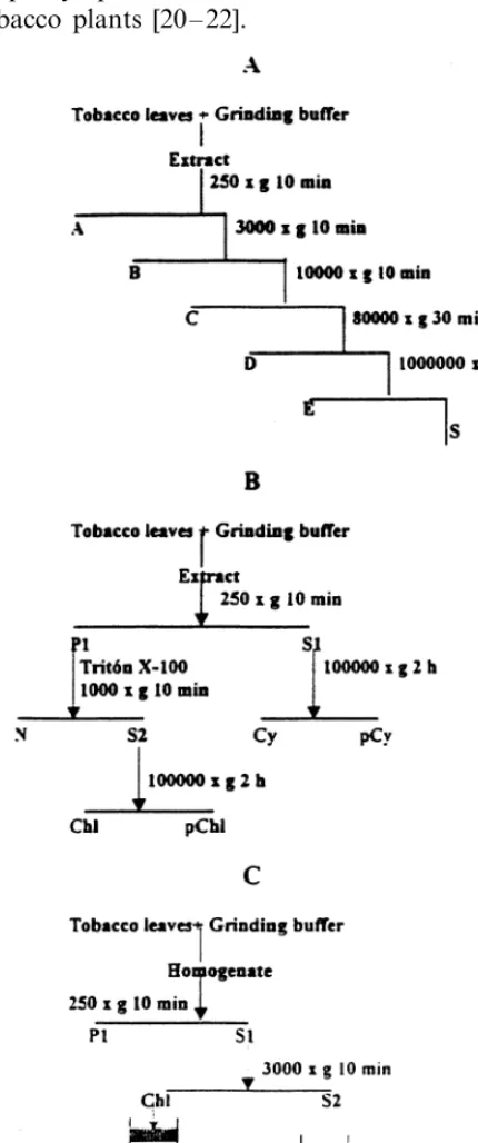
g for 10 min in each case in a swinging-bucket rotor (Beckman JS-13.1). The 10 000×g superna-tant was centrifuged at 80 000×g for 30 min and then at 100 000×g for 2 h in a fixed angle rotor (Beckman 50.2Ti). By using this differential cen-trifugation procedure (Fig. 1A), five pellets (frac-tions A, B, C, D and E) and a final supernatant (S) were obtained. All of the fractions were stored at −20°C for later nucleic acid extraction.
The morphology of each subcellular fraction was also analysed by transmission electron mi-croscopy (TEM). Each fraction was basically pro-cessed as described in Diaz-Ruiz et al. [26] except for the embedding resin used which was Epon. Ultrathin sections were cut with a diamond knife (Diatome) using an Ultracut E microtome (Leica) and mounted on copper grids. The sections were contrasted with uranyl acetate and lead citrate and visualised using a TEM-10 Zeiss microscope.
A second procedure of subcellular fractionation was performed in order to separate more selec-tively the nuclear and the chloroplastic fractions (Fig. 1B). Five grams of CLRV-infected and mock-inoculated tobacco plants were ho-mogenised at 4°C in a mortar with 2 vol. of the grinding buffer described above. The homogenate was filtered through four layers of cheesecloth and the filtrate was centrifuged at 250×g for 10 min in a swinging-bucket rotor (Beckman JS-13.1). The nuclear and chloroplastic fractions were selec-tively separated by resuspending the 250×g pellet in 0.5 ml/g of tissue of the grinding buffer plus 1%
(v/v) of Triton X-100 and incubation for 10 min at 4°C with occasional shaking. After centrifugation at 1000×g for 10 min the supernatant (S2) was centrifuged at 100 000×g for 2 h. All the frac-tions, including the pellet (Cy) and the supernatant (pCy) obtained after centrifugation of the 250×g
supernatant (S1) at 100 000×g for 2 h, were pro-cessed to determine their nucleic acid composition.
2.3. Chloroplast purification
Chloroplasts were isolated following the proce-dure previously described [25,27] with some mod-ifications (Fig. 1C). Five grams of CLRV-infected and mock-inoculated tobacco leaves were gently homogenised at 4°C in a mortar with 2 vol. of a grinding buffer containing 0.35 M sucrose, 3 mM EDTA, 0.1% (w/v) BSA, 50 mM Tris – HCl pH 7.2 and 10 mM mercaptoethanol. The homogenate was filtered through four layers of cheesecloth and the filtrate was centrifuged at 250×g for 10 min. The supernatant (S1) was then centrifuged at 3000×g for 10 min. The pellet, containing par-tially purified chloroplasts (Chl) was gently resus-pended in 2 ml of grinding buffer and the chloroplasts were further purified in a discontinu-ous gradient (20, 40, 60 and 80% (v/v)) of Percoll. Two bands corresponding to intact and broken chloroplasts were obtained after centrifugation at 10 000×g for 20 min. Each band was mixed with 4 vol. of the grinding buffer and the chloroplasts were recovered by centrifugation at 3000×g for 10 min. All the centrifugation steps were carried out at 4°C in a swinging-bucket rotor (Beckman JS-13.1). The quality of the chloroplasts was analysed by phase contrast light microscopy (LM) with a Leica DMRB-Flu microscope. The unbro-ken chloroplasts were easily distinguishable by their bright refractive outline. The chloroplasts were then processed to determine their nucleic acid composition.
2.4. Control experiments
In order to rule out a possible artefactual bind-ing of the viral RNAs to the subcellular fractions or the purified organelle, two types of control experiments were performed. In reconstruction ex-periments, 100 ml of purified virion (5 mg/g of
de-scribed above. A second type of control experi-ments was performed incubating 0.5 vol. of the fraction corresponding to unbroken chloroplasts with RNase A (10mg/ml) for 1 h on ice. The pellet
obtained after centrifugation at 1000×gfor 3 min was resuspended in 2 ml of grinding buffer plus 0.5 M CaCl2 and thermolysin (200 mg/ml)
(protease type X, Sigma). After incubating the solution for 30 min on ice, the reaction was stopped by adding 1 mM EDTA and the chloro-plasts were washed twice with 5 ml of grinding buffer.
2.5. Nucleic acid extraction and Northern hybridisation
All the pellets and supernatants obtained after each centrifugation were processed by adding 1/5 vol. 0.2 M Tris – HCl pH 8.9, 1/20 vol. 0.1 M EDTA pH 7.0, 1/20 vol. 5% (w/v) SDS and 10 mM mercaptoethanol. The nucleic acids were ex-tracted with phenol/chloroform and precipitated from the aqueous phase using ethanol. The nucleic acids were electrophoresed in non-denaturing 1% (w/v) agarose gel in TBE (TBE 1×: 90 mM Tris Borate, 2 mM EDTA), visualised by ethidium bromide staining (Fig. 2A) and transferred to ny-lon membranes in 10×SSC (1.5 M NaCl, 0.15 M sodium citrate pH 7.00) according to standard procedures [28]. The synthesis of the digoxigenin-labelled RNA probe that recognises both genomic viral RNAs, prehybridisation, hybridisation with the digoxigenin-RNA probe (dig-viral RNA probe) and chemiluminescent detection (Fig. 2B), were performed as described previously [29,30]. The RNA probe is the same described in Ref. [29] and corresponds to the 3%-UTR of CLRV RNA2, that has a high homology with the same region of CLRV RNA1.
2.6. Western blot analysis
Proteins were separated by denaturing SDS polyacrylamide gel electrophoresis (SDS-PAGE) [31] and then transferred onto nitrocellulose mem-branes (Schleicher and Schuell), using MiniPROTEAN II and MiniTransblot cell sys-tems (BioRad). Electrotransfer was conducted in 10 mM CAPS pH 11.0, 3 mM DTT, 0.02% SDS, and 20% methanol. Primary antibodies against wCLRV were a polyclonal antiserum kindly
pro-vided by Dr A. Rowhani (University of California, Davis, CA), diluted 1:900. Antibody binding was detected by immunoreaction to anti-rabbit IgG coupled to alkaline phosphatase and subsequent chemiluminescent detection using CDP-Star™ substrate (Boehringer Mannheim).
2.7. Tissue fixation and embedding for LM studies
Typical symptomatic areas [20] of systemically infected leaves were cut at 10 days post-inocula-tion (dpi) with a punch of 0.7 cm diameter. The samples were immediately immersed in freshly pre-pared fixation solution (2.5% glutaraldehyde, 4%
p-formaldehyde in 0.1 M cacodylate buffer pH 7.2), postfixed in osmium tetroxide, dehydrated in ethanol series and embedded in Spurr resin. Semithin sections (1 mm) were cut with glass
knives on a Reichert Ultracut microtome and visu-alised by LM in a Leica DMRB-Flu microscope after staining with toluidin blue for 2 min (Fig. 4).
2.8. Immunogold labelling
Primary antibodies against wCLRV were the same described in Section 2.6, but at a different concentration. The semithin sections were mounted on TESPA (3-amino-propyl-trietoxi-silane) (Sigma) precoated glass slides. The samples were incubated with PBS (135 mM NaCl, 1.5 mM KH2PO4, 8 mM Na2HPO4, 2.7 mM KCl, pH 7.0)
for 5 min, with 0.1 M ClNH4 in PBS for 3 min
and with 5% BSA in PBS for 30 min. After three washes (20 min each) in PBS, 3% BSA in PBS and 3% BSA+0.1% Nonidet P-40 in PBS, the sections were incubated for 3 h in a 40 ml drop of the
antibody solution containing polyclonal antiserum against CLRV CP diluted 1:1000 in 3% BSA in PBS. The sections were then washed three times as above and incubated in a 40 ml drop of the
sec-ondary antibody IgG-gold conjugate (10 nm, Bio-Cell Research laboratories Cardiff, UK) diluted 1:100 in 3% BSA in PBS. Silver enhancement was performed by incubating the sections with 40 ml of
2.9. In situ hybridisation
For in situ hybridisation studies, semithin sec-tions, collected on TESPA precoated glass slides, were pre-treated with xylene (2×5 min),
dehy-drated with series of alcohol (30 – 100%) and incu-bated with proteinase K (1 mg/ml) for 10 min at
37°C. To prevent non-specific binding of the probe to positively-charged amino groups, the sections were treated with 0.1 M triethanolamine for 10
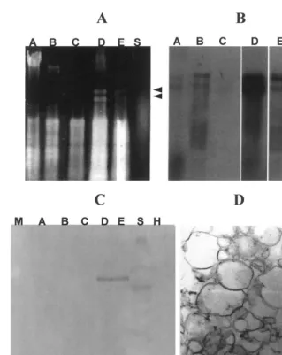
Fig. 2. Subcellular localisation of cherry leaf roll nepovirus (CLRV) by differential centrifugation of tobacco infected leaves. (A) Electrophoretic analysis of the nucleic acids from the subcellular fractions of CLRV-infected tobacco leaves. The samples were analysed in non-denaturing agarose gels and visualised after ethidium bromide staining. Lanes A – E fractions corresponding to the pellets obtained after centrifugation at 250×g10 min, 3000×g10 min, 10 000×g10 min, 80 000×g30 min and 100 000×g
2 h, respectively. Lane S fraction corresponding to the final supernatant after centrifugation at 100 000×g2 h. (B) The nucleic acids were transferred to nylon membranes and hybridised with the dig-viral RNA probe that recognises both viral RNAs. The membranes were developed using the chemiluminescent substrate AMPPD. The viral signal, showed in A, B, C, E and S, corresponds to a 30 min exposition, while in D, the signal corresponds to 10 min exposition. The two arrows in (A) and (B) indicate the location in the gel of CLRV RNAs 1 and 2. (C) Western blotting analysis of the CLRV CP in the different subcellular fractions. The samples were analysed in denaturing SDS polyacrylamide gels (SDS-PAGE) and then electrotransferred onto nitrocellulose membranes. Lanes A – E fractions correspond to the same pellets just describe for (A), and lane S fraction corresponds to the final supernatant after centrifugation at 100 000×g2 h, lane M corresponds to the marker, and lane H to fraction D obtained from a healthy plant. Notice that CP is only present in fractions D and E. (D) TEM micrograph of the microsomal fraction (fraction D) obtained after centrifugation at 80 000×g30 min, showing mostly membranous vesicles. Bar 1
min and 0.1 M triethanolamine plus 0.25% acetic anhydride for 10 min. The non-isotopic ribo-probe was the same as the one used in the Northern blot analysis (see above). The hybridis-ation was performed in a moist chamber overnight at 55°C with 1 mg/ml of the dig-viral
RNA probe in 40 ml of the hybridisation
solu-tion containing 50% deionised formamide, 2×
SSC and 10% dextran sulphate. The sections were then washed in 2×SSC for 10 min and incubated with RNase A (20 mg/ml) in 2×SSC
for 10 min. After several washes, with 1 mM DTT in 2×SSC at room temperature and with 1 mM DTT in 0.1×SSC at 55°C, the samples were incubated in PBS for 5 min, in 0.1 M ClNH4 in PBS for 5 min and 5% BSA in PBS
for 30 min. The incubation with the antibody solution (1:25 dilution of the sheep anti-digoxi-genin antibodies coupled to gold particles, 10 nm, BioCell Research laboratories Cardiff, UK) was performed for 1 h at room temperature. The sections were then washed several times in PBS for 10 min. Silver enhancement, stain and visual-isation of the samples were performed as de-scribed above. Control experiments were performed by in situ hybridisation on sections taken from healthy or mock-inoculated tobacco plants as well as by not including the probe in the hybridisation solution.
3. Results
3.1. CLRV RNAs and coat protein accumulate in the microsomal fraction of infected tobacco cells
To determine the pattern of subcellular accu-mulation of CLRV in infected tobacco cells, leaf extracts of mock-inoculated and CLRV-infected tobacco plants were processed by differential centrifugation as described in Section 2. The EM analyses of these fractions showed that each one was enriched in a subcellular component corre-sponding to nuclear, chloroplastic, mitochon-drial, microsomal and ribosomal fractions (fractions A, B, C, D and E, respectively in Fig. 1A). Fig. 2D shows a representative TEM mi-crograph of fraction D revealing that this frac-tion mainly contained endoplasmic reticulum membranous vesicles of different sizes. Analysis of the nucleic acids extracted from each fraction
(Fig. 2A,B) revealed that the viral RNAs accu-mulated mainly in fraction D (Fig. 2A,B lane D) obtained after centrifugation at 80 000×g for 30 min (see Fig. 1A). Northern hybridisation analy-sis of the same extracts revealed a weaker hy-bridisation signal in the chloroplastic and ribosomal fractions (Fig. 2A,B, lanes B and E, respectively). No signal was observed in subcellu-lar fractions of mock-inoculated plants (data not shown). Moreover, analysis of the proteins ex-tracted from each fraction (Fig. 2C) revealed that the CLRV Coat protein (CP) was present in fractions D and E.
In reconstruction experiments (see Section 2 for details), the viral RNAs appeared to equally accumulate in all subcellular fractions (data not shown) supporting the idea that viral RNA accu-mulation in fraction D is not due to an artefac-tual association produced during the processing of the samples.
3.2. A minor percentage of CLRV RNAs is associated with purified chloroplasts of infected tobacco cells
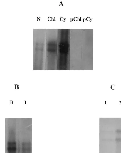
Fig. 3. Northern hybridisation analysis of the viral RNA accumulation in chloroplasts of cherry leaf roll nepovirus (CLRV)-infected tobacco leaves. (A) Viral RNA detection in cytoplasmic, nuclear and chloroplastic fractions obtained af-ter differential centrifugation using Triton X-100. Lanes N, Chl and Cy, correspond to nuclear, chloroplastic and cyto-plasmic fractions obtained after centrifugation at 250×gfor 10 min and 100 000×g for 2 h. Lanes pChl and pCy, supernatants obtained after centrifugation of the non-purified chloroplastic fraction and the cytoplasmic fraction at 100 000×g for 2 h, respectively. The hybridisation signal observed in N and Chl corresponds to 30 min exposition while in D, the signal corresponds to 10 min exposition. (B) Viral RNA detection in intact purified chloroplasts (lane I) and broken (lane B) from CLRV-infected tobacco leaves. The film was exposed for 1 h. (C), Viral RNA detection in intact purified chloroplasts (lane 2) and after incubation with RNase A (lane 1). Note the low percentage of viral RNA that accumulates inside the chloroplasts. In all cases, the mem-branes were developed using the chemiluminescent substrate AMMPD. Lanes were not adjacent in the gel.
hybridisation was performed, a higher amount of CLRV RNAs were detected in the fraction cor-responding to the broken chloroplasts (Fig. 3B, lane B) than observed in association with the intact chloroplasts (Fig. 3B, lane I).
To determine whether CLRV virions or RNAs could artefactually be attached to the surface of the chloroplasts as a result of the isolation pro-cedure, control experiments were performed in which the intact chloroplasts were treated with RNase A and thermolysin. After treatment, Northern hybridisation of the extracted nucleic acids (Fig. 3C) showed that only a minimal amount of viral RNAs was actually maintained in this fraction (Fig. 3C, lane 1) compared with the viral RNAs detected in non-treated intact chloroplasts (Fig. 3C, lane 2). These results seem to indicate that during the chloroplast isolation procedure, CLRV RNAs were attached to the external surface of the chloroplasts. The weak hybridisation signal observed inside the treated intact chloroplasts (Fig. 3C, lane 1) might, how-ever, have a biological function in the CLRV infection cycle.
3.3. LM analysis of a typical ringspot induced by CLRV in tobacco lea6es re6eals three areas with different cytopathological alterations
CLRV invades tobacco plants inducing ne-crotic ringspots distributed throughout the leaves [21]. LM observations of a typical symptomatic area revealed at least three clearly distinguishable areas based on their cytopathological alterations (Fig. 4). At the LM level, the peripheral tissue surrounding the symptom appeared non-dam-aged, without manifest alterations (Fig. 4A). A second area, closest to the necrotic ring showed some plasmolysed, damaged cells (Fig. 4B, ar-rows) with no visible subcellular components and abundant starch granules immersed in an amor-phous cytopathological structure (paramural body, Pb), as can be observed in the enlarge-ment of this area shown in Fig. 4D. In a third cytopathological area, corresponding to the ne-crotic ring, the cells appeared completely col-lapsed and intensively stained (Fig. 4C). Neither of these alterations was observed in asymp-tomatic areas of systemically infected leaves or in cells of mock-inoculated plants identically processed (not shown).
3.4. Immunocytochemical localisation of CLRV CP in infected tobacco cells
In order to obtain a microscopic view of the CLRV CP accumulation in infected tobacco cells and to correlate this accumulation with the cyto-pathological alterations induced by the viral infec-tion, immunocytochemical studies at LM level were performed on semithin sections from typical symptomatic areas of systemically infected leaves (Fig. 5A,B). Strong brilliant silver-enhanced sig-nals (shown by epipolarized light in Fig. 5B) were observed in the cytoplasm and in the cytopatho-logical structures (Pb) present in the cells adjacent to the necrotic ring. Phase contrast microscopy allowed a contrasted visualisation of these cells
and a clear identification of the cytopathological structures induced by the viral infection (Fig. 5A). No other subcellular components of infected tis-sues appeared labelled, including chloroplasts, nu-clei, nucleoli or intercellular space (compare Fig. 5A,B). A similar pattern of CP distribution was observed in all cellular types (epidermis, meso-phyll, parenchyma and vascular tissues) localised in this area. No signal was observed in the periph-eral cells surrounding the ringspot, in mock-inocu-lated tissues or in asymptomatic areas of systemically infected leaves (data not shown). No signal was found when the immunocytochemistry was done on mock-inoculated or healthy plants, and when the primary or secondary antibodies were not used for the experiment (data not
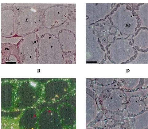
Fig. 5. Light microscopy (LM) immunolocalisation of cherry leaf roll nepovirus (CLRV) CP by immunogold silver enhancing (A,B), and in situ hybridisation (ISH) of CLRV RNAs (C,D). (A,B,C), symptomatic areas of systemically infected leaves processed at 10 d.p.i. (A) Phase contrast. (B) Epipolarization. Bright spots show the silver precipitate that corresponds to the immunolocalisation of CLRV CP, visualised under epipolarized light. Both micrographs (A,B) show part of the epidermis (E) and parenchyma tissue (P) from exactly the same area. The localisation is cytoplasmic in the different tissues that constitute the leaf. Notice the high concentration of CLRV CP present in the paramural body (Pb). There is a very low background as shown by the non-labelled nucleus (N), nucleolus ( ) and cytoplasmic vacuole (V), chloroplasts ( ). (C,D) The visualisation was done by a combination of phase contrast and epipolarization to show the exact localisation of the bright spots that correspond to the RNA hybridisation signal. (C) ISH performed on sections from healthy tobacco leaf. (D) ISH performed on sections from infected tobacco leaf. CLRV RNAs localisation is cytoplasmic in the different tissues that constitutes the leaf. Notice the low background level inside the section as shown by the non-labelled nucleus (N) and nucleolus ( ). Moreover, the low background level can be seen between preparations as shown by a comparison with the ISH done on the healthy leaf (C), where the absence of silver precipitate in any of the cells is shown. (E) epidermis; (P) parenchyma; (BS) bundle sheath; (V) cytoplasmic vacuole; (W) cell wall;
shown). All the results presented here are repre-sentative examples of replicated data based on different experiments.
3.5. Localisation of CLRV RNAs in infected tobacco cells by in situ hybridisation
Thin sections of symptomatic ringspots induced by CLRV in systemically infected leaves were hy-bridised as described in Section 2 with the dig-viral RNA probe in order to analyse the subcellular distribution of the viral RNAs. Similarly to CP distribution, in areas adjacent to the necrotic ring, the signal was preferentially observed in the cyto-plasm of the infected cells (illustrated in Fig. 5D by a combination of epipolarization and phase contrast microscopy). Note that the nucleus of the cell located at the left upper part of Fig. 5D is free of label. The cytopathological structures induced by the infection, previously shown to contain CLRV CP were also labelled in these hybridisation experiments (not shown). In addition, the hybridi-sation signal was also observed in cells of the vascular system such as the sieve elements (se) and bundle sheath cells (BS) (Fig. 5C,D). The labelling
was detected at the same places on successive serial sections and occasionally, appeared associ-ated to the abundant starch granules induced by the infection (compare the different amount of starch present in Fig. 5C,D). Cell labelling of mock-inoculated or healthy plants was very low and not associated with any subcellular compo-nent (Fig. 5C). Similarly, no signal was obtained when the sections were processed without the probe or without gold conjugate antibodies (not shown). All the results presented here are repre-sentative examples of replicated data based on different experiments.
The in situ hybridisation studies clearly suggest a preferential localisation of CLRV RNAs in the cytoplasm of the infected cells with a distribution pattern similar to that observed for the CP. These results are also consistent with those described above obtained by biochemical subcellular fractionation.
4. Discussion
In this paper we have studied the subcellular localisation of CLRV components (genomic
RNAs and CP) by using two different approaches: biochemical fractionation and cell biology tech-niques. The results obtained showed a cytoplasmic accumulation of both, CP and genomic RNAs in CLRV-infected tobacco cells. The cytopathologi-cal alterations induced by CLRV infection in to-bacco cells and their correlation with the pattern of CLRV accumulation have also been analysed. Classical cell fractionation experiments followed by Northern blot hybridisation and Western blot analysis clearly showed an accumulation of the viral RNAs and CP in the microsomal fraction, mainly composed of endoplasmic reticulum vesi-cles. Cytoplasmic membranous inclusions contain-ing vesicles are characteristic of como and nepoviruses [2]. Moreover, it has been suggested that the replication of these types of viruses is likely to be associated with viral-induced vesicles [32,33]. Replication of most positive-strand RNA viruses takes place in association with membranes [34]. It is reasonable to think that this vesiculated fraction could be the site where CLRV replicates and/or accumulates.
Chloroplasts are one of the main targets in the infection cycle of plant viruses, especially in those infections causing stunting and/or chlorosis [see [1], for a review]. In addition, in some well-docu-mented examples, chloroplasts have been associ-ated with viral replication [3,35].
In the case of CLRV, several types of evidence suggested a possible association of CLRV with chloroplasts. Firstly, infected ash trees [18] and N.
The cytopathological alterations induced by CLRV were mainly localised in the symptomatic areas distributed throughout the leaves. The sever-ity of these cellular alterations was evident in the necrotic regions while no cell damage was ob-served in asymptomatic areas of infected leaves or in cells from mock-inoculated plants. In the area closely surrounding the necrotic tissue, CLRV in-duced the formation of cytopathic structures visi-ble by the light microscope (Fig. 4B,D). A similar variety of subcellular alterations was observed in leaf beans infected with another nepovirus, to-bacco ringspot virus (TRSV) [37].
Immunolocalisation of CLRV CP in the cells within symptomatic areas revealed a homogeneous distribution of the CP in all cellular types. The CP was mainly localised in the cytoplasm of infected cells (Fig. 5A,B) suggesting that CLRV CP is synthesised by cytoplasmic ribosomes. This is con-sistent with the results described by Rezaian et al. [38] who demonstrated, by subcellular fractiona-tion and antibiotic inhibifractiona-tion experiments, cyto-plasmic synthesis of the TRSV CP. In situ hybridisation studies also showed a cytoplasmic localisation of CLRV RNAs (Fig. 5D). CLRV RNAs, as well as CP, strongly accumulated in the cytopathic structures induced by viral infection. The significance of the presence of the high signal detected in the Pbs is unclear, since virions were not found here when they were analysed by elec-tron microscopy (not shown). They could be re-garded as cellular regions for the sequestration and eventual degradation of the potential excess of CP and/or viral products in a function similar to the one proposed for the inclusion bodies induced in TMV-infected plants [39]. Non-structural CPMV proteins have been immunolocalised in electron dense material of cytopathological struc-tures [40].
In systemic infection, viruses use the vascular system to invade distant parts of the plant [1]. In vascular tissues, CLRV CP and RNAs accumu-lated in the sieve elements (se) (Fig. 5) suggesting that the virus is transported with the net photo-synthetic flow. Furthermore, the presence of CP in the sieve elements seemed to indicate that CLRV requires the CP for long-distance movement, as previously suggested by Ma´s and Palla´s [21]. Im-munocytochemistry and in situ hybridisation tech-niques can provide a powerful tool to understand the mechanisms, by which viruses enter in the
vascular system, identifying the intercellular barri-ers to which viruses are restricted in defective phloem long-distance transport system.
The use of biochemical and cell biology tech-niques described here have allowed us to deter-mine the pattern of subcellular distribution of CLRV in infected tobacco cells. These approaches in combination with ultrastructural studies at the electron microscopy level can provide important insight into the mechanisms of virus infection and its interactions with the plants.
Acknowledgements
We thank Dr E. Olmos for helpful technical advice and stimulating discussions and P. Sa´nchez for revising the English grammar. This research was supported by Grants PB91-0058 and BIO96-0459 from the Spanish granting agency DGYCIT. P. Ma´s was the recipient of a fellowship from the Consejerı´a de Educacio´n, Cultura y Turismo from Comunidad de Murcia.
References
[1] R.E.F. Mathews, Plant Virology, third ed., Academic Press, New York, 1991.
[2] G.P. Martelli, M. Russo, Plant virus inclusion bodies, Adv. Virus Res. 21 (1977) 175 – 266.
[3] D. Lafle`che, C. Bove´, G. Dupont, C. Mouche`s, T. Astier, M. Garnier, J.M. Bove´, Site of viral RNA replication in the cells of higher plants; TYMV-RNA synthesis on the chloroplast outer membrane system, Proc. FEBS Meet. Amsterdam, RNA Viruses/Ribosomes 27 (1972) 43 – 71. [4] M. Nakayama, M. Horikoshi, K. Mise, N. Yamaoka, P. Park, I. Furusawa, J. Shishiyama, Replication of brome mosaic virus RNA in chloroplasts, Ann. Phytopath. Soc. Jpn. 53 (1987) 301 – 309.
[5] I.P.S. Gadh, V. Hari, Association of tobacco etch virus related RNA with chloroplasts in extracts of infected plants, Virology 150 (1986) 304 – 307.
[6] N.S. Lin, W.G. Langenberg, Spherical vesicles in pro-plastids of barley stripe mosaic virus infected-wheat cells contain double-stranded RNA, Virology 142 (1985) 291 – 298.
[7] B.D. Harrison, D.J. Robinson, Tobraviruses, in: M.H.V. van Regenmortel, H. Fraenkel-Conrat (Eds.), The Plant Viruses, Plenum Press, New York, 1986, pp. 339 – 369. [8] G.O. Adejare, R.H.A. Coutts, Ultrastructural studies on
Nicotiana benthamiana tissue following infection with a virus transmitted from mosaic-diseased Nigerian cassava, Phytopathol. Z. 103 (1982) 87 – 92.
[9] K.S. Kim, T.L. Shock, R.M. Goodman, Infection of
[10] J.G. Gao, A. Nassuth, Alteration of major cellular or-ganelles in wheat leaf tissue infected with Wheat streak mosaic rymovirus (potyviridae), Phytopathology 83 (1993) 206 – 213.
[11] G.P. Martelli, M. Russo, Nuclear changes in mesophyll cells of Gomphrena globosa L. associated with infection by Beet mosaic virus, Virology 38 (1969) 297 – 308. [12] N.S. Lin, Y. Hsu, R.J. Chiu, Identification of viral
structural proteins in the nucleoplasm of potato yellow dwarf virus-infected cells, J. Gen. Virol. 68 (1987) 2723 – 2728.
[13] F. Motoyoshi, R. Hull, The infection of tobacco proto-plasts with pea enation mosaic virus, J. Gen. Virol. 24 (1974) 89 – 99.
[14] R. Goldbach, G.P. Martelli, R.G. Milne, Family Co-moviridae, in: F.A. Murphy, C.M. Fanquet, D.H.L. Bishop, S.A. Ghabrial, A.W. Jarvis, G.P. Martelli, M.A. Mayo, M.O. Summers (Eds.), Virus Taxonomy Classifi-cation and Nomenclature of Viruses, I.C.T.V., Springer Verlag, New York, 1995, pp. 341 – 347.
[15] A.T. Jones, CLRV. CMI/AA Descriptions of Plant Viruses, number 306B (1985).
[16] C.U.T. Hellen, J.I. Cooper, The genome-linked protein of cherry leaf roll virus, J. Gen.Virol. 68 (1987) 2913 – 2917.
[17] J.I. Cooper, P.R. Massalki, M.L. Edwards, Cherry leaf roll virus in the female gametophyte and seed of birch and its relevance to vertical virus transmission, Ann. Appl. Biol. 105 (1984) 55 – 64.
[18] J. Hamacher, A. Quadt, Light-and electron microscopic studies of cherry leaf roll virus (CLRV) on European ash (Fraxinus excelsiorL.), J. Phytopathol. 131 (1991) 215 – 226.
[19] A.T. Jones, A.M. Kinninmonth, I.M. Roberts, Ultra-structural changes in differentiated leaf cells infected with cherry leaf roll virus, J. Gen. Virol. 18 (1973) 61 – 64. [20] P. Ma´s, V. Palla´s, Non-isotopic tissue-printing
hybridisa-tion: a new technique to study long-distance plant virus movement, J. Virol. Methods 52 (1995) 317 – 326. [21] P. Ma´s, V. Palla´s, Long-distance movement of cherry
leaf roll virus in infected tobacco plants, J. Gen. Virol. 77 (1996) 531 – 540.
[22] J.M. Balsalobre, P. Ma´s, M.A. Sa´nchez-Pina, V. Palla´s, Spatial distribution of acidic chitinases and their messen-ger RNA in tobacco plants infected with cherry leaf roll virus, Mol. Plant-Microbe Interact. 10 (1997) 784 – 788. [23] A. Rowhani, S.M. Mircetich, R.J. Sepherd, J.D.
Cu-cuzza, Serological detection of cherry leaf roll virus strains, Phytopathology 75 (1985) 48 – 52.
[24] J. Galindo, D.R. Smith, T.O. Diener, Etiology of planta macho, a viroid disease of tomato, Phytopathology 72 (1982) 49 – 54.
[25] J.F. Marcos, R. Flores, Subcellular location of avocado sunblotch viroid in avocado leaves, Plant Sci. 67 (1990) 237 – 244.
[26] J.R. Dı´az-Ruiz, M.J. Avila-Rincon, I. Garcia-Luque, Subcellular localization of cucumovirus-associated satel-lite double-stranded RNAs, Plant Sci. 50 (1987) 239 – 248.
[27] G.A. Berkowitz, M. Gibbs, Chloroplasts as a whole, in: H.F. Linskens, J.F. Jackson (Eds.), Modern Methods of Plant Analysis, New Series, Springer-Verlag, Berlin, 1985.
[28] J. Sambrook, E.F. Fritsch, T. Maniatis, Molecular Cloning: A Laboratory Manual, Cold Spring Harbor Laboratory Press, New York, 1989.
[29] P. Ma´s, J.A. Sa´nchez-Navarro, M.A. Sa´nchez-Pina, V. Palla´s, Chemiluminescent and colorigenic detection of cherry leaf roll virus with digoxigenin-labeled RNA probes, J. Virol. Methods 45 (1993) 93 – 102.
[30] V. Palla´s, P. Ma´s, J.A. Sa´nchez-Navarro, Detection of plant RNA viruses by non-isotopic dot-blot hybridisa-tion, in: G. Foster, S. Taylor (Eds.), Plant Virus Proto-cols: From Virus Isolation to Transgenic Resistance, Humana Press, Totowa, NJ, 1998, pp. 461 – 468. [31] U.K. Laemmli, Cleaveage of structural proteins during
the assembly of the head of the bacteriophage T4, Na-ture 227 (1970) 680 – 685.
[32] G.A. de Zoeten, A.M. Assink, A. van Kammen, Associa-tion of cowpea mosaic virus-induced double-stranded RNA with a cytopathological structure in infected cells, Virology 59 (1974) 341 – 355.
[33] T. Hatta, R.I.B. Francki, Enzyme cytochemical identifi-cation of single-stranded and double-stranded RNAs in virus-infected plant and insect cells, Virology 88 (1978) 105 – 107.
[34] T.C. Hall, W.A. Miller, J.J. Bujarski, Enzymes involved in the replication of plant viral RNAs, in: D. Ingram, P. Williams (Eds.), Advances in Plant Pathology, Academic Press, London, 1982, pp. 179 – 189.
[35] M. de Graaf, L. Coscoy, E.M.J. Jaspars, Localization and biochemical characterization of alfalfa mosaic virus replication complexes, Virology 194 (1993) 878 – 881. [36] J.E. Schoeltz, M. Zaitlin, Tobacco mosaic virus enters
chloroplasts in vivo, Proc. Natl. Acad. Sci. USA 86 (1989) 4496 – 4500.
[37] J.A. White, O.P. Sehgal, Ultrastructure of tobacco ringspot virus-induced local lesions in lima bean leaves, J. Phytopathol. 138 (1993) 177 – 188.
[38] M.A. Rezaian, R.I.B. Francki, P.W.G. Chu, T. Hatta, Replication of tobacco ringspot virus. III. Site of virus synthesis in cucumber cotyledons, Virology 74 (1976) 481 – 488.
[39] K.J. Reinke, G.A. de Zoeten, In situ localisation of plant viral gene products, Phytopathology 81 (1991) 1306 – 1314.
[40] J. Wellink, J.W.M. van Lent, R.W. Goldbach, Detection of viral proteins in cytopathic structures in cowpea pro-toplasts infected with cowpea mosaic virus, J. Gen. Virol. 69 (1988) 751 – 755.