DETECTION OF HPV TYPE 45 L2 GENE IN CERVICAL CANCER PATIENTS BY POLYMERASE CHAIN REACTION METHOD
YUFRI ALDI1, AYU NOVITA TRISNAWATI1, ANDANI EKA PUTRA2, AKMAL D1,
MARLINA1
1Faculty of Pharmacy, Andalas University, Post Code 25163, Padang, Indonesia, 2 Faculty of Medicine, Andalas University, Post Code 25163, Padang, Indonesia.
Email: [email protected]
ABSTRACT
Objective: Cervical cancer is the second common type of cancer among women in the Indonesian population. Persistent infection with high risk Human papillomavirus (hrHPV) has been considered as the most significant risk factor of cervical cancer. Research on the identification of HPV type 45 L2 gene as the third cause of cervical cancer in Indonesia is still small even has not been done in West Sumatera and Riau, whereas HPV gene identification by molecular biology techniques can be used as an initial screening for cervical cancer.
Methods: DNA was isolated from 43 samples of cervical smears and cervical cancer biopsies obtained from M. Djamil General Hospital Padang and Arifin Achmad General Hospital Pekanbaru. HPV DNA detection was conducted using polymerase chain reaction (PCR) using GP5+/6+ primers. HPV type 45 L2 gene was identified by PCR method with HPV type 45 L2 gene specific primers.
Results: The concentration of the isolated DNA varied from 2.8 to 326.3 ng/µl. Isolates DNA from tissue samples had good value of purity and the purity of isolated DNA from cervical smears samples was not too good. Amplification of HPV DNA showed that 20 tissue samples and 6 cervical smear samples were positive HPV DNA. There was only one sample (Hpv.J.14) which L2 genes of HPV
type 45.
Conclusion: Results of DNA amplification by PCR method showed that 60.47% of cervical cancer patients in this study infected with HPV DNA and 3.85% sample had gene L2 of HPV type 45.
Keywords: Cervical cancer, HPV type 45, L2 gene, PCR.
INTRODUCTION
Cervical cancer is the fourth most common cancer in the world. The second most cancer incidence in Indonesian women is cervical cancer [1]. Infection of high risk Human papillomavirus (HPV) known as the main factor that causes cervical cancer.
International Agency for Research on Cancer (IARC) of the 1000 samples from 22 countries found the 99.7% of the cervical cancer's presence of HPV infections [2]. HPV contributes to the development of cancer by their E6 and E7 genes. Based on their oncogenicity against cervical cancer, HPV can be divided into high risk HPV and low risk HPV [3].
In Indonesia, the identification of HPV genes focused on HPV types 16 and 18. Meanwhile, research on the identification of genes of HPV type 45 is still small even have not been done in West Sumatera and Riau. The aim this study was to detect the L2 genes of HPV types 45 from cervical cancer patients in west Sumatera and Riau used PCR method.
MATERIALS AND METHODS
Sample collection
A total of 43 samples used in this study. Formalin-fixed paraffin-embedded samples of cervical cancer biopsy (4 samples), fresh tissue samples cervical cancer biopsy (20 samples) was collected from Dr M. Djamil general hospital in Padang, West Sumatera. Cervical smear samples (19 samples) from cervical cancer patients Arifin Achmad general hospital in Pekanbaru, Riau also used in this study. The samples were taken from patients who were treated during the period of April- July 2015. Consent of the patient for the use of their samples for research purpose was taken.
Isolation of Human papillomavirus DNA
The tissue samples were isolated using DNA Extraction Kit gSYNCTM and cervical smear samples were isolated using Genomic DNA Mini LinkTM Pure Kit (InvitrogenTM). Isolation of DNA was done according to the protocol of the kit being used.
Concentration and purity of DNA isolated
Quantification was made using a Nanodrop spectrophotometer at 260 and 280 nm. DNA purity is determined based on the value ratio A260 / 280 with units of ng/µl. First the optical hole cleaned with a tissue. Nuclease free water was used as blank. 1 ml nuclease free water put in into the optical hole. Then, the optical hole was cleaned again before the sample is introduced. A total of 2 ml of sample DNA is inserted into optical hole. Absorbance value measured at 260 and 280 nm.
Detection of HPV DNA
HPV DNA detection had done by using HPV GP5+/6 + primer (forward 5'-TTT GTT GTA GTG GAT ACT ACT AC-3'dan reverse 5'-GAA AAA TAA ACT CAT AAT GTA ATT C-3 ') with product length 150 bp [6]. Prepared 25 µl of the PCR mix solution, consist of 1.5 µ l DNA template, 0.5 µl primer Foward, 0.5 µl Reverse Primer, and 22.5 µl Platinium® PCR Super Mix. The PCR conditions were hot start 94 0C (5 min), denaturation 94 0C (30 s), annealing 50 0C (60 s), extension 72 0C (60
s) and final extension 72 0C (10 min). The amplified products were analyzed on a 1.5% agarose gel, stained with SYBR Safe and visualized in gel documentation system.
Detection of HPV type 45 L2 gene
The identification had done by using specific primer of HPV types 45 L2 gene with product length 298 bp. Prepared 25 µl of the PCR mix solution, consist of 1.5 µl DNA template, 0.5 µl primer Foward (TAC ACC CAC CAT GGA CGT GGA C), 0.5 µl Reverse Primer (CTG ACG CAC TGA GGA GAC GGT C), and 22.5 µl Platinium® PCR Super Mix. The PCR conditions were hot start 94
0C (5 min), denaturation 95 0C (30 s), annealing 59 0C (30 s), extension 72 0C (60 s) and final
extension 72 0C (10 min). The amplified products were analyzed on a 1.5% agarose gel, stained with
SYBR Safe and visualized in gel documentation system.
RESULT AND DISCUSSION
The concentration of isolated DNA
had DNA concentration more than 10 ng/ml. Only samples Hpv.J.9, Hpv.J.12, and Hpv.J.16, which
[image:3.595.128.472.98.487.2]has a DNA concentration of less than 10 ng/µl (Table 1).
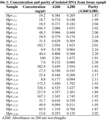
Table 1: Concentration and purity of isolated DNA from tissue samples Sample Concentration
(ng/µl)
A260 A280 Purity
(A260/A280)
Hpv.J.1 19.2 0.384 0.205 1.87
Hpv.J.2 18.7 0.374 0.188 1.99
Hpv.J.3 18.5 0.371 0.182 2.04
Hpv.J.4 104.3 2.086 1.033 2.02
Hpv.J.5 48.3 0.966 0.468 2.06
Hpv.J.6 18.9 0.379 0.174 2.18
Hpv.J.7 31.5 0.629 0.305 2.06
Hpv.J.8 102.7 2.054 1.023 2.01
Hpv.J.9 6.9 0.138 0.064 2.16
Hpv.J.10 40.4 0.808 0.368 2.20
Hpv.J.11 160 3.201 1.672 1.91
Hpv.J.12 7.6 0.152 0.066 2.30
Hpv.J.13 282.9 5.659 2.954 1.92
Hpv.J.14 17.5 0.350 0.193 1.81
Hpv.J.15 22.4 0.448 0.206 2.17
Hpv.J.16 8.8 0.177 0.084 2.11
Hpv.J.17 172.5 3.450 1.792 1.93
Hpv.J.18 326.3 6.525 3.427 1.90
Hpv.J.19 217.9 4.357 2.301 1.89
Hpv.J.20 167.3 3.345 1.745 1.92
Hpv.J.21 31.7 0.634 0.329 1.93
Hpv.J.22 49.9 0.999 0.511 1.95
Hpv.J.23 65.9 1.318 0.706 1.87
Hpv.J.24 12.6 0.253 0.125 2.02
A260: Absorbance on 260 nm wavelengths A280: Absorbance on 280 nm wavelengths
On 19 cervical smear samples, only samples Hpv.A.18, Hpv.A.7, Hpv.A.1had good concentration ≥ 10
ng/µl (Table 2). DNA concentrations less than 10 ng/µl can reduce the efficiency of DNA amplification using PCR methods [7]. DNA isolated from cervical smear had a smaller concentration of DNA compared to DNA derived from the tissue sample.
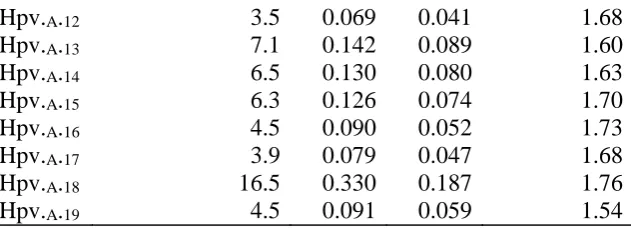
Table 2: Concentration and purity of isolated DNA from cervical smear samples Sample Concentration
(ng/µl)
A260 A280 Purity
(A260/A280)
Hpv.A.1 11.6 0.231 0.120 1.93
Hpv.A.2 7.7 0.153 0.069 2.22
Hpv.A.3 6.3 0.125 0.062 2.02
Hpv.A.4 4.2 0.083 0.035 2.37
Hpv.A.5 7.3 0.146 0.068 2.15
Hpv.A.6 3.1 0.061 0.024 2.54
Hpv.A.7 10.0 0.200 0.109 1.83
Hpv.A.8 3.4 0.069 0.049 1.41
Hpv.A.9 3.5 0.070 0.049 1.43
Hpv.A.10 2.8 0.056 0.053 1.06
[image:3.595.109.501.585.782.2]Hpv.A.12 3.5 0.069 0.041 1.68
Hpv.A.13 7.1 0.142 0.089 1.60
Hpv.A.14 6.5 0.130 0.080 1.63
Hpv.A.15 6.3 0.126 0.074 1.70
Hpv.A.16 4.5 0.090 0.052 1.73
Hpv.A.17 3.9 0.079 0.047 1.68
Hpv.A.18 16.5 0.330 0.187 1.76
Hpv.A.19 4.5 0.091 0.059 1.54
A260: Absorbance on 260 nm wavelengths A280: Absorbance on 280 nm wavelengths
The purity of isolated DNA
The Purity of isolated DNA also could affect DNA amplification by PCR. DNA contamination can cause errors such as the unwanted and non-specific amplification products [7]. DNA purity determined by comparing the absorbance value at 260 nm wavelengths and 280 nm wavelengths. In molecular analysis, DNA is pure if A260/A280 ranged from 1.8 to 2.0. The Purity <1.8 means that there are still a protein contamination or reagents used in the process of DNA isolation [8].
Isolates DNA from tissue samples had good value of purity. Purity of isolated DNA from tissue samples Hpv.J.3, Hpv.J.4, Hpv.J.5, Hpv.J.6, Hpv.J.7, Hpv.J.8, Hpv.J.9, Hpv.J.10, Hpv.J.12, Hpv.J.15, Hpv.J.16
and Hpv.J.24 was > 2.0 (Table 1). But the great value of purity that this will not affect the results of
DNA amplification because the value of purity 2.0 does not indicate any fault in the process of isolation [8]. The purity of isolated DNA of cervical smear samples was not good, because only a sample Hpv.A.7 and Hpv.A.1 that meet the range of purity for molecular analysis (Table 2). Low DNA
purity grades can be caused by proteins and other contaminants in the isolated DNA.
Detection of HPV DNA
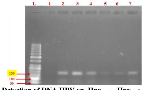
Results of DNA amplification using PCR method for the detection of HPV DNA shown 26 of the 43 samples in this study had HPV DNA. In the gel photograph the results of DNA amplification using GP5+/6+ primer, samples Hpv.J.2, Hpv.J.3, Hpv.J.4, Hpv.J.5, Hpv.J.6, Hpv.J.7, Hpv.J.8, Hpv.J.9, Hpv.J.10,
Hpv.J.11, Hpv.J.13, Hpv.J.14, Hpv.J.15, Hpv.J.16, Hpv.J.17, Hpv.J.18, Hpv.J.19, Hpv.J.22, Hpv.J.23, Hpv.J.24,
Hpv.A.2, Hpv.A.3, Hpv.A.4, Hpv.A.5, Hpv.A.6, Hpv.A.7 appear on the band at 150 bp, shown in (Fig.
1-5). This indicates that the sample is positive for HPV DNA.
Fig 1: Detection of DNA HPV on Hpv.J.1 - Hpv.J.12 Samples
L: DNA Ladder, 1: Hpv.J.1, 2: Hpv.J.2,3: Hpv.J.3 ,4 : Hpv.J.4, 5 : Hpv.J.5 , 6: Hpv.J.6, 7: Hpv.J.7, 8:
Fig 2: Detection of DNA HPV on Hpv.J.13 - Hpv.J.22 Samples
L: DNA Ladder, 1: Hpv.J.13, 2: Hpv.J.14,3: Hpv.J.15 4 : Hpv.J.16, 5 : Hpv.J.17 , 6: Hpv.J.18, 7:
Hpv.J.19, 8: Hpv.J.20, 9: Hpv.J.21, 10: Hpv.J.22
[image:5.595.242.353.328.466.2]From this study, note that 60.47% of cervical cancer patients in West Sumatera and Riau infected by Human papillomavirus. While the results of research by the International Agency for Research on Cancer (IARC) of the 1000 samples shown 99.7% of 22 HPV infection in cervical cancer patients [2]. This suggests that HPV infection in cervical cancer patients in West Sumatra and Riau has a smaller percentage than HPV infection in cervical cancer patients in the world.
Fig 3: Detection of DNA HPV on Hpv.J.23 - Hpv.J.24 Samples
L: DNA Ladder, 1: Hpv.J.23, 2: Hpv.J.24
The difference in the prevalence of HPV in cervical cancer patients in West Sumatra and Riau within the world can be due to differences in the number of samples studied and because of cultural factors [9]. Cervical cancer suffered by patients in this study were not infected with HPV can be caused by other risk factors such as smoking causes cervical cancer, use of oral contraceptives, weak immune system and Chlamydia and HSV infection. These risk factors may not directly cause cervical cancer, but it happens gradually.
Fig 4: Detection of DNA HPV on Hpv.A.1 - Hpv.A.7 Samples
[image:5.595.175.407.592.740.2]Fig 5: Detection of DNA HPV on Hpv.A.8 - Hpv.A.19 Samples
L: DNA Ladder, 1: Hpv.A.8, 2: Hpv.A.9, 3: Hpv.A.10,4 : Hpv.A.11, 5 : Hpv.A.12 , 6: Hpv.A.13, 7:
Hpv.A.14 , 8 : Hpv.A.15, 9: Hpv.A.16 , 10: Hpv.A.17, 11: Hpv.A.18 , 12: Hpv.A.19
Detection of HPV type 45 L2 gene
HPV types 45 consist of 7858 base pairs with 8 ORF is E1, E2, E4, E5, E6, E7 and L1 and L2. HPV types 45 L2 gene arranges by 1,392 base pairs that are at the base of 4,236 to 5,627 on the HPV type 45. L2 gene serves as a minor constituent viral capsid. Structural studies indicate that the new L2 genes are expressed in a complex stage of virus infection. So that these genes can only be identified in patients with cervical cancer. L2 gene identification is important as a first step to designing a prophylactic vaccine for HPV infection [10].
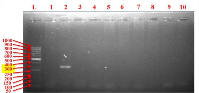
Identification of HPV type 45 L2 gene was performed using specific primers of HPV types 45 L2 gene by PCR showing only positive samples containing genes Hpv.J.14 HPV L2 45 (Fig. 6) Where the
band appeared on 298 bp. Other samples did not show band on the 298 bp, so it concluded that these samples did not have L2 genes of HPV type 45.
Fig 6: Identification of HPV Type 45 L2 gene on Hpv.J.13 - Hpv.J.22Samples
L: DNA Ladder, 1: Hpv.J.13, 2: Hpv.J.14,3: Hpv.J.15 4 : Hpv.J.16, 5 : Hpv.J.17 , 6: Hpv.J.18, 7:
Hpv.J.19, 8: Hpv.J.20, 9: Hpv.J.21, 10: Hpv.J.22
[image:6.595.131.465.447.604.2]CONCLUSION
DNA amplification by PCR method showed that 60.47% of cervical cancer patients in this study infected with HPV DNA and there is only one sample (Hpv.J.14) which L2 genes of HPV type 45.
ACKNOWLEDGEMENTS
We gratefully acknowledge Dr. M. Djamil General Hospital, Padang and Arifin Achmad General hospital, Pekanbaru for providing us cervical cancer tissue biopsies and cervical smears. We would like to thank to university superior research grant from the Ministry of Research, Technology and Higher Education of Republic Indonesia for supporting this study.
REFERENCES
[1] Bruni L, Barrionuevo-Rosas L, Serrano B, Brotons M. Human papillomavirus and related diseases report in Indonesia. Barcelona: ICO Information Center on HPV and Cancer; 2015.
[2] Parkin DM & Bray, F. The burden of HPV-related cancers. Vaccine 2006;24:1-25.
[3] Munoz N, Bosch FX, de Sanjose S. Epidemiologic classification of Human papillomavirus types associated with cervical cancer. The New Engl J Med 2003;348: 8-27.
[4] Guan P, Howell-Jones R, Li N, Bruni L. Human papillomavirus types in 115,789 HPV-positive women, a meta analysis of cervical infection to cancer. Int J Cancer 2012;131:2349-59.
[5] Chan PKS, Picconi MA, Cheung TH , Giovannelli L, Park JS. Laboratory and clinical aspects of human papillomavirus testing. Clinical Laboratory Sciences 2012;49:117-36.
[6] Abreu ALP, Souza RP, Gimenes F, Consolaro, MEL. A review of methods for detect Human papillomavirus infection. Virology Journal 2012;9:262.
[7] Surzycki, S. Human molecular biology laboratory. Malden: Black Well Publishing; 2003.
[8] Calladine CR, Drew HP, Luisi BF, Travers AA. Understanding DNA the molecule & how it works. 3rd ed. London: Elsevier; 2004.
[9] Siddiqa A, Zainab M, Qadri I, Bhatti MF, Parish JL. Prevalence and genotyping of high risk Human papillomavirus in cervical cancer samples from Punjab, Pakistan. Viruses 2014;6:2762-77. [10] Tyler M, Tumban E, Chackerian B. Second-generation prophylactic HPV vaccines: successes and challenges. Vaccines 2014;1:1-9.