J OU R NA L O F N AT U RA L R E ME D IE S
* Corresponding author Email: [email protected]
Ethanolic extract of
Moringa oleifera
increased
cytotoxic effect of doxorubicin on HeLa cancer cells
Adam Hermawan*, Kholid Alfan Nur, Sarmoko, Dyaningtyas Dewi, Pamungkas Putri, Edy Meiyanto
Cancer Chemoprevention Research Center, Faculty of Pharmacy, Universitas Gadjah Mada,Yogyakarta, Indonesia Sekip Utara II, Yogyakarta 55281
Abstract
Moringa oleifera is a strong chemopreventive agent in several cells. The aim of this study was to investigate cytotoxic activities of ethanolic extract of Moringa oleifera (EMO) alone and in combination with doxorubicin in HeLa cancer cells. Cell viability assay of EMO, doxorubicin and combination treatments were carried out by using MTT assay. Apoptosis assay was done by double staining method using Ethidium Bromide-Acridine Orange. EMO showed cytotoxic effect in HeLa cells with IC50 >250 µg/mL. MO (5, 50 and 250 µ g/mL) increased cytotoxic effect of doxorubicin compared to doxorubicin alone 100 and 200 nM. The strongest cytotoxic activity was showed by the combination of 250 nM doxorubicin and 250 µ g/mL EMO. Single treatment of 250 µg/mL EMO showed weak apoptosis induction, while combination of 250 nM doxorubicin and 250 µg/mL EMO increased apoptosis induction of doxorubicin compared to 250 nM doxorubicin single treatment. Moringa oleifera is potentially to be developed as co-chemotherapeutic agent for cervical cancer, while molecular mechanism need to be explored.
Keywords: Moringa oleifera, doxorubicin, co-chemotherapy, cervical cancer, apoptosis.
1. Introduction
Cervical cancer is one of the most death-causing cancers in the world [1]. Cervical cancer treatment with chemotherapeutic agents is limited because of drug resistance problem and toxic effect on normal tissue which leads to immunosuppression and cardiotoxicity [2] Combination of chemotherapeutic agents with chemopreventive agents is an interesting issue to be studied [3].
Moringa oleifera L. has been used as traditional medicine and food source in various parts of
the world. M. oleifera also has been proven to inhibit proliferation of some cancer cells. It showed chemopreventive activity by modulating hepatic carcinogen metabolizing enzymes on cancer mice model [4]. Chemical properties of
Another study concluded that BITC showed apoptotic activity on ovarian cancer and G2/ M cell cycle arrest on pancreatic cancer cells [6]. Previous study showed EMO enhanced anti-tumor activity of 5-Fluorouracil (5-FU) through increased cytotoxicity and apoptosis induction [7].
Those researches showed the potency of EMO as a chemopreventive agent and are the basis for our effort to develop EMO as a co-chemotherapeutic agent to increase the cytotoxic activity and reduce the side effects of doxorubicin. Therefore, the aim of this study is to examine the effect of MO singly and in combination with doxorubicin on cytotoxicity and apoptosis of HeLa cells.
2. Methods
2.1 Plant Material
M. oleifera L. leaves were obtained from nature in the Yogyakarta area, which was then officially determined by the Department of Pharmaceutical Biology, Faculty of Pharmacy, Universitas Gadjah Mada, Indonesia.
2.2 Preparation of Extract (EMO)
The extract was prepared by macerating dried leaves powder in 95% ethanol for 7 days. The macerate was then concentrated under vacuum rotary evaporator continued with freeze drying to concentrate the extract. The concentrated extract was then prepared in DMSO (Sigma) for treatment. The final DMSO concentration was set at less than 0.1 %.
2.3 Cell Lines
HeLa cells were cultured in Dulbecco’s Modified Eagle’s Medium (DMEM) containing Fetal
Bovine Serum (FBS) 10% (v/v) (FBS qualified, Gibco, InvitrogenTM USA) and
penicillin-streptomycin 1 % (v/v) (Gibco, Invitrogen Corporation, Grand Island, NY, 14072, USA). Those cell lines were kindly provided by Prof. Tatsuo Takeya (Nara Institute of Science and Technology, Nara, Japan).
2.4 Drugs
Doxorubicin Ebewe ((vial 10 mg/5 ml) purchased from P.T. Ferron Par Pharmaceutical (Cikarang, Indonesia) was diluted directly in culture medium.
2.5 Cytotoxic Assay-MTT Method [8]
HeLa cells (5x10³ cells/well) were transfered
into 96-well plate and incubated for 24 hours (70-80% confluent). Cells were treated by EMO, doxorubicin (Ebewe), and their combination, then incubated for 24 hour. At the end of the treatment incubation, MTT [3-(4,5-dimethylthiazol-2-yl)-2,5-diphenyl tetrazolium bromide] 0.5 mg/ml were added to each well followed by 4 hours incubation in 37°C chamber. Viable cells react with MTT to form purple formazan crystals. After 4 hours, stopper sodium dodesil sulphate (SDS) 10% in 0,1 N HCl solution were added to dissolve the formazan crystals. Following overnight incubation (with protection from light exposure), the cells were shaken for 10 minutes before being read by ELISA reader at 595 nm. The obtained absorbance of each well converted to percentage of viable cells:
2.6 Apoptotic Assay (Double Staining Method)
Cells (5x10⁴ cells/well) were seeded on
coverslips (Nunc) in a 24-well plate (Iwaki) and incubated for 24 hours (50-60% confluent). Cells were then treated by EMO, doxorubicin, and their combination, followed by incubation for 15 hours. At the end of the incubation, coverslips containing cells were moved to object glass and a mixture solution of etidium bromide-acridine orange (Sigma, Sigma-Aldrich Corp, St. Louis, MO, USA) were added to the cells to form fluorescent cells. The fluorescent cells were examined by fluorescence microscope (Zeiss MC80) immediately. Green fluorescent cells showed viable cells, while red fluorescent cells showed dead cells.
2.7 Statistical Analysis
Statistical analysis of combinational assay was performed using analysis of variance (ANOVA) with Bonferroni’s test (SPSS release 17.0; SPSS Inc.,).
3. Results
3.1 Cytotoxic Assay
This study explored the effect of EMO singly and in combination with doxorubicin on cytotoxicity and apoptosis induction of HeLa cells. EMO and its chemical properties have been proven to have anticancer activity in vitro and
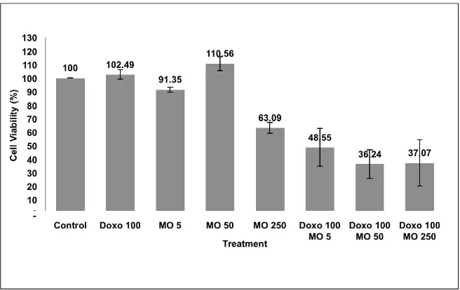
in vivo. Our study showed that single treatment of EMO showed weak cytotoxic effect on HeLa cells with IC50 >250 µg/mL (Fig 1). We did not check the cytotoxicity of EMO at concentrations higher than 250 µg/mL. Previous study mentioned that potent plant derived anticancer agent must have IC50 value less than 100 µg/mL, otherwise it remains potential to be developed as a chemopreventive agent [9].
Combination of 100 nM doxorubicin with EMO 5, 50, and 250 g/mL showed higher inhibition of proliferation than single treatment of doxorubicin on HeLa (Fig 2). At the higher combination concentration of 250 nM doxorubicin with EMO 5, 50, and 250 µg/mL,
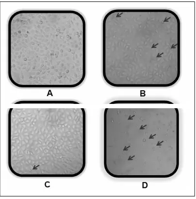
higher inhibition of proliferation was also observed compared to single treatment of 250 nM doxorubicin on HeLa cells (Fig 3). Morphology of the treated cell (Fig 4) also showed single treatment of EMO 250 µ g/mL did not induce extensive cell death, neither did doxorubicin 100 nM, while combination of EMO 250 µ g/mL and doxorubicin 100 nM changed cell morphology extensively compared to control which is shown cell death in the most. Probably, this increased of cytotoxic effect may be due to apoptotic induction.
A previous study of doxorubicin cytotoxicity on HeLa cells showed that doxorubicin was weakly cytotoxic on HeLa with IC50 value of 1000 nM [10]. This is due to the characteristic of HeLa cells that is resistant to doxorubicin, of which the p53 protein had already been degraded by E6 expressed by the Human Papilloma Virus (HPV) [1]. Protein p53 is a regulator of cell cycle and apoptosis due to DNA damage [12]. Our next experiment also confirmed that the increase in doxorubicin cytotoxicity is as a result of induction of apoptosis.
3.2 Apoptosis Assay
Fig 1. Effect of EMO to the viability of HeLa cells. The assay performed by incubating 5x10³ of HeLa cells/well with EMO (5-250 g/mL) for 24 hours. After 24 hours, cells were added by MTT reagent to calculate the absorbance which represent viable cells.
Fig 3. The effect of EMO and doxorubicin to the viability of HeLa cells. The assay performed by incubating 5x10³ HeLa cells/well with EMO (5, 50 and 250 µ g/mL) and doxorubicin (250 nM) on HeLa cells for 24 hours. After 24 hours, cells were added by MTT reagent to calculate the absorbance which represent viable cells. Cell viability profile was shown from average ± standard of error (SE) of 3 experiment. *combination was considered significant to doxorubicin single treatment (p< 0.05).
Fig 5. Apoptotic effect of EMO and doxorubicin on HeLa cells. Cell control (A) 100 nM doxorubicin (B), 250 µ g/mL EMO (C), and combination of 100 nM doxorubicin and 250 µ g/mL EMO (D). The assay performed by incubating 5x10⁴ of HeLa cells/well with doxorubicin, EMO and their combination on HeLa cells for 15 hours and stained by etidium bromide-acridine orange. Viable cells give green fluorescence, apoptotic cells give orange fluorescence (pointed by red arrow). Examination of apoptosis was done by using fluorescent microscope with magnification 400 x.
4. Discussion
This present study explored the effect of EMO alone and in combination with doxorubicin on cytotoxicity and apoptosis induction of HeLa cells. Single treatment of EMO did not show potent cytotoxic effect, while combination with doxorubicin increased cytotoxic effect of doxorubicin. These results are interesting to be evaluated. Combination of EMO and doxorubicin probably increased concentration of intracellular EMO. Previous study reported doxorubicin induced cell membrane peroxidation leads to membrane leakage and increased transport of EMO into the cells [13]. The proposed mechanism must be explored in the future study.
Transmembrane transporter is one of the cause of cancer cell resistances to chemotherapy. Overexpression of transmembrane transporter decreases intracellular concentration of chemotherapeutic and conferring multidrug resistance [14]. Chemotherapeutic agents such
as doxorubicin induce expression of transmembrane transporter via activation of NF-kB, a transcription factor of transmembrane transporter protein [15]. Another reason is due to resistance to induction of apoptosis. Apoptosis pathway in HeLa cells does not occur via p53 pathway because of degraded p53 in these cells [11]. Resistance to apoptosis on HeLa cells is also mediated by overexpression of Bcl-2, an antiapoptosis protein [16]. Expression of Bcl-2 is regulated by NF-kB [17]. Thus, inhibition of NF-kB activation is an important role in reversing chemotherapeutic resistance.
by down-regulating Bcl-2/XIAP on MCF-7 cells [18]. Probably compounds in EMO are able to inhibit NF-êB activation and downregulate Bcl-2 on HeLa cells. Another possible way is that compounds in EMO may also able to stabilize p53 in HeLa cells, so that cells would take p53 pathway in their apoptotic pathway. The increase of post combination treatment could also be due to the p53 independent apoptosis induction mechanism of isothiocyanates found in EMO. Both PEITC and BITC can induce apoptosis through a p53 independent pathway [19,20]. The above proposed mechanism above needs to be explored further.
The present study, showed the potency of
Moringa oleifera to be delevoped as a co-chemotherapeutic agent for doxorubicin. The use of doxorubicin together with EMO is expected to increase the activity and reduce the side effects of doxorubicin. However, the molecular mechanism of apoptotic induction by this combination need to be explored in detail.
5. Conclusion
This research showed that combination of EMO and doxorubicin increased the effect of doxorubicin through apoptotic induction. Based on this result, EMO is potential to be delevoped as a co-chemotherapeutic agent for doxorubicin in cervical cancer therapy.
1. Jemal A, Siegel R, Xu J, Ward E. (2010) CA Cancer J Clin, doi: 10.3322/caac.20073.
2. Tyagi AK, Agarwal C, Chan DCF, Agarwal R.
(2004) Oncology Reports, 11, 493-499.
3. Fimognari C, Nusse MN, Lenzi M, Sciuscio D, Cantelli-Forti G, Hrelia P. (2006) Mutation Research, 601:92–101.
4. Bharali R, Tabassum J, Azad, MRH. (2003) Asia Pacific Journal of Cancer Prevention, 4: 131 – 139.
5. Hetch SS. (2009) Journal of Nutrition, 129: 768s – 774s.
6. Srivastava SK, Singh SV. (2004) Carcinogenesis, 25(9): 1701-1709.
7. Nur KA, Putri H, Cahyani FM, Katarina A, Susidarti RA, Meiyanto E. (2010) Indonesian Journal of Cancer Chemoprevention, 1(2):124-128.
8. Mosmann T. (1983) J. Immunol Methods 65:55-63.
9. Prayong P, Barusrux S, Weerapreeyakuleda N. (2008) Fitoterapia 79: 598–601
10. Kusharyanti I, Larasati, Susidarti RA, Meiyanto E. (2011) Indonesian Journal of Cancer Chemoprevention, 2(2):267-273.
11. Desaintes C, Goyat S, Garbay S, Yaniv M, Thierry F. (1999) Oncogene, 18:4583-4545.
12. Foster I. (2008) Radiography, 14:144-149.
13. Minotti G, Menna P, Salvatorelli E, Cairo G, Gianni L. (2004) Pharmacol Rev 56: 185-228.
14. Valeria P. (2005) Cancer cell International, 5 (20).
15. Olivier S, Robe P, Bours V. (2006) Biochemical Pharmacology, 72:1054-1068.
17. Shishodia S, Amin HM, Lai R, Aggarwal BB. (2005) Biochemical Pharmacology, 70:700-713.
18. Lee JW, Cho MK. (2008) Arch. Pharm. Res., 31(12): 1604-1612.
19. Kim Su-Hyeong, Singh SV. (2011) Cancer Prev Res (Phila)., 3(6):718-726.
20. Xiao D, Singh SV. (2010) Pharmaceutical Research, Vol. 27, No. 4, p:722-731.