The journal homepage www.jpacr.ub.ac.id p-ISSN : 2302 – 4690 | e-ISSN : 2541 – 0733
Stability of Silver Nanoparticles as Imaging Materials
Umi Nur Sholikhah1*, Anung Pujiyanto2, Enny Lestari3, Endang Sarmini4, Triani Widyaningrum5, Kadarisman6,
Triyanto7, Putri Puspitasari8
1-7
Center for Radioisotope and Radiopharmaceutical Technology, National Nuclear Energy Agency, Puspiptek Area, Build 11, South Tangerang, 15314
8
Chemistry Department, Mathematics and Natural Sciences Faculty, Sebelas Maret University, Surakarta
*
Corresponding email : [email protected]
Received 20 July 2016; Revised 24 August 2016; Accepted 17 October 2016
ABSTRACT
Determining the stability of silver nanoparticles is a very important process. It was associated with unwanted metal charge and materials properties. Therefore, we studied to synthesis and stability of silver nanoparticles (AgNPs). The synthesis was performed by reduction method used sodium borohydride (NaBH4). Silver nitrate solution 0.0005 M in 1 mL was reduced using 1 mL 0.002 M of NaBH4. Then a 40.0 L of
polyvinylpyrrolidone 0.3% and 20 L of 1.5 N NaCl was added to the mixture. Characterization of silver nanoparticles is undertaken using spectrophotometer UV-Vis, transmission electron microscopy, particle size analyzer and zeta potential. The stability of products is observed for 5 times using spectrophotometer UV-Vis. The product was characterized by determining its surface plasmon resonance (SPR) of AgNPs and the result was obtained at 403 nm. The size of AgNPs was 20 nm using tomography emission microscopy analysis and the particle size distribution give 5.8 nm. The dielectric charge was 53 mV. The stable AgNPs showed no significant SPR shift at 402±0.89 nm wavelength during 5 days observation. Based on the size and stability, it was suitable for imaging materials.
Key word: silver nanoparticles, Surface Plasmon Resonance, Imaging
INTRODUCTION
shapes. The characteristic of Ag NPs formation can be determined by surface plasmon resonance (SPR) absorption at about 406 nm using UV-Vis spectrophotometer [16]. In addition, zeta potential analysis refers to measuring the stability of the nanoparticles systems. Its value can be negative or positive indicates the repulsion between adjacent or charged particles in the system which can give impact to nanoparticles stability [17].
In the present study, the synthesis of silver nanoparticles by NaBH4 reduction method was carried out. It was characterized by UV-Vis spectra, TEM, PSA and zeta potential analysis. The aims were to synthesis silver nanoparticles below 50 nm and stable during storage.
EXPERIMENT
Chemicals and instrumentation
The chemicals used were silver nitrate (AgNO3), sodium chloride (NaCl) and pH universal indicator (pH 1-14) which were obtained from Merck. Sodium borohydride (NaBH4) and polyvinylpyrrolidone (PVP) were obtained from Sigma Aldrich. Demineralized water was used as solvent from Center for Radioisotope and Radiopharmaceutical Technology. It also used aluminum foil to seal the vial samples. The tools used include glassware, micropipette, disposable cuvette and vials. Some instruments operated for analysis were Acculab analytical balance, UV-Vis spectrophotometer (Jasco32), TEM (JEOL) of Gadjah Mada University, particle size analyzer and zeta potential (Nanoplus) of PT Nanotech Herbal Indonesia.
Procedure reaction
Silver Nanoparticle Synthesis
Synthesis of Ag NPs made by reduction method following Chrastina and Schnitzer (2010) [1] using sodium borohydride (NaBH4). Silver nitrate solution was 0.0005 M in 1 mL was reduced by adding dropwise 1 mL of 0.002 M sodium borohydride (NaBH4) without stirring. Then, 40 L of polyvinylpyrrolidone 0.3% (w/v) and 20 L of 1.5 M NaCl were added to the mixture. The color changes and pH value of the solution were observed. Samples were stored in sealed bottles by aluminum foil and stored in the refrigerator. The Ag NPs characterization was performed using UV-Vis spectrophotometer, transmission electron microscopy (TEM), particle size analyzer (PSA) and zeta potential.
Stability of Silver Nanoparticles
The stability testing was carried out by measuring the SPR used a UV-Vis spectrophotometer for 5 days and also visual observation.
RESULT AND DISCUSSION
Silver nanoparticles were successfully prepared by the reduction method. Stabilizer and reducing agent played an important role on synthesis. This research used sodium borohydride as a strong reducing agent, PVP as polymeric stabilizer and NaCl as an ionic stabilizer. Silver nanoparticle was reduced by NaBH4 as reaction below:
AgNO3 + NaBH4 + 3H2O Ag0 + B(OH)3 + NaNO3 + 3.5 H2
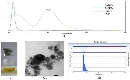
Figure. 1 UV-Vis spectra of Ag NPs and raw materials (a), Ag NPs solution (b) TEM of Ag NPs (c), particle size analyzer result of Ag NPs (d)
UV - Vis spectrophotometer analysis also can be used to predict the size and shape of the nanoparticles. Moreover, this analysis is also the quickest and easiest type of analysis to determine whether the nanoparticles have been formed. Identification of silver nanoparticles formation based on the position of surface plasmon resonance in the UV-Vis spectrum shown in Figure 1.a. From the UV-Vis spectra show there is no evidence of AgNO3, NaBH4 and PVP absorption in the range of 400–800 nm but Ag NPs show a distinct absorption at around 403 nm which corresponds to SPR band of silver nanoparticles. The SPR band position is dependent on nanoparticles size. Particles size was analyzed using TEM shown in Figure 1.c, their size was 20 nm formed with a round shape. Distribution of particles was measured using PSA, and the result of PVP-capped Ag NPs have average diameters 5.8 ± 1.5 nm and polydispersity index was 0.504 shown in Figure 1.d. It means the uniformity of nanoparticles size. Besides nanoparticles size, we also analyzed zeta potential. Zeta potential value of this Ag NPs in demineralized water was -53.77 mV. Silver nanoparticles in demineralized water gave negative zeta potential means the adsorption of various anions onto the Ag NPs surface. High zeta value means there is probability to aggregate. The higher of zeta value was showed the higher chance to aggregate.
Stability of Silver Nanoparticles
stabilizing Ag NPs. The stability of silver nanoparticles was measured using Spectrophotometer UV-Vis for 5 days. The measurement data are shown in Table 1.
Table 1. Silver nanoparticle data measurements
Sample AgNPs max (nm) Absorbance FWHM (nm)
1st day (18-01-2016) 403 1.51 89.1258
2nd day (19-01-2016) 401 1.92 76.8949
3rd day (20-01-2016) 403 1.24 86.2404
4th day (21-01-2016) 403 2.02 74.1463
5th day (22-01-2016) 402 2.45 74.1224
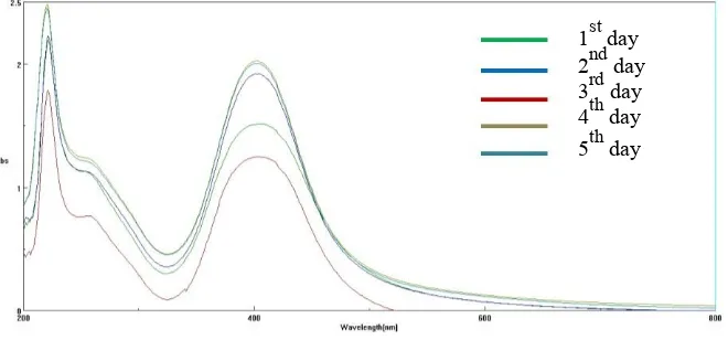
Wavelength maximum measurements over 5 times did not show significant changes shown in Figure 2. The retrieved wavelength of maximum was average 402±0.89 nm. As UV–vis surface plasmon resonance band showed significant changes, Figure 2 indicated good stability of Ag NPs in demineralized water. The absorbance showed increasing over 5 times observation mean the silver reaction still on the process but full width at half maximum decreases. The full width at half maximum (FWHM) corresponding peaks determines dispersity of the nanoparticles, where a large FWHM is attributed to peak broadening and hence, polydispersity. The average of FWHM is 80.1059 nm as the size of Ag NPs were 20 nm. The Full width at half maximum gives information about the particle size distribution. The position of the peak depends on the size of nanoparticles directly. The SPR band position shifts to longer wavelengths as the particle size increases. Aggregation of nanoparticles can change the physical and chemical properties of nanoparticles. Moreover, it also can reduce the ratio of the surface area of the nanoparticles. To overcome this, a stabilizer used to get the size of the nanoparticles stable because of uncontrolled aggregation process. Poly Vinyl pyrrolidone (PVP) is used as a stabilizer polymer and NaCl as stabilizer that serves as a deterrent ionic interaction of charged so as to avoid the aggregation process. The observation of the day to 5 showed still clear yellow solution without sediment.
1st day 2nd day 3rd day 4th day 5th day
Figure 2. UV-Vis Spectra on AgNPs stability measurement
CONCLUSION
Silver nanoparticles were very stable in demineralized water at pH 9. Nanoparticles sizes were 20 nm and spherical shapes. The SPR of silver nanoparticles exhibited at 403 nm wavelength. The average diameters were 5.8 ± 1.5 nm and polydispersity Index was 0.504. The dielectric charge was -53 mV. The stable Ag NPs showed there was no significant SPR shift at 402 ± 0.89 nm wavelengths during 5 days observation. Based on the size and stability, it was suitable for imaging material.
ACKNOWLEDGMENT
This research was funded by DIPA-BATAN 2015.
REFERENCES
[1] Chrastina and J. E. Schnitzer, Int.J. Nanomed., 2010, 5, 653-659.
[2] Devaraj, P., Arun, R., Aarti, C., Prachi, K., Int. .J Pharm. Pharm. Sci., 2014, 6(8), 1-5. [3] Vera V. Pinto, Maria José Ferreiraa, Ricardo Silva, Hélder A. Santosc, F. Silva and
Carlos M. Pereira, Colloid Surf. A., 2010, 364,19-25.
[4] S. Iravani, H. Korbekandi, S.V. Mirmohammadi, and B. Zolfaghari, Res. Pharm. Sci., 2014, 9(6), 385-406.
[5] Kamyar, S., Mansor, B. A., Mohsen, Z., Wan, M. D. Z. Y., Abdolhossein, R. and K. Shameli, Int.J. Nanomed., 2011, 6, 581–590.
[6] Bin Ahmad, M. J.J., Lim, K., Shameli, N.A., Ibrahim, M.Y., Tay, Molecules, 2011, 16, 7237-7248.
[7] Mavani, K., Shah, S., Int. J. Eng. Res. Technol. (IJERT), 2013, 2, 1-5.
[8] Rashid, M.U., Bhuiyan, M.K.H., Quayum, M. E., J. Pharm,. Sci., 2013, 12(1), 29-33. [9] Rodríguez-León, E., Ramón, I., Rosa, E. N., Ronaldo, H., Judith, T., Claudia, I. P., and
Amir, M., Nanoscale Res. Lett., 2013, 8, 318-326.
[10] Dong, P. V., Chu, H. H. and Jörn, K., Int. Nano Lett., 2012, 2(1), 1-9.
[11] Flavio, P., Colloidal stability of silver nanoparticles and their interactions with the alga Chlamydomonas reinhardtii, Environmental Sciences, ETH Zurich, Switzerland, 2012. [12] Doolette, C. L., Mike, J. M., Jason K. K., Damien, J. B., Hugh, H. H., Huoqing G.,
and Geert C., Chem.Cent. J., 2013, 7(46), 1-18
[13] Ahmed, T., S., Imdad, S., Ashraf, N.M., Butt, IJTAN, 2012, 1(1), 111-116.
[14] Malina, D., Agnieszka, S., Zbigniew, W., and Zygmunt, K., Dig. J. Nanomater. Bios., 2012, 7(4), 1527-1534.
[15] Pamies, R., Cifre, J.G.H., Espin, V.F., Collado-González, M., Baños, F., and Torre, J.G., J. Nanopart. Res., 2014, 16, 2376,1-11.