e-ISSN: 2278-3008, p-ISSN:2319-7676. Volume 8, Issue 5 (Nov. – Dec. 2013), PP 58-65 www.iosrjournals.org
Secondary metabolites, antimicrobial, brine shrimp lethality & 4
thinstar
Culex quinquefasciatus
mosquito larvicidal screening of
organic & inorganic root extracts of
Microcos paniculata.
Md. Abdullah Aziz
1*, Mohammad Mahfuz Ali Khan Shawn
2,
Shahariar Rahman
1, Tariqul Islam
1, Mohoni Mita
3,
Abdullah Faruque
1,
Md. Sohel Rana
11
Laboratory of Natural Products Research, Department of Pharmacy, Jahangirnagar University, Savar, Dhaka, Bangladesh.
2
Department of Biochemistry & Molecular Biology, Jahangirnagar University, Savar, Dhaka, Bangladesh.
3
Department of Pharmacy, Atish Dipankar University of Science & Technology, Dhaka, Bangladesh.
Abstract: The present investigation highlights the antimicrobial, toxic & larvicidal activities of the root extracts of Microcos paniculata using various organic & inorganic solvents (methanol, chloroform and water). Antimicrobial, toxicity & larvicidal outcomes were studied by means of agar disc diffusion, brine shrimp lethality bioassay (BSLB) & standard WHO protocol with slight modification. The susceptibility of the microorganisms to the plant extracts was compared with standard antibiotic ciprofloxacin. Root chloroform extract (RCE) revealed a broad spectrum of antimicrobial activity in contrast to other extracts, particularly significant against gram negative bacteria, Vibrio cholerae being the most (zone of inhibition 27 mm).Moreover, the RCE was found to be the most toxic to brine shrimp nauplii, with LC50 of 19.4 μg/ml,
whereas anticancer drug vincristine sulphate (VS) proved LC50 value 1.7 μg/ml indicating that there is a
possibility of finding potent toxic compounds in this plant have affinity relatively for non- polar solvents. 4th instar Culex quinquefasciatus larval mortality was observed and recorded after 24 h exposure period. Both the root methanolic extract (RME) & RCE exhibited larvicidal effects with LC50 of 257.3 μg/ml & 267.6 μg/ml
respectively. Taken together, these results suggest that the organic fractions, RME & RCE could be potential sources of natural antimicrobial, toxic & larvicidal agents.
Keywords:Microcos paniculata, Zone of inhibition, Brine shrimp lethality bioassay, Culex quinquefasciatus, LC50.
I. Introduction
Microcos paniculata L. (English Name: Microcos; Family: Tiliacece; Synonym: Microcos nervosa
(Lour.) S. Y. Hu, Grewia nervosa (Lour.) Panigrahi) locally known as ‘Kathgua or Fattashi’ in Bangladesh. It is also known as Bu zha ye, Po bu ye (Transcribed Chinese);Kaphla (Transcribed Thai). It is a herbaceous plant which looks like a shrub or small tree that widely distributed and naturally grown throughout Bangladesh. It is also native and distributed more or less throughout India, Andaman and Nicobar (Andaman Islands), Sri Lanka, China, Cambodia, Myanmar, Thailand, Vietnam, Indonesia and Malaysia. Microcos paniculata L. is added in Chinese herb tea. It's claimed to have medicinal values as well. The taste of it is mildly sour. Traditional beliefs claim it services the digestive system to work better and it is additionally employed for other health conditions inclusive of colds ,diarrhea, hepatitis, heat stroke and dyspepsia. This plant traditionally used in wound healing, fever and as an insecticide in Bangladesh. Literature study reveals that the stem bark of Microcos paniculata
contained a new alkaloid, N-Methyl-6 beta-(deca-1',3',5'-trienyl)-3 beta-methoxy-2 beta-methylpiperidine, which showed good insecticidal activity against Aedes aegypti second instar larvae. Another study claims that two new piperidine alkaloids, microcosamines A (1) and B (2), were isolated from the leaves of Microcos paniculata. Both new compounds showed significant larvicidal activity against Culex quinquefasciatus. On the other hand, the free-radical-scavenging assay of various solvent extracts of stem of Microcospaniculata yielded five compounds: a new triterpene named methyl 3beta-O-p-hydroxy-Ecinnamoyloxy- 2alpha,23-dihydroxyolean-12-en-28-oate (1), epicatechin (2), 3-trans-feruloyl maslinic acid (3), maslinic acid (4) and sucrose (5). Among them, compound 2 displayed significant free-radical-scavenging activity which is similar to that of standard antioxidant ascorbic acid and therefore may be a promising natural antioxidant. Beides Rahman et al., 2011, investigated ethanolic extract of leaves of M. paniculata for cytotoxic activity[13].. From the existing information it is evident that the plant may possess some important biological activities.
Therefore, the main objective of this study was to evaluate the secondary metabolites, antimicrobial, brine shrimp lethalityand larvicidal activities against the 4th instar larvae of Culex quinquefasciatus filarial vector employing the RME, RCE & RWE(Root water extract) of Microcos paniculata
.
II. Materials And Methods 2.1. Collection and Identification of Plant Materials
Fresh rootsof M. paniculata were collected from the Jahangirnagar University campus, Savar, Dhaka, Bangladesh in January, 2013 that were identified at the Bangladesh National Herbarium having the accession number 35348. A dried specimen was deposited in the herbarium.
2.2. Preparation of the Plant Extracts
Dried plant material (180g) was used for extraction procedure. The extracts were all made with analytical grade solvents (Merck). The roots were washed thoroughly 2-3 times with running water and once with sterile distilled water to remove the dust particles and then dried under shade for a period of 7 days. The dried plant materials were then ground into fine powders using a laboratory grinding mill. Plant samples were extracted with 900 ml of methanol, chloroform and water separately using a soxhlet apparatus following hot extraction procedure. Then the extract was filtered using Whatman No.1 filter paper. The filtrates were then dried in hot air oven at 400C. Then the extracts were stored under refrigeration at 40C for further studies.
2.3. Preliminary Secondary Metabolites Screening
For preliminary secondary metabolites screening all the extractives were subjected to various tests (Table-1) for determination of chemical nature of the extractives[14].
2.4.1. Test Microorganisms and Preparation of Stock Culture
The activity of plant extracts was tested on thirteen different organisms: four gram positive bacteria (Bacillus subtilis, Bacillus cereus, Bacillus megaterium, Staphylococcus aureus) and nine gram negative bacteria (Salmonella typhi, Vibrio cholerae, Proteus mirabilis, Escherichia coli, Salmonella typhi, Serratia spp., Erwinia spp., Pseudomonas spp., Salmonella spp., Shigella boydii) were kindly provided by the Department of Microbiology, Jahangirnagar University, Savar, Dhaka, Bangladesh and reconfirmed by gram staining and sub culturing in appropriate selective media.
2.4.2. Preparation of Standard Culture Inoculum of Test Microorganisms
Three or four isolated colonies were inoculated in the 2 ml nutrient broth and incubated till the growth in the broth was equivalent with Mac-Farland standard (0.5%) as recommended by WHO.
2.4.3. Antimicrobial Assay
And they were allowed to cool under Laminar air flow. Aseptically transfer about 20 ml of media into each sterile Petri dishes and allowed to solidify. 1 ml inoculum suspension was spread uniformly over the agar medium using sterile glass rod to get uniform distribution of bacteria. The readily prepared sterile discs were loaded. The paper diffuse discs were placed on the medium suitably apart and the plate were incubated at 5°C for 1 hour to permit good diffusion and then transferred to an incubator at 37°C for 24 hours. The antimicrobial activity was recorded by measuring the width of the clear inhibition zone around the disc using zone reader (mm).
2.5. Brine Shrimp Lethality Bioassay
The experiment was carried out using the method described by Meyer et al. (1982) [16]. In brief,
Artemia salina Leach (brine shrimp eggs) was allowed to hatch and matured as nauplii (Larvae) in seawater for 48 hrs at 250C. Serially diluted test solutions were added to the seawater, containing 10 nauplii. After incubation for 24 h at 250C, the number of survivors was counted. Vincristine sulfate was used as positive control.
2.6. Larvicidal Bioassay
The Larvicidal bioassay was carried as per standard WHO protocol with slight modification [17]. Various extracts of Microcos paniculata were investigated for their larvicidal activity against 4th instar larvae of
Culex quinquefasciatus. In the present study, for treatment of larvae with the root extracts of M. paniculata, 100 ml of tap water was kept in a series of glass beakers (of 200 ml capacity). Required quantity of stock solution was added into each beaker (containing 100 ml of tap water) to obtain a particular concentration of the extracts. Control medium was also maintained with 100 ml of tap water. Separate series of exposure medium with desired concentrations of extracts were kept for C. quinquefasciatus. The larval mortality of fourth instar larvae of C. quinquefasciatus were observed separately incontrol, 50, 100, 150, 200, 250, 300, 350, 400, 450 and 500 ppmconcentrations. Twenty numbersof 4th instar larvae of C. quinquefasciatus were separately introduced into control and different concentrations of root extracts. The number of alive and dead larvae at the end of 24 h , response %, corrected response %, linear response %, linear probit, LC50, LC90, lower limit & upper limit and chi-square values were recorded.
III. Statistical Analysis
The LC50 (50% lethal concentration, μg/ml) of root extracts were determined from duplicate experiments. LC90 and other statistics at 95 percent fiducial limits of upper confidence limit and lower confidence limit and Chi-square values were calculated by using LDP Line software (trial version).
IV. Results
The crude extractives when tested with various chemical reagents demonstrated the presence of alkaloids, glycosides, glucosides, saponins, tannins, steroids and carbohydrates shown in Table 1.
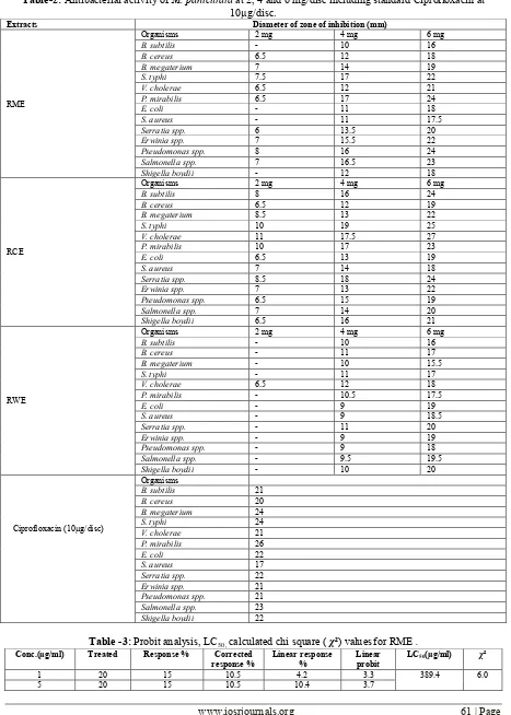
The antibacterial activity of specific concentrations of different extracts of M. paniculata is given in Table 2. The crude extracts of plant showed potent antibacterial activity. All the plant extracts showed dose dependent inhibition. Among the extracts, RCE showed maximum efficacy against all the bacteria and satisfactory inhibition against gram negative bacteria selected, V. cholera being the most. The standard, ciprofloxacin, exhibited significant zone of inhibition against all the test organisms. Among them, gram negative bacteria were inhibited more than gram positive bacteria by ciprofloxacin, E.coli being the most.
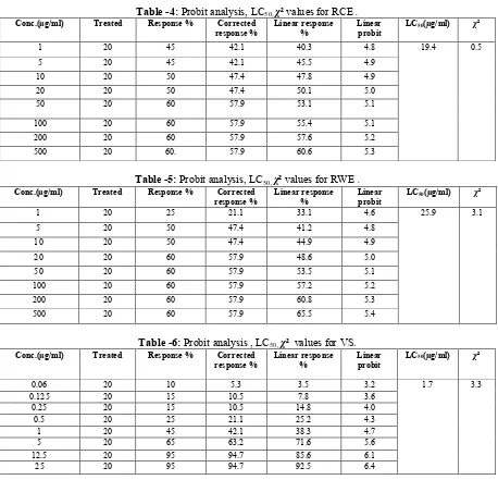
In case of BSLB, RCE was found to be the most toxic to Brine Shrimp nauplii, with LC50 of 19.4 μg/ml, whereas the anticancer drug VS showed LC50 value 1.7 μg/ml. The order at which brine shrimp lethality potential of the test samples decreased was as follows:
Vincristine sulphate > RCE > RWE > RME.
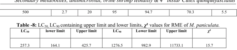
The results of various extracts of M. paniculata were screened for their larvicidal activity against 4th instar larvae of Culex quinquefasciatus at the end of 24 h are represented in (Table 7-12). Among the three extracts screened, RME showed promising larvicidal effects. Remaining extracts also showed considerable larvicidal activities. Based on the Probit analysis, the 24 h LC50 value of RME for Culex quinquefasciatus was found to 257.3 ppm (Table 8).
Table-1: Secondary metabolites screening of root extracts of M. paniculata.
Compounds Observation of various extracts
Methanol Chloroform Water
Alkaloid + + +
Saponin + - +
Tannin - + +
Flavonoid - - -
Carbohydrate + + +
Glucoside - + -
Glycoside + + + or -
(+) = Present; (-) = absent
Table-2: Antibacterial activity of M. paniculata at 2, 4 and 6 mg/disc including standard Ciprofloxacin at 10µg/disc.
Extracts Diameter of zone of inhibition (mm)
RME
Conc.(µg/ml) Treated Response % Corrected
10 20 15 10.5 14.5 3.9
Conc.(µg/ml) Treated Response % Corrected
response %
Conc.(µg/ml) Treated Response % Corrected
response %
Conc.(µg/ml) Treated Response % Corrected
response %
Table -7: Response %, corrected response % , linear response % and linear probit of 4th instar larvae of Culex quinquefasciatus exposed for 24 hours to different concentrations for RME of M. paniculata.
Concentration(PPM) Log conc. Treated Response
Table -8: LC50, LC90 containing upper limit and lower limits, χ² values for RME of M. paniculata.
LC50 lower limit Upper limit LC90 Lower limit Upper limit χ²
257.3 164.1 425.7 1276.5 982.9 11733.1 15.7
Table -9: Response %, corrected response % , linear response % and linear probit of 4th instar larvae of Culex quinquefasciatus exposed for 24 hours to different concentrations for RCE of M. paniculata.
Concentration(PPM) Log conc. Treated Response
%
Corrected response %
Linear response % Linear
probit
50 1.7 20 20 15.8 3.9 3.2
100 2.0 20 20 15.8 15.1 4.0
150 2.2 20 20 15.8 27.2 4.4
200 2.3 20 30 26.3 38.0 4.7
250 2.4 20 40 36.8 47.1 4.9
300 2.5 20 40 36.8 54.8 5.1
350 2.5 20 60 57.9 61.1 5.3
400 2.6 20 70 68.4 66.3 5.4
450 2.7 20 90 89.5 70.7 5.5
500 2.7 20 90 89.5 74.4 5.7
Table -10: LC50, LC90 containing upper limit and lower limits, χ² values for RCE of M. paniculata.
LC50 lower limit Upper limit LC90 Lower limit Upper limit χ²
267.6 181.0 409.2 908.1 766.6 4009.1 19.4
Table -11: Response %, corrected response % , linear response % and linear probit of 4th instar larvae of
Culex quinquefasciatus exposed for 24 hours to different concentrations for RWE of M. paniculata.
Concentration(PPM) Log conc. Treated Response
%
Corrected response %
Linear response % Linear
probit
50 1.7 20 10 5.3 3.2 3.2
100 2.0 20 10 5.3 7.6 3.6
150 2.2 20 15 10.5 11.8 3.8
200 2.3 20 20 15.8 15.6 4.0
250 2.4 20 20 15.8 19.1 4.1
300 2.5 20 30 26.3 22.2 4.2
350 2.5 20 30 26.3 25.1 4.3
400 2.6 20 30 26.3 27.7 4.4
450 2.7 20 30 26.3 30.2 4.5
500 2.7 20 40 36.8 32.4 4.5
Table 12: LC50, LC90 containing upper limit and lower limits, χ² values for RWE of M. paniculata.
LC50 lower limit Upper limit LC90 Lower limit Upper limit χ²
1061.1 596.8 8838.9 8807.7 2327.9 1776985.2 1.2
V. Discussion
Plants used in Ayurveda can provide biologically active molecules and lead structures for the development of modified derivatives with enhanced activity and /or reduced toxicity of the 2,50,000 higher plant species on earth, around 5000 species have specific therapeutic value[18].
M. paniculata is one of the medicinal plants that are commonly used by the local practitioners for various human ailments, but no attempt has been made to study its antimicrobial activity.
Polyphenols have been reported to exhibit antibacterial activities with distinguish characteristics [19].The inhibition of microorganisms by phenolic compounds may be due to iron deprivation or hydrogen bounding with vital proteins such as microbial enzymes [20].The RCE of Microcos paniculata has been shown potent antibacterial activity. This may be due to the presence of alkaloid, steroid, tannin and some polyphenolic compounds present in this extract (Table 1). The present study showed that RCE of Microcos paniculata
inhibited the gram negative bacteria better than gram positive bacteria. Phytochemical constituents such as alkaloids, flavonoids, tannins, phenols, saponins, and several other aromatic compounds are secondary metabolites of plants that serve a defense mechanism against prediction by many microorganisms, insects and
other herbivores [21]. Analysis of plant extracts revealed the presence of alkaloids, glycosides, saponins, steroids and tannins which could be responsible for the observed antimicrobial property. These bioactive compounds are known to act by different mechanism and exert antimicrobial action.Tannins bind to proline rich proteins and interfere with the protein synthesis [22].Flavonoids are hydroxylated phenolic substance known to be synthesized by plants in response to microbial infection and it should not be surprising that they have been found in vitro to be effective antimicrobial substances against a wide array of microorganisms. Their activity is probably due to their ability to complex with extracellular and soluble proteins and to complex with bacterial cell walls [23]. Antimicrobial property of saponin is due to its ability to cause leakage of proteins and certain enzymes from the cell [24]. Steroids have been reported to have antibacterial properties, the correlation between membrane lipids and sensitivity for steroidal compound indicates the mechanism in which steroids specifically associate with membrane lipid and exerts its action by causing leakages from liposomes [25]. The higher antimicrobial activity of the chloroform extract as compared to aqueous & methanolic extracts might be due to the lack of solubility of active constituents in aqueous solution. It is clear that the effectiveness of the extracts largely depends on the type of solvent used. The organic extracts provided more powerful antimicrobial activity as compared to aqueous extract. This observation clearly indicates that the existence of non-polar residues in the extracts which have higher antimicrobial abilities.
Brine shrimp lethality bioassay is widely used in the bioassay for the bioactive compounds [16, 26].In toxicity evaluation of plant extracts by Brine shrimp lethality bioassay LC50values lower than 1000 μg/ml are considered bioactive [16].The Brine Shrimp Lethality Bioassay also indicates antifungal effects, pesticidal effects, teratogenic effects, toxicity to environment and many more [27].Tables 3,4,5 show the lethality of different extracts of M. paniculata to the Brine Shrimp nauplii. The degree of lethality shown by the extractives was found to be directly proportional to the concentration of the extractives ranging from the lowest
concentration (1 μg/ml) to the highest concentration (500 μg/ml). This concentration dependent increment in percent mortality of Brine Shrimp nauplii produced by the M. paniculata indicates the presence of cytotoxic principles in these extractives.Preliminary phytochemical screening revealed the presence of alkaloids and steroids. So the observedcytotoxic action may be due to the presence of such compounds. Again,reports exist onthe role of alkaloids and steroids in cytotoxic activity of plant extracts [28, 29, 30].Moreover, this significant lethality of the plant extracts (LC50 values less than 390 µg/ml) to brine shrimp is indicative of the presence of potent cytotoxic and probably insecticidal compounds which warrants further investigation.
Vector-borne diseases constitute the major cause of morbidity in most of the tropical and subtropical countries and have always been a challenge to the medical professionals struggling for the welfare of humanity. Mosquitoes are the most deadly vector for several of these disease causing organisms. In many parts of the world, plant-derived natural products have traditionally been used against mosquitoes [31]. In the search for safer insecticide technologies, more selective modes of action and reduced risks for non-target organisms and the environment, progress has been made in the last twenty years with the development of natural compounds capable of interfering with the processes of growth, development and metamorphosis of the target insects [32]. Phytochemicals may serve as suitable alternatives to synthetic insecticides as they are relatively safe and are readily available in many areas of the world. According to Bowers et al. (1995) [33] the screening of locally available medicinal plants for mosquito control would reduce dependence on expensive imported products and stimulate local efforts to enhance public health and crude extracts of many plants showed larvicidal activity against Culex quinquefasciatus [34].
The findings of the current investigation revealed that the RME part of M. paniculata contained alkaloid and RCE showed the presence of alkaloid, steroid and tannin that may be responsible for their strong larvicidal activities against the 4th instar larvae of Culex quinquefasciatus.
VI. Conclusion
In conclusion, the study clearly point outs that the extracts of Microcos paniculata exhibit potent antimicrobial, toxic & larvicidal properties. Agar disc diffusion, BSLB & larvicidal results may be used to guide the researchers for further experiments on RME & RCE parts of M. paniculata to isolate the bioactive compounds. Other antimicrobial, toxicity & larvicidal tests and specific bioassays may be done on the isolated bioactive compounds later.
Acknowledgement
The authors would like to thank professor Dr. Md. Sohel Rana & professor Abdullah Faruque, department of pharmacy, Jahangirnagar University, Savar, Dhaka, Bangladesh for providing facilities and encouragement.
[1] Krishnaraju, A.V., Rao, T.V.N., Sundararaju, D., Vanisree, M., Tsay, H.S., Subbaraju, G.V., 2006. Biological screening of medicinal plants collected from eastern Ghats of India using Artemia salina (Brine shrimp test). Int. J. Appl. Sci. Eng. 4(2), 115-125.
[2] Cohen, M.L., 1992. Epidemiology of drug resistance: implications for a post antimicrobial era. Science. 257, 1050-1055.
[3] Vermani, K., Garg, S., 2002. Herbal medicines for sexually transmitted diseases and AIDS. J. Ethnopharmacol. 80, 49-66.
[4] Apu, A.S., Bhuyan, S.H., Khatun, F., Liza, M.S., Matin, M., Hossain, M.F., 2013. Assessment of cytotoxic activity of two
medicinal plants using brine shrimp (Artemia salina) as an experimental tool. Int J Pharm Sci Res. 4(3), 1125-1130.
[5] Das, M.K., Ansari, M.A., 2003. Evaluation of repellent action of Cymbopogan martinii martinii Stapf var Sofia oil against
Anopheles sundiacus in tribal villages of Car Nicobar Island, Andaman & Nicobar Islands, India. J Vect Borne Dis.40, 101-4.
[6] Das, P.K., Rajagopalan, P.K., 1981. Role of stimulated migration of mosquitoes in development and reversal of malathion
resistance in Culexpipensfatigans. Indian J.Med. Res. 73, 139-143.
[7] Brown, A.W.A., 1986.Insecticide resistance in mosquitoes: a plagmatic review. J. Am. Mosq. Control. Assoc. 2, 123- 140.
[8] Jeeshna, M. V., Mallikadevi, T., Paulsamy, S., 2010. Screening of the weed plant species, Croton bonplandianumBaill. for
larvicidal activity of Aedes aegypti. JBiopest. 3(1 Special Issue) 192 – 194.
[9] Holder, P.W., Brown, G., Bullians, M., 1999. The mosquitoes of New Zealand and their animal disease significance. Surveillance.
26(4), 12-15.
[10] Samuel, T., Arivoli, S., Martin J. J., 2011. Larvicidal efficacy of Vernonia cinerea (L.) (Asteraceae) leaf extracts against the filarial
vector Culex quinquefasciatus Say (Diptera: Culicidae). JBiopest. 4 (1), 37 – 42.
[11] Jacobson, M., 1958. Insecticides from plants: a review of the literature, 1941-1953. U.S. Dept. Agricultural Hand Book. p. 154.
[12] Alkofahi, A., Ruppercht, J., Anderson, J.E., McLaughlin, J.L., Mikolajezak, K.L., Scott, B.A., 1989. Search for new pesticides from
higher plants. In: Insecticides of plant origin. JAm Chem Soc.p. 25-43.
[13] Rahman, M.A., Sampad, K.S., Hasan, H.N., Saifuzzaman, M., 2011. Analgesic and cytotoxic activities of Microcos paniculata L.
Pharmacologyonline. 1, 779-785.
[14] Ghani, A., 2003. Medicinal Plants of Bangladesh. 2nd Ed. The Asiatic Society of Bangladesh. Dhaka, Bangladesh. pp. 31, 39-40,
418, 500-504, 589-580.
[15] Bayer, A.W., Kirby, W.M.M., Sherris, J.C., Turck, M., 1966. Antibiotic susceptibility testing by a standardized single disc method.
Am. J. Clin. Pathol. 45,493-496.
[16] Meyer, B.N., Ferrigni, N.R., Putnam, J.E., Jacobsen, L.B., Nichols, D.E., Mclaughlin, J.L., 1982. Brine shrimp: a convenient general bioassay for active plant constituents. Planta Med. 45, 31-34.
[17] WHO. Report of the WHO informal consultation on the evaluation and testing of insecticides. Geneva: WHO, 1996.
[18] Saba, I., Maryum, M., Farzana, P., 2012. In-vitro anti-bacterial activities of three medicinal plants using agar well diffusion method. Res. J. Biol. 2(1), 1-8.
[19] Haslam, E.,1996. Natural polyphenols (vegetable tannins) as drugs : possible mode of action. J. Natl. Prod. 5, 205-215.
[20] Scalbert, A., 1991. Antimicrobial properties of tannins. Phytochem. 30, 3875-3883.
[21] Bonjar, G.H.S., Nik, A.K., Aghighi, S., 2004. Antibacterial and antifungal survey in plants used in indigenous herbal -medicine of
south eastregions of Iran. J Biol Sci. 4, 405-412.
[22] Shimada, T., 2006. Salivary proteins as a defense against dietary tannins. J. Chem. Ecol.32 (6), 1149-1163.
[23] Marjorie, C., 1999. Plant products as antimicrobial agents. Clin. Microbiol. Rev.12, 564-582.
[24] Zablotowicz, R.M., Hoagland, R.E., Wagner, S.C., 1996. Effect of saponins on the growth and activity of rhizosphere bacteria. Adv
Exp Med Biol. 405, 83-95.
[25] Raquel, F.E., 2007. Bacterial lipid composition and the antimicrobial efficacy of cationic steroid compounds. Biochimica et
Biophysica Acta. 2500–2509.
[26] Zhao, G.X., Hui, Y.H., Rupprecht, J.K., McLaughlin, J.L., Wood, K.V., 1992. Additional bioactive compounds and trilobacin, a
novel highly cytotoxic acetogenin, from the bark of Asimina triloba. J. Natl. Prod. 55, 347-356.
[27] Vanhaecke, P., Persoone, G., Claus, C., Sorgeloos, P., 1981. Proposal for a short-term toxicity test with Artemia nauplii. Ecotoxicol.
Environ.Safety.5, 382-387.
[28] Badami, S., Manohara, S. A., Kumar, E. P., 2003. Antitumor activity of total alkaloid fraction of Solanum pseudocapsicum
leaves. Phytother. Res. 17, 1001-1004.
[29] Dhar, M. L., Dhar, M. N., Dhawan, B. N., 1973. Screening of Indian medicinal plants for biological activity. Ind. J. Expt. Biol. 11,
43-45.
[30] Vijayan, P., Rreethi, V., Prashanth, S. H., Raghu, H., 2004. Cytotoxicity activity of the total alkaloids isolated from different parts
of Solanum pseudocapsicum. Biol. Pharm. Bull. 24, 528-530.
[31] Curtis, C.F., Lines, J.D., Baolin, L., Renz, A., 1991. Natural and synthetic repellents. In: Curtis, C.F. (Eds.) Control of disease
vectors in the community. Wolf, London, P. 75–92.
[32] Altstein, M., Aharonson, N., Menn, J.J., 1993. Overview: New targets for insect management in crop protection. Arch. Insect Biochem. and Physiol. 22, 5-12.
[33] Bowers, W.S., Sener, B., Evans, P. H., Bingol, F., Erdogan, I., 1995. Activity of Turkish medicinal plants against mosquitoes Aedes
aegypti and Anopheles gambiae. Insect Sci Appl. 16(3/4), 339-342.