NTRODUCTION
Mucous membrane pemphigoid (MMP) is an autoimmune, chronic inflammatory, subepithelial blistering diseasepredominantly affecting mucous membranes that is characterized by linear deposition of IgG, IgA, or C3 along the epithelial basal membrane zone (BMZ).1
The oral mucosa is almost always affected, followed by other mucosa (ocular, nose, pharynx, larynx, esophagus, genitalia, and anus) and rarely skin (5-10%).2 Gingiva is most commonly involved, but oral involvement can also be seen on palate, buccal mucosa, lips, posterior part of pharynx, tongue, and floor of the mouth.3
Clinical diagnosis and management of mucous
membrane pemphigoid: Case Report
Nurdiana*, Bagus S.**
* Resident of Oral Medicine, Faculty of Dentistry Airlangga University, Surabaya – Indonesia ** Department of Oral Medicine, Faculty of Dentistry Airlangga University, Surabaya – Indonesia
Correspondence: Nurdiana,Resident of Oral Medicine, Faculty of Dentistry Airlangga University, Surabaya – Indonesia. Email: [email protected]
Abstract
Background: Mucous membrane pemphigoid (MMP) is a chronic, recurrent autoimmune blistering disease that primarily affects mucous membranes, particularly in oral mucosa. Definite diagnosis of MMP based on anamnesis of disease history, clinical features, and biopsy. The treatment of “low-risk” patients is topical or systemic corticosteroid. The treatment of “high-risk” patient is systemic corticosteroid and/or systemic immunosuppressant (such as, dapsone or azathioprine).Purpose: The purpose of this case report is to discuss clinical diagnosis and management of MMP. Case: This paper reports a case of 60 year-old woman with chronic MMP. Case Management: Diagnosis of this case only based on anamnesis of disease history and clinical features. The biopsy was not done because the patient refused it. The patient was treated with systemic corticosteroid (prednisone). Result: After about 11 weeks of treatment the lesion showed improvement. Conclusion: Definite diagnosis of MMP must be based on biopsy because clinical features of MMP resemble other vesicobullous diseases. MMP does not respond well to the treatment due to its autoimmune nature but the therapy with systemic prednisone give a quite good result.
Keywords:mucous membrane pemphigoid, autoimmnune disease, corticosteroid
The typical lesion is a small or large, clear–fluid bulla, which breaks fairly rapidly leading to pseudomembrane-covered, irregularly shaped erosions.4 Lesions may present as intact bulla of gingival or other mucosal surfaces, but more frequently appear as nonspecific-appearing erosions.5
The lesion usually recur, persist for along time, and occasionally lead to atrophy or scarring.2 Precise data on the incidence of MMP are not known; however, MMP is uncommon.3 MMP found in 2-5 people per 100,000 population a year.6
at around 70 years.7 It appears to be twice as common in women than men.6-7
Oral corticosteroids (prednisone or prednisolone) are the cornerstone of therapy for MMP. The initial dose varies from 30 to 60 mg/day depending on the severity of the disease. It usually takes 2-3 weeks to stop new bulla formation and for old ones to heal. The dose is subsequently tapered by 20% every 2-3 weeks until the dose of 10 mg/day is reached. This dose is subsequently maintained on alternate days and reduced by 5 mg every 2 weeks until it is completely stopped.2
The purpose of this case report is to discuss MMP in 63 year-old female patient that was diagnosed clinically as well as the management of this disease.
CASE
On July 29, 2008, a 63 year-old female patient came to Dental Department of Dr. Soetomo Hospital with chief complain of painful wounds in her mouth. According to anamnesis was revealed that since 2 months ago she had painful wounds resembled large ulcers in her lips, cheeks, and tongue and never healed. This wound made her difficult to eat and speak. Patient then went to the dentist and referred to Dental Department of Dr. Soetomo Hospital.
General condition of the patient was weak. Medical history examination revealed that patient had accident about 30 years ago and diagnosed with contusion cerebri. Since that she often had vertigo and used antivertigo (dimenhydrinate 50 mg, vitamin B6 50 mg). Patient also had gastritis, stomach ulcer,
and occasionally melena. She had to use antacid and antiulserative (omeprazol) everyday. In family medical history there’s no known disorder.
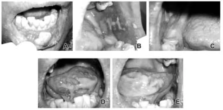
Extra oral examination of lower lips showed long brown crust size 5 x 2 mm (Figure 1, A). Intra oral examination of lower labial mucosa showed irregular bulla size 15 mm (Figure 1, A). Left buccal mucosa showed long irregular bulla size 3-5 mm (Figure 1, B). Right buccal mucosa showed irregular bulla size 12 mm (Figure 1, C). Left lower gingiva showed oval bulla size 15 mm (Figure 1, B). Right lower gingiva showed irregular bulla size 12 mm (Figure 1, C). Left and right lateral tongue showed irregular bulla (Figure 1, D and E).
CASE MANAGEMENT
At 1st
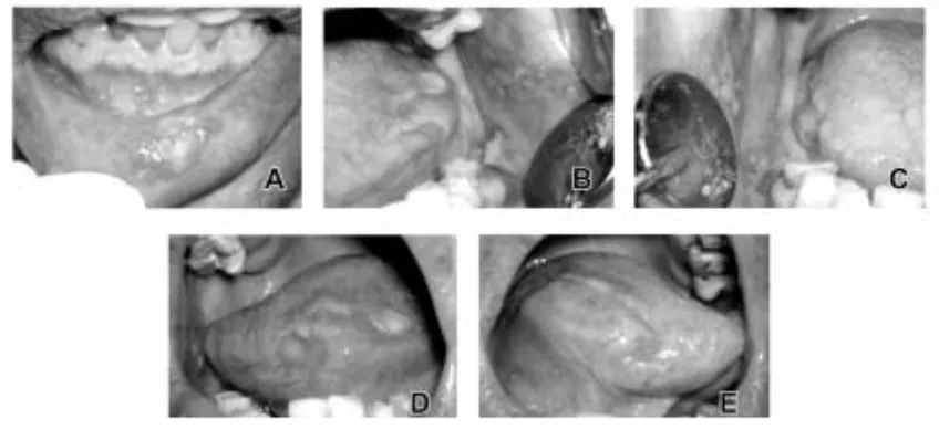
visit, according to anamnesis and clinical examination, clinical diagnosis this case was MMP with pemphigus vulgaris as the differential diagnosis. Patient was instructed to do complete blood, SGPT, SGOT, and blood glucose examination, but she already done it and the result was normal. Patient was treated with prednisone 5 mg 3 times 4 tablets daily, hepatic protector once daily, and benzydamin HCl oral rinse 3 times daily for 2 weeks. At 2nd visit,2 weeks later (August 12, 2008), based on anamnesis it was known that the pain was greatly reduced. Crust on lower lips was healed (Figure 2, A). Lower labial mucosa showed irregular bulla size 10 mm (Figure 2, A). Left buccal mucosa showed irregular bulla size 3 x 10 mm (Figure 2, B). Right buccal mucosa showed three long bullas size
8 x 3 mm, 8 x 4 mm, and 8 x 2 mm (Figure 2, C). Left lower gingiva showed irregular bulla size 12 x 4 mm (Figure 2, B). Bulla on right lower gingiva was healed (Figure 2, C). Left and right lateral tongue showed irregular bulla (Figure 2, D and E). Patient was treated with prednisone 5 mg 3 times 4 tablets daily, hepatic protector once daily, and benzydamin HCl oral rinse 3 times daily for 1 week.
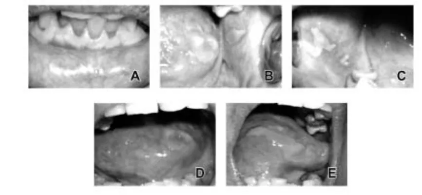
At 3rd visit, 8 days later (August 20, 2008), anamnesisrevealed that the pain was greatly reduced. Lower labial mucosa showed round bulla size 7 mm (Figure 3, A). Left buccal mucosa showed irregular bulla size 3 x 8 mm (Figure 3, B). Right
buccal mucosa showed two irregular bullas size 12 x 10 mm and 10 x 2 mm (Figure 3, C). Left lower gingiva showed long bulla size 12 x 3 mm (Figure 3, B). Right lower gingiva showed oval bulla size 5 x 3 mm (Figure 3, C). Left and right lateral tongue showed irregular bulla (Figure 3, D and E). Patient was treated with prednisone 5 mg 4 times 2 tablets daily, hepatic protector once daily, and benzydamin HCl oral rinse 3 times daily for 2 weeks.
At 4th visit, 2 weeks later (September 3, 2008), according to anamnesis known that the pain was still the same. Lower labial mucosa showed round bulla size 4 mm (Figure 4, A). Left buccal mucosa Figure 2. Visit II (August 12, 2008): bulla on lower labial mucosa, left and right buccal mucosa, left gingiva and left and right lateral
tongue showed improvement, crust on lower lips and bulla on right gingiva was healed.
Figure 4. Visit IV (September 3, 2008): bulla on lower labial mucosa labial and left buccal mucosa showed improvement, bulla on right buccal mucosa, right lower buccal fold mucosa, and left and right lateral tongue became worse, and bulla on left gingiva was healed.
showed oval bulla size 4 x 3 mm (Figure 4, B). Right buccal mucosa showed oval bulla size 15 x 8 mm (Figure 4, C). Right lower buccal fold mucosa showed irregular bulla size 15 x 8 mm (Figure 4, C). Bulla on left lower gingiva was healed (Figure 4, B). Left and right lateral tongue showed irregular bulla (Figure 4, D and E). Patient was treated with prednisone 5 mg 3 times 2 tablets daily, hepatic protector once daily, and benzydamin HCl oral rinse 3 times daily for 2 weeks.
At 5th
visit, from anamnesis 2 weeks later (September 17, 2008), it was known that the pain was still the same. Lower labial mucosa showed round
bulla size 2 mm (Figure 5, A). Left buccal mucosa showed irregular bulla size 10 x 4 mm (Figure 5, B). Right buccal mucosa showed irregular bulla size 10 x 8 mm (Figure 5, C). Bulla on left lower gingiva was healed (Figure 5, B). Left and right lateral tongue showed irregular bulla (Figure 5, D and E). Patient was treated with prednisone 5 mg 4 times daily, hepatic protector once daily, and benzydamin HCl oral rinse 3 times daily for 2 weeks.
At 6 th
visit, 4 weeks later (October 15, 2008), anamnesis revealed that the mouth felt more comfortable. Lower labial mucosa showed round bulla size 1 mm (Figure 6, A). Left buccal mucosa
showed oval bulla size 10 x 6 mm (Figure 6, B). Right buccal mucosa showed two irregular bullas size 12 x 10 mm and 5 x 4 mm (Figure 6, C). Left lateral tongue showed round bulla size 5 x 3 mm (Figure 6, D). Right lateral tongue showed irregular bulla (Figure 6, E). Patient was treated with prednisone 5 mg twice daily, hepatic protector once daily, and benzydamin HCl oral rinse 3 times daily for 2 weeks.
DISCUSSION
The diagnosis of MMP is mainly based on history, clinical examination and biopsy of the lesions.4 Patients with MMP typically present with persistent, painful erosions on the mucous membranes.3 Oral involvement is characterized by erythematous patches, bullas,erosions, and ulcerations which can be seen on palate, buccal mucosa, lips, posterior part of pharynx, tongue, and floor of the mouth, and often begin on gingiva, particularly near the teeth.1,3,7 Intact bullas are rarely seen, but they may appear flaccid or tense.3
When performing a biopsy, it had to include vesicle and perilesional tissue not the erosion itself.4 Biopsy done for routine histopathology and direct immunofluorescent study.5 Histologically there is loss of attachment and separation of the full thickness of epithelium from the connective tissue at BMZ. Epithelium, though separated, remains for a time intact and forms the roof of a bulla. The floor of bulla is formed by connective tissue alone, infiltrated with inflammatory cells.8 Direct immunofluorescent of perilesional mucosa reveals linear deposisition of IgG,
C3, or less commonly IgA along BMZ. The use of salt split skin or mucosal epithelium for indirect immunofluorescence studies may increase the sensitivity of this technique. Indirect immunofluorescence utilizing salt split skin may reveal binding to the roof and/or floor depending upon the antigen targeted.9
In this case, diagnosis was based on history and clinical examination. From anamnesis known that the lesion was persistent and painful. Clinical examination showed bullas on mucous membranes of the mouth (gingiva, buccal mucosa, lips, and tongue). Biopsy was not done because the patient refused to it with the reason that this lesion was very painful and she wanted to wait until the lesion was healed.
Differential diagnosis of this case was pemphigus
vulgaris (PV). Many mucosal diseases appear very
similar; therefore, a complete diagnosis cannot be made without utilizing several clinical tests such as viewing the tissue histologically and using immunofluorescence to confirm the diagnosis.10 Clinical feature of MMP can be differentiated with PV. Classical lesion of PV is a thin-walled bulla on a noninflamed base arising on otherwise normal mucosa. More frequently, the clinician sees shallow irregular ulcers because the bulla rapidly breaks. A thin layer of epithelium peels away in an irregular pattern, leaving a denuded base. The edges of the lesion continue to extend peripherally over a period of weeks until they involve large portions of the oral mucosa.5
This case shown intact bullas on gingiva, Figure 6. Visit VI (October 15, 2008): bulla on lower labial mucosa and left and right lateral tongue showed improvement, and bulla
buccal mucosa, lips and tongue, and also there was no erosion; therefore we diagnosis this case as MMP. There is no gold standard therapy for MMP.11 MMP is a particularly difficult disease to treat.3-4,7 The response of MMP to treatment modalitieshas been variable.11
The primary aim of treatment is to stop bulla formation, promote healing and prevent scarring.3-4,7
Oral care includes appropriate dental hygiene, referred for dental care, monitoring for candidiasis, and topical anesthetics for pain.9
Candida infections
often result with the long-term use of corticosteroids when patients are prone to these infections, so nystatin and other antifungal treatment are often needed as well. 10 Patients with MMP should avoid foods high in acid (e.g., tomatoes and orange juice) and foods with hard surfaces that may mechanically traumatize oral mucosa (e.g., chips and nuts).3 Patients also should identify factor that may cause exacerbations of the disease and avoid these precipitants.7
In this case the patient was treated with prednisone 60 mg/day as the initial dose for 3 weeks. After that the dose was reduced gradually every 2 weeks, except for the dose of 20 mg/day because the patient did not come for the next control. She came 4 weeks after her last control and said that she continued the therapy with dose of 20 mg/day. The patient instructed to eat soft foods and avoid acidic foods. She also instructed to eat high calorie and high vitamin food (e.g. fruit juice)
It can be concluded that diagnosis of MMP based on history, clinical examination and biopsy of the lesion. Definite diagnosis of MMP must be based on biopsy because clinical features of MMP resemble other vesicobullous diseases. MMP is a disorder that extremely difficult to treat which does not respond well to the treatment due to its autoimmune nature, but the therapy with systemic prednisone give a quite good result. The success of treatment also depends on the ability to identify and avoid factors that may cause exacerbations of the disease.
REFERENCES
1. Heffernan MP, Bentley DD. Successful treatment of mucous membrane pemphigoid with infliximab. Arch Dermatol 2006; 142: 1268-70.
2. Laskaris G. Treatment of oral diseases. A concise textbook. 1st
ed. Stuttgart: Thieme; 2005. p. 38-9. 3. Freiman A. Cicatricial pemphigoid. Available from:
http://www.emedicine.com/derm/topic79.htm. Accessed August 10, 2008.
4. Uppoor AS, Humagain M, Nayak DG, et al. Mucous
membrane pemphigoid: case report with review of literature. Available from: http://www.priory.com/ den/pemphigoid.pdf. Accessed February 1, 2009. 5. Greenberg MS. Ulcerative, vesicular, and bullous
lesions of the oral musoca. In: Greenberg MS, Glick M. Burket’s oral medicine diagnosis & treatment. 10th
ed. Hamilton: BC Decker; 2003. p. 69, 72-4.
6. Darling MR, Daley T. Blistering mucoctaneous diseases of the oral mucosa – A review: part 1. mucous membrane pemphigoid. J Can Dent Assoc 2005; 71(11): 851-4.
7. Ngan V. Cicatricial pemphigoid. Available from: http:/ /dermnetnz.org.immune.cicatricial-pemphigoid.html. Accessed August 10, 2008.
8. Cawson RA. Odell EW. Cawson’s essentials of oral pathology and oral medicine. 7th
ed. Edinburgh: Churchill Livingstone; 2002. p. 204-5.
9. Neff AG, Turner M, Mutasim DF. Treatment strategies in mucous membrane pemphigoid. Therapeutics and Clinical Risk Management 2008; 4(3): 617-26.
10. Burkhart N. Mucous membrane pemphigoid. Available from: http://www.rdhmag.com.display_ article.288168.56.ARTCL.none.none.Muc.htm. Accessed November 16, 2007.
11. Canizares MJ, Smith DI, Conners MS, et al. Successful