Vol. 24 No. 3 September - December 2016
pISSN 0854-0381
CONTENT
1. Human pappilomavirus genotype in cervical tissue of patients with Cervical Intraepithelial Neoplasia (CIN) 1, CIN 2, and CIN 3
Gondo Mastutik, Rahmi Alia, Alphania Rahniayu, Anny Setijo Rahaju, Renny I’tishom, Suhartono Taat Putra ...
74 – 78
2. Parity as failure determinants of labor induction in Bangka Belitung
Dina Delvin Anggriani, Lilik Herawati, Ernawati ... 79 – 83
3. The comparison of creatinine and cystatin C value in preeclampsia severity and neonatal outcome
John Wantania, Abraham Winarto ... 84 – 89
4. Theurapeutic effectiveness of rat bone marrow stem cells in Poly Cystic Ovary Syndrome Mice Model on folliculogenesis, TGF-β, GDF-9 expression, and estrogen, TNF-and androgen Levels
Budi S, Agus S, Salmon Charles S, Widjiati ... 90 – 93
5. Influence of prenatal class to the practice of P4K (Birth Planning and Prevention of Birth Complication)
Robitha Faiza, Hari Basuki Notobroto, Bambang Trijanto, Oedojo Soedirham,
Ah. Yusuf, Kusuma Halim ...
94 – 99
6. Three years survival rate of epithelial ovarian cancer patients in Dr. Kariadi Hospital, Semarang, Central Java
Indah Nur Damayanti, Indra Yulianti, Etty Hary Kusumastuti ... 100 – 104
7. Characteristics overview of mother with perinatal death at Dr. Soetomo Hospital in 2015
Yulisa Haslinda, Budi Prasetyo... 105 – 108
Cover :
Cervical Intraepithelial Neoplasia (CIN) 2
with hematoxylin eosin staining (400X magnification).
Mastutik et al. : Human pappilomavirus genotype in cervical tissue of patients with CIN 1, CIN 2, and CIN 3
74
Human pappilomavirus genotype in cervical tissue of patients
with Cervical Intraepithelial Neoplasia (CIN) 1, CIN 2, and CIN 3
Gondo Mastutik1, Rahmi Alia2, Alphania Rahniayu1, Anny Setijo Rahaju1, Renny I’tishom3,
Suhartono Taat Putra1
1Department of Anatomic Pathology, Faculty of Medicine, Universitas Airlangga 2Soekandar Public Regional Hospital, Mojokerto,
3Department of Medical Biology, Faculty of Medicine, Universitas Airlangga
ABSTRAK ABSTRACT
Tujuan: mengetahui genotipe HPV pada jaringan serviks penderita lesi prekanker.
Bahan dan Metode:Penelitian observasional deskriptif dengan pendekatan cross sectional dari blok parafin penderita CIN1, CIN2, CIN3 di RSUD Dr Soetomo. DNA HPV diekstraksi dari blok paraffin, kemudian dilakukan PCR dan genotiping HPV. Sampel terdiri dari 28 blok parafin jaringan serviks penderita CIN1, CIN2, dan CIN3. Penderita berusia antara 26-74 tahun (standart deviasi 10,12).
Hasil: Genotipe HPV yang menginfeksi penderita CIN1 yaitu HPV16 dan 18, CIN2 yaitu HPV16 dan 52, dan CIN3 yaitu HPV16, 67, dan infeksi gabungan HPV16/67 dan HPV52/67. Genotipe HPV pada infeksi tunggal terdapat 26/28 yaitu HPV16, HPV18, HPV52, dan HPV67, dan infeksi gabungan terdapat 2/28 yaitu HPV16/67 dan HPV52/67.
Simpulan: Genotipe HPV yang paling dominan menginfeksi penderita lesi prekanker serviks yaitu HPV16, HPV67, HPV52, dan HPV18.
Kata kunci:lesi prekanker serviks, CIN1, CIN2, CIN3, genotipe HPV
Objectives:to determine the genotype of HPV in patients with precancerous lesions of cervical tissue.
Materials and Methods: An observational study with cross sectional study of patients paraffin block CIN1, CIN2, CIN3 was conducted in Dr Soetomo Hospital. HPV DNA was extracted from paraffin blocks, then performed PCR and genotyping of HPV. The sample consisted of 28 patients with cervical tissue paraffin blocks CIN1, CIN2 and CIN3. Patients aged between 26-74 years (standard deviation 10,12).
Results: HPV genotypes that infect patients with CIN1 were HPV16 and 18, CIN2 were HPV16 and 52 and CIN3 were HPV16, 67, and combined infection HPV16/67 and HPV52/67. HPV genotypes in a single infection were 26/28 (HPV16, HPV18, HPV52 and HPV67), and multiple infections were 2/28 (HPV16/67 and HPV52/67).
Conclusion:The most dominant HPV genotypes infect patients with precancerous lesions of the cervix were HPV16, HPV67, HPV52, and HPV18.
Key words:cervical precancer lession, CIN1, CIN2, CIN3, HPV genotype
Correspondence:Gondo Mastutik, Department of Anatomic Pathology, Faculty of Medicine, Universitas Airlangga, Jl. Prof. Dr. Moestopo 47, Surabaya 60131, Phone: 62-31-5020251 psw 182, Fax 62-31- 5026333, E-mail: [email protected]
INTRODUCTION
Cervical cancer is the second most cancer of women in the world. World Health Organization (WHO) estimates that in Indonesia there are 13.762 Indonesian women who are diagnosed with cervical cancer and 7.493 die from this disease.1The etilogy of cervical cancer is Hu-man Pappilomavirus (HPV) infection. There are two groups of HPV types, the high-risk HPVs, which are associated with the development of cervical cancer, which are HPV types 16, 18, 26, 31, 33, 35, 39, 45, 51, 52, 53, 56, 58, 59, 61, 73 and 81, and low-risk HPVs which are associated with the incidence of benign warts in the oral and urogenital epithelium in adults and children, and is rarely found in malignant tumors ie types 6, 11, 40, 42, 43, 44, 54, 61, 72 and 81.3
Cervical cancer is preventable because they have fairly long period of precancer. The pathological process from HPV infection to cancer takes about 3 to 20 years.4 HPV infection causes precancerous lesion starting from
mild dysplasia or interepithelial cervical cervix (CIN) 1, moderate dysplasia (also called CIN 2) and severe dysplasia (also called CIN 3), laterly causes invasive malignancy. The precancerous lesions period is a period of silence, in which the disorder does not cause symp-toms in patients, and therefore, regular examination should be done to detect precancerous lesions as early as possible. The detection of high-risk HPV type can be used as an additional biomarkers for routine cytology examination in precancer lesion screening, manage-ment of abnormal border line cytology (ASCUS), follow-up in women who have received treatment for HSIL, and to categorize women based on precancerous lesions and cancer risk factors.5,6HPV genotypes exam-ination during precancerous lesions is needed to predict the outcome of disease in patients.
Materia Obstetrics & Gynecology, Vol. 24 No. 3 September - December 2016 : 74 - 78
75
MATERIALS AND METHODS
This was an observational descriptive study with cross sectional approach to explore the types of HPV infect-ing patients with CIN 1, CIN 2, CIN 3, usinfect-ing the paraf-fin block preparations in Dr Soetomo Hospital. The population of this study were patients with CIN 1, CIN 2, CIN 3 who were performed histopathology examin-ation using HE staining in the Pathology Department of the Faculty of Medicine, Universitas Airlangga, Dr Soetomo Hospital from January 1st to December 31st 2013. The samples were taken from cervical biopsies procedure done towards patients with CIN 1, CIN 2 and CIN 3, and.then processed to be paraffin blocks.
The inclusion criteria of this study were 1) histopatho-logical examination should be performed at the Anatomical Pathology Installation of Dr Soetomo Hospital, 2) microscopic appearances are in accordance with the CIN 1, CIN 2 and CIN 3 criteria, 3) tissues in
paraffin blocks are still representative for the research’
purposes. The exclusion criterias were insufficient sam-ples and damaged tissue in the paraffin blocks.
Paraffin block numbers were firstly recorded, then the paraffin blocks and slides which were stained by Hematoxillin eosin (HE) were collected. The HE slides were used for histopathological examination to deter-mine whether or not the microscopic appearances match the criteria in the study. Paraffin blocks which were still representative were later used as randomy taken sam-ples using simple random sampling technique.
HPV DNA was extracted from paraffin blocks using Nucleospin Tissue (Macherey Nagel) reagents. The paraffin blocks were cut using microtome, resulting 5 pieces of slides, each was 5µm thick, then tube ependrof was inserted for deparafination process using xylol. DNA extraction was done according to the manual instructions for reagents usage.
Extraction product was used as a template to perform the Polymerase Chain Reaction (PCR) technique in the temperature of 950C for 10 minutes, 950C for 30 seconds, 500C for 30 seconds, 720C for 30 seconds in 50 cycles, and 720C for 5 minutes in 1 cycle. HPV geno-typing is done using HPV-Type Express Ampliquality (AB analitica) with reverse line blot technique. The results were in the form of ribbons on a blot membrane which is in accordance with the type of HPV. The collected data were calculated and analyzed using percentages and were presented in the form of images and tables.
RESULTS AND DISCUSSION
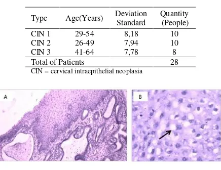
The sample used in the study consisted of 28 paraffin blocks of cervical tissue derived from patients with CIN 1 (10 blocks), CIN 2 (10 blocks) and CIN 3 (8 blocks). The patients aged between 26-74 years old (standard deviation of 10:12) (Table 1). Cervical Intraepithelial Neoplasia (CIN) 1 was characterized by the presence of mild dysplasia in one third layer of squamous epithelial cells whose nucleus were about to swell and more hyperchromatic, or the presence of cells infected with HPV with slightly enlarged nucleus, more hyperchro-matic, and irregular nucleus edge. The core was surrounded by perinuclear hallo (koilositosis) (Figure 1). CIN 2 was characterized by moderate dysplasia in two-third layer of squamous epithelial cell whose nucleus are about to swell, and more hyperchromatic (Figure 2). CIN 3 was characterized by severe dysplasia in the whole layers of squamous epithelial cell whose nucleus were enlarged, hyperchromatic, with thinner cytoplasm (Figure 3) .10
Table 1.Research patient’s data
Type Age(Years) Deviation Standard
Quantity (People)
CIN 1 29-54 8,18 10
CIN 2 26-49 7,94 10
CIN 3 41-64 7,78 8
Total of Patients 28
CIN = cervical intraepithelial neoplasia
Figure 1. Cervical Intraepithelial Neoplasia (CIN) 1 with hematoxylin eosin staining (A 200x magnification, and B 400X).
CIN 1, CIN 2, and CIN 3 respectively represents mild, moderate, and severe dysplasia. Based on the
management of patients’ treatment, a new classification
Mastutik et al. : Human pappilomavirus genotype in cervical tissue of patients with CIN 1, CIN 2, and CIN 3
76 Figure 2. Cervical Intraepithelial Neoplasia (CIN) 2
with hematoxylin eosin staining (400X magnification).
Figure 3. Cervical Intraepithelial Neoplasia (CIN) 3 with hematoxylin eosin staining (A 200x magnification, and B 400X).
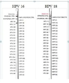
HPV genotyping in this study was conducted by reverse line blot technique that can be used to detect 40 types of HPV, including high risk and low risk groups. HPV types that can be detected are 6, 11, 16, 18, 26, 31, 33, 35, 39, 40, 42, 43, 44, 45, 51, 52, 53, 54, 55, 56, 58, 59, 61, 62, 64, 66, 67, 68 (AEB), 69, 70, 71, 72, 73, 81, 82, 83, 84, 87, 89, 90. The result of this HPV genotyping test using this technique showed the ribbons that are visible in the blot membrane (Figure 4).
Figure 4. HPV genotyping’s result withreverse line blot technique
These results indicated that HPV genotypes that infect patients with CIN 1, namely HPV 16 and 18, patients with CIN 2 (HPV 16 and 52), and patients with CIN 3 (HPV 16, 67, and combined HPV 16/67 and 52/67 infection) (table 2). HPV genotypes found in patients with single infection precancerous lesions, which was 92.9% (26/28), are HPV 16, HPV 18, HPV 52, and HPV 67. Combining HPV genotypes, which was 7.1% (2/28) are HPV 16/67 and 52/67 (Table 3). The most dominant HPV genotypes in infecting patients with
cervix’ precancerous lesions in this studywere HPV 16 (86%), HPV 67 (11%), HPV 52 (8%), HPV 18 (4%) (Table 3).
Table 2. HPV genotype distribution in cervical precancer lession
Type Samples Amount
HPV Genotipe
Quantity
CIN 1 10 HPV 16 9
HPV 18 1
CIN 2 10 HPV 16 9
HPV 52 1
CIN 3 8 HPV 16 5
HPV 67 1
HPV 16, 67 1
HPV 52, 67 1
Patients Amount 28
Table 3. HPV genotype profile in cervical precancer lession
HPV Genotipe Single infection
Combined infection
Total %
HPV 16 23 1 24 86
HPV 18 1 0 1 4
HPV 52 1 1 2 8
HPV 67 1 2 3 11
HPV 16/67 1 1 4
HPV 52/67 1 1 4
Total of Samples 28
CIN 1 is included to LSIL, CIN 2 and CIN 3 are includ-ed to HSIL in Bethesda classification. All CIN1 samples in this study are HPV positive. The prevalence of HPV that infect HSIL patients in the world is 85% (ranging from 78% in Asia and 85% in Europe).13 HPV geno-types that present in LSIL cases in this research were HPV 16 and 18, in HSIL are single infection of HPV 16, HPV 52 , HPV 67, and combined infection of HPV 16/67 and 52/67. The genotypes that infect patients with HSIL are HPV 16, 58, 52, 18, and 51. Genotype 31, 33,35, 39, 52, 56, 58, 68, 73 are more often to be found in HSIL than in invasive cervical carcinoma, as well as low risk HPV such as 6, 11, 52, 66, 70, and 82 are more frequently found in cervical carcinoma HSIL than invasive.13
Materia Obstetrics & Gynecology, Vol. 24 No. 3 September - December 2016 : 74 - 78
77 of a research done in Thailand, which found that 50% of HPV types 16 and 18 are the type that infects people in all cases of LSIL and HSIL. HPV 16 is the most frequent and its prevalence increases with the increasing degree of lesion, 73.4% in LSIL, and 87.2% in HSIL. In other hand, the prevalence of low-risk HPV types (HPV 6, 11, 40, 43, 70 and 82) is decreased or there is no increase in the disease stage.14
HPV types that are identified in normal cervix and LSIL in Japan are HPV11, 16, 18, 30, 31, 33, 35, 39, 42, 44, 45, 51, 52, 53, 54, 55, 56, 58, 59, 61, -62, -66, -67, -68, -72, and -73. HPV types that have been identified on HSIL and SCC in Japan are HPV-16, -18, -31, -33, -35, -45, -51, -52, -56, -58, and -67. In HSIL and SCC, HPV-16 is the most infecting type, followed by HPV-51, -52, and -58. HPV type -39, -59, and -68 are not detected.15
This analysis showed that HPV types 16, 18, 31, 51, 52, and 58 are often associated with the incidence of SCC, and HPV types 16, 31, 33, 35, 45, 51, 52, 56, and 58 are often associated with HSIL, therefore HPV-16, -18, -31, -51, -52, -58, and possibly HPV-45 and -67 are often considered as high risk type in Japanese women.15HPV types 33 and 55 are not detected as single type in invasive cervical cancer, although they are related to HSIL. HPV types 11, 39, 42, 44, 53, 54, 59, 61, 62, 73 are associated with the incidence of HSIL, but not related to the HSIL and invasive cervical cancer and therefore, these type are associated with LSIL and low risk cancer. LSIL HPV 39, 59, and 68 are not identified on HSIL and SCC although they are related to cancer.15
HPV types that often infect normal cervix in Asia are HPV 16, 52, 58, 51, 18, 56, 35, 33, 39, 31. LSIL are HPV 16, 58, 52, 18, 51, 56, 39, 31 , 68, and 35. While HPV 16, 58, 52, 18, 33, 51, 31, 56, 35, and 45 are HSIL.16
HPV testing in patients with CIN2 is helpful to diagnose borderline cases or cases of normal cytology, as well as to follow up patients with CIN 2. HPV testing in CIN 3 are needed to monitor the cervix of patients with CIN 3 who are treated surgically. Cytological examination during the treatment follow-up is less sensitive, since the smear results are difficult to evaluate, while 5-10% of patients usually experience relapse. HPV testing is useful to follow up the those cases and is more sensitive. Negative results imply that there is perfect healing so that the woman can follow a routine screen-ing .5
Detection of HPV types are generally useful to predict the cervix of infected women. Based on the clinico-pathology grading consensus, HPV testing can be used
to justify ASCUS clinically. Determining HPV type and detection of multiple HPV in clinical samples can be used as an secondary marker for cervix cell abnormal-ities.15High-risk types of HPV infection can trigger the development of cervical cancer, while low-risk HPV can lead to genital warts cases.
HPV genotypes found in LSIL cases of this research are HPV 16 and 18. Eventhough in LSIL patients there is a possibility of complete healing and disappearance of the virus because of increased immunity, it is still necessary to monitor the health of the cervix for the next 6 months. LSIL can either evolve to HSIL or recover completely. The results of this study shows that in LSIL, the HPV infecting cervix are the high risk types. Therefore, it is possible to progress towards HSIL. HPV genotypes of HSIL cases in this study are single infections such as HPV 16, HPV 52, HPV 67, and com-bined infections such as HPV 16/67 and 52/67. HPV types 16, 52, and 67 are high-risk types. Combined infection of two high-risk types of HPV can accelerate the development towards invasive cervical carcinoma. Patients should get conication procedure with loop electrical excition procedure (LEEP) to recover. HPV testing still need to be done in patients with HSIL after the procedure to monitor the healing progress.
High-risk HPV types are the mostly found types in HSIL and almost all cervical cancers, while in LSIL, obly 60% were infected with high risk HPV types. Single or combined, high-risk HPV types infections are more often found in HSIL and SCC compared with the LSIL and normal cervix.15 High-risk HPV infection infects longer than the low-risk HPV types. The immune response in the cervix can easily beat the low-risk HPV types, where high-low-risk HPV types are resistant to immunity and infect longer. High-risk types of HPV infection appears to be related to persistent infection and were able to develop into HSIL and SCC.15This study found that the combined HPV infection is a factor in persistent HPV infection in young women with a normal cervix. Persistent HPV infection indicates the degree of cervical dysplasia development. Multiple HPV infection appears to be related to cervical neoplasia development. Multiple infections produce conditions that alter the immune protection to be a persistent infection.15
There are several combined infections in clinical samples. The combined infection seems to be typical in women with cervical abnormalities and was significant-ly associated with the risk of LSIL, HSIL and SCC. The combined infection is more common in LSIL compared to malignant lesions (HSIL and SCC).15
Mastutik et al. : Human pappilomavirus genotype in cervical tissue of patients with CIN 1, CIN 2, and CIN 3
78 cervical abnormalities in this study were HPV 16 (86%), HPV 67 (11%), HPV 52 (8%), HPV 18 (4%). Most types of HPV that cause cervical cancer are HPV 16 and 18. The detection of high risk HPV types can be used as an additional biomarker for routine cytologic examina-tions in screening for precancerous lesions, management of abnormal borderline cytology result (ASCUS) and follow-up in women who has gone through the treat-ments for HSIL and to categorize women based on precancerous lesions and cancer risk factors (Meijer CJ, et al, 2006, Youens KE, et al, 2011). These HPV types are associated with protection against cervical cancer. Vaccines which are available today are HPV 16/18 bivalent and quadrivalent HPV 16/18 and 6/11 vaccine. HPV 16/18 vaccination can provide protection against cervical cancer caused by HPV 16 and 18 infection. The second-generation of HPV vaccines, with the addition of HPV types, is expected to provide higher protection against progression toward malignancy.
CONCLUSION
The most dominant HPV genotypes infecting patients with cervical precancerous lesions in this study were HPV 16, HPV 67, HPV 52 and HPV 18, and therefore, HPV types 52 and 67 should be considered to be a second-generation HPV vaccine for protection against cervical cancer incidence in Indonesia.
REFERENCES
1. WHO. WHO/ICO Information Centre on HPV and Cervical Cancer (HPV Information Centre). Human Papillomavirus and Related Cancers in Indonesia.
Summary Report 2010. Available at
http://apps.who.int/hpvcentre/Statistics/dynamic/ico /country _pdf/IDN.pdf. 2010. Accessed 10 May 2013.
2. Zaravinos A, Mammas IN, Sourvinos G, Spandi-dos DA. Molecular Detection methods of Human Papillomavirus. The International Journal of Biological Markers. 2009;24(4):215-22.
3. Gutiérrez-Xicoténcatl L, Plett-Torres T, Madrid-González CL, Madrid-Marina V. Molecular diagnosis of human papillomavirus in the development of cervical cancer. Cuernavaca Jan. 2009;51(3).
4. Ellenson LH, 2010. Ellenson LH, Pirog EC. The Female Genital Tract Chapter 22, In Robbins and
Cotran Pathologic Basis of Disease. 8th Eds. Philadelphia: Saunders Elsevier; 2010. p. 1017-24. 5. Meijer CJ, Snijders PJ, Castle PE. Clinical utility of
HPV genotyping. Gynecologic Oncology.
2006;103:12-7.
6. Youens KE, Hosler GA, Washington PJ, Jenevein EP, Murphy KM. Clinical Experience with the Cervista HPV HR Assay Correlation of Cytology and HPV Status from 56,501 Specimens. The Journal of Molecular Diagnostics. 2011;13(2):160-6.
7. Andrijono. Vaksinasi HPV merupakan Pencegahan Primer Kanker serviks. Majalah Kedokteran Indonesia. 2007;57(5):153-8.
8. Djauzi S dan Rengganis I. Imunisasi Human Pappilomavirus (HPV) dalam Infeksi Human Pappilomavirus, Editor Andrijono dan Indriatmi W, Badan Penerbit Fakultas Kedokteran Universitas Indonesia. 2013;189-98.
9. Crosbie EJ, Einstein MH, Franceschi S, Kitchener HC. Human papillomavirus and cervical cancer http://dx.doi.org/10.1016/S0140-6736(13) 60022-7. 2013.
10. Kumar V, Abbas A, Fausto N. Neoplasia. In : Robbins and Contrans Pathologic Basis of Diseas-es. 8th Eds. Philadelphia: Saunders Elsevier; 2010. 11. Berek JS (ed). Berek & Novak’s Ginecology. 14th
Eds. New York: Lippincot Williams and Wilkins; 2007. p. 1404-50.
12. Montag A, Kumar V. The Female Genital System and Breast Chapter 19 in Robbin Basic Pathology. 8th Eds. Philadelphia: Saunders Elsevier; 2010. p 716-20.
13. Smith JS, Lindsay L, Hoots B, Keys J, Franceschi S, Winer R, Clifford GM. Human papilloma-virus type distribution in invasive cervical cancer and high grade cervical lesions. 2007.
14. Suthipintawong C, Siriaunkgul S, Tungsinmunkong K, Pientong C, Ekalaksananan T, Karalak A, Kleebkaow P, Vinyuvat S, Triratanachat S, Khunamornpong S, Chongsuwanich T. Human Papilloma Virus Prevalence, Genotype Distribution and Pattern of Infection in Thai Women. Asian Pacific J Cancer Prev. 2011;12:853-6.
15. Sasagawa T, Basha W, Yamazaki H, Inoue M. High-Risk and Multiple Human Papilloma-virus Infections Associated with Cervical Abnor-malities in Japanese Women. Cancer Epidemiol Biomarkers Prev. 2001;10:45-52.