THE RELATIONSHIP BETWEEN BIRTH WEIGHT AND ENAMEL DEFECT PROBABILITY IN CHILDREN WITH PRENATAL MALNUTRITION
Presented in 6th FDI — Indonesian Dental Association Joint Meeting & Balikpapan International Dental Exhibition 2010, 12-14 November in Gran Senyiur,
Balikpapan-Indonesia.
Willyanti Soewondo Syarif NIP. 19541218 198002 2 001
FACULTY OF DENTISTRY PADJADJARAN UNIVERSITY
CHAPTER 1 INTRODUCTION
1.1 Background
The growth and development of the teeth is a part of the whole body growth, and it might be affected by the interaction of genetic and environmental factors. The growth and development of the teeth is a complex and long lasting process, and it is a sensitive period against intrinsic as well as extrinsic factors of disturbances.1,2 The disturbances might happen in various phase/stage of teeth development with various intensity and period. This might result as the defect of teeth development i.e. enamel or dentin defects according to the disturbed tissues. Enamel defect means incompleted enamel that was caused by systemic environmental factors during prenatal or postnatal period. The critical growth and development of deciduous teeth occurs in perinatal period, while the critical phase of permanent teeth is in the postnatal period.2,4 Enamel defects include enamel hypoplasia and hypocalcification. Hypoplasia is caused by the disturbance of enamel matrix, while hypocalcification is caused by the disturbance disturbance of mineralization, and enamel maturation. According to ADA (Australian Dental Association), there are several factors that are related to the defects such as maternal factors, drugs during pregnancy, premature low birth weight, infection, malnutrition, and trauma.5 Prenatal factors that might cause defect of the teeth development are mostly maternal such as severe infection during pregnancy, malnutrition of the mother, mother's metabolic disorders, besides child's factor such as low birth weight.2,3,6-8
Systemic condition such as Intrauterine Growth Retardation (IUGR) means the disturbances that occur in prenatal period that might cause intra uterine malnutrition of the fetus that might result as baby with prenatal malnutrition or might be call as Small For Gestational Age/ SGA, i.e. birth weight of lower than the normal -2 SD.9 It might result as a disruption of the development that cause defect of the organ development especially deciduous teeth. A good growth of fetus shows the mean weight gain in each phase/stage according to gestational age. Birth weight and gestational age indicate the growth of fetus; SGA neonates have birth weight of -2 SD beyond the normals (<2500 g).
CHAPTER 2
1 Enamel Defect
Teeth development begins as early 4-5 weeks intrauterine, and in 2 months gestation deciduous dentition begins to develop. Prenatal period is the critical stage for any disturbances. Teeth are formed by mineralization of protein matrix and the process begins at about 4 months gestation and completed after birth. Enamel is hard tissue in the body that is not remodeled, changes in its structure could be happened in developing period. Enamel formation or Amelogenesis is very sensitive to any disturbances. Changes in its structure become a defect, it could be happened both in deciduous and permanent dentition. Genetic and environmental factor play the role in this condition. Environmental factor Could cause the defect, that are; prenatal infection, nutritional disturbances, systemic illness. Prematurity and low birth weight infant have been associated with defect in both permanent and
deciduous teeth( 1,10,11,12)
Enamel defect means incompleted enamel, could be manifest as enamel hypoplasia and enamel hypocalcification, depend on disturbances and stage of growth and development of the teeth.
There are more 120 Risk factors have been linked to enamel defects, the most common are; Severely nutritional deficiency in pregnancy mother, Prematurity, Birth difficulties especially with hypoxia and disorders of the blood, trauma, and certain medications which given in pregnancy. For deciduous teeth enamel defects may result in from disturbances in prenatal period, and for permanent teeth enamel defects may result from disturbances in postnatal period(5,10-2)
Several nutritions are important for amelogenesis and osteogenesis; calcium, Phosfor, Magnesium Fluor, and some vitamis such, vitamin A, C, D, K. Deficiency in calcium can disturb amelogenesis and osteogenesis, 73 % children with calcium deficiency show enamel hypoplasia (14). Deficiency in vitamin C can cause disturbances in amelogenesis because vitamin C is important for prolin building in matrix formed.14
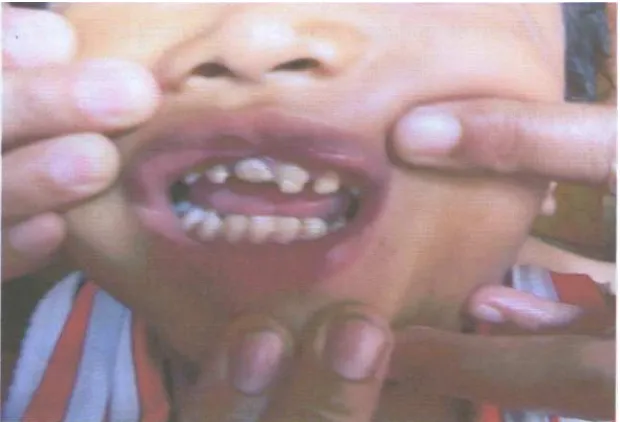
2.2 Clinical manifestation
Enamel Hypocalcification is deficiencies in mineralization during mineralization phase, there are opaque, white, or yellow area of enamel on smooth surfacace.2,5,8
Clinical manifestation due to the cause of disrturbances. Systemic disturbances show that the defect is generally, bilaterally. Local disturbances such as used of laryngoscope in managing baby's hypoxia, birth trauma show that the defect is locally, unilaterally and only 1 or more teeth affected.
Figure 2.1. Mild Enamel Hypoplasia12
Figure 2.3. Enamel Hypocalsification
2.3 Mechanism of Enamel Defect
Amelogenesis contains of matrix secretion and maturation of the matrix. Ameloblast is very sensitive to any disturbances resulting from genetic and environmental factor. Disturbances in secretion phase can disturb elongation of enamel crystal, it will reduction protein secretion resulting insufficiency of crystal elongation. This condition resulting reduction of the matrix cause thin enamel formed, part of enamel formed, or no enamel formed. This condition call enamel Hypoplasia.
If there is malnutrition during amelogenesis, there is reduction of ameloblast and resulting reduction of matrix forming and hypoplasia formed. Maturation of the enamel shown by reduction of water and calcium deposition. If there are nutritional/calcium deficiency, may resulting hypocalcification.
The size of defect depend on, instensity of etiological factor, time of disturbances, and period during crown formation.10-3
2.4 Prenatal Malnutrition
average 5 gram/days, and 30-35 gram/day in 34 week gestation. In the third trimester there is nutrition and hormonal influence rather than genetic.9'15'16.
The etiology of prenatal mal nutrition devided on 3 groups; maternal factor, foetus factor, and placenta. Maternal factor such as; maternal TORCH infection, diabetes, hypertension preelampsia, maternal malnutrition, maternal infections in long period, age of the mother more 35 years, all that condition might be caused prenatal malnutrition. Foetus factor such as chromosom anomaly, syndromes also could be caused prenatal malnutrition. All that condition resulting failure of the placenta to transfer nutrition to the foetus, so prenatal malnutrition happened. Placenta's factor such, anomaly of the placenta. Placental infark and Placentitis could be caused prenatal malnutrition.9,15,16,18
Birth weight show fetal growth rate, and the baby with mal nutrition shows birth weight lower or, much lower than normal commonly below 2500 gram. According to Fanaroff baby with malnutrition is shown that their birth weight is below than 2 SD normal birth weight. It could be Low birthweight /LBW (below 2500 gram), very low birth weight /VLBW (1500-2000 gram) and Very-very low birth weight /VVLBW (below 1500 gram). Birth weight could be as indicator of the condition of baby's nutrition, and also indicate if there is deviation of baby's growth.9,15-6
CHAPTER 3
3.1 Subject and Method
Subject were 150 children born with prenatal malnutrition aged 4-48 months and 300 normal children as control. We had complete data of mother's pregnancy (mother's age at birth, disease during pregnancy, and smoking habits) and children's birth including birth weight, gestational age, birth length, head circumference, hypoxia, and gender.
This was a clinical epidemiology study. Enamel defects were examined using DDE scoring modification and FDI index and determinate; mild defect score <12, severe defect; >12band Interviews were done to collect data on birth weight and length, head circumference, gestational age, gender, and hypoxia in neonates. Enamel defects were recorded three times with 2 months intervals. We recorded the defects of deciduous teeth enamel and confounding variable factors of the child such as gender/sex, gestational age, birth weight and length, head circumference, and hypoxia. All data were analysed using multivariate analysis.
3.2 Operational Definition
1. Children with prenatal malnutrition means a neonate with birth weight beyond -2SD of intrauterine growth curve. Birth weight usually below 2500 gram, some children are 2600 gram
2. The defect of deciduous teeth including hypoplasia and hypocalcification and the severity was measured using DDE index and FDI modification and determinate with mild defect with score <12 dan severe with score >1217
CHAPTER 4
Figure 1 shows that the lower the birth weight, the more the probability of the severity. The higher the birth-weight the lower the probability of severity. Prenatal malnutrional. Children had less nutritional intakes that occur during the first trimeter of pregnancy, and further during the second or third trimester that might occur in a short till a long time.
A healthy pregnant woman with adequate nutritional status will deliver a healthy neonate with normal birth weight, while a sick or unhealthy pregnant woman with low socio-economic condition will deliver a low birth weight neonate. About 70% of the low birth weights are SGA babies with birth weight lower than -2SD normal weight or the 10th percentile of Lubchenco intra uterine growth curve (<2500 g), or even has a birth weight of 2600 grams.9,15-6
This condition could be caused by systemic condition such intra uterine growth retardation (IUGR) as a result of maternal factors such as severe infection during pregnancy, preeclampsia hypertension, maternal diabetes, smoking, alcohol, and mother's age at delivery of >35 years.9,11 Other causes of IUGR are placental abnormalities, and child's factors such as genetic abnormalities, syndromes, multiple gestation, that might cause intrauterine malnutrition.9,15-6,19
the enamel matrix decrease and hypoplasia occur to become a further permanent defect. Nutritional deficiency caused hypoplasia and hypocalcification or both, according to the period and intensity of the defect.5-8,14
Systemic condition such as Intra Uterine growth retardation (IUGR) might cause SGA children and defects in the development of organs, and the abnormalities depend on the time of occurence and intensity of the abnormalities.9 The abnormalities of organ developments might cause hypoplasia or enamel hypocalcification, or both, of the teeth.10," The risk of SGA children to have enamel defect is 79%.18
A good growth of a fetus is described as a mean weight gain in each phase/stage, i.e. 5 g/day up to 15 weeks, 15-20 g/day up to 24 weeks and 30-35 g/day up to 34 weeks pregnancy. Low nutritional status of the fetus might result in low birth weight, and this mightcause a defect of the organ growth and development might be including the teeth.18
Birth weight and gestational age reflect the Fetal Growth Rate that means the birth weight reflects the nutritional status of the fetus, the better the nutritional status, the better the birth weight, and that means the intrauterine growth and development restriction did nor occur or only in a mild intensity.14 Birth weight is the reflection of the intrauterine growth and development, so that the decrease of birth weight due to malnutritrition might be a risk to become a growth defect.9,15-6,19Mal nutrition is caused by lack of absorption of the placenta.
CHAPTER 5
5.1 Conclusion
Birth weight in children with prenatal malnutrition reflect the severity of the defect of deciduous teeth, the lower the birth weight, the more severe the probability defect of the enamel.
5.2 Acknowledgement
REFERENCES
1. Moyers RE. 1988. Handbook of Orthodontics.4thed. Chicago: Yearbook Medical
Publisher Inc.
2. Stewart RE, Witkop CJ, Bixler D. The dentition. In: Stewart RE, Barber TK, Troutman KC,
Wei SHY. Pediatric dentistry, scientific foundation and clinical practice. Editors. ST. Louis:
The C.V. Mosby Co., 1982. p. 87-94.
3. Laskaris G. Color atlas of oral diseases in children and adolescents. Stuttgart: Thieme, 2000.
p. 20-3.
4. Magnusson BO. Pedodontic: a systematic approach. Munksgaard: PJ. Schmidt Vojens,
1981. p. 79-85.
5. Australian Dental Association. Tooth enamel defects. Mi-tec Medical Publishing, 2007
Jan; 1.
6. Seow WK. Effect of preterm birth on oral growth and development. Australian Dental
Journal. 1997;42(2):85-91.
7. McDonald RE, Avery DR. Dentistry for the child and adolescent. 6thEd. St Louis: CV.
Mosby Year-Book Inc, 1994. p. 53-9.
8. Welburry RR. Pediatric dentistry. 2ndEd. Oxford University; 2001. P. 294,394-5.
9. Fanaroff AA, Martin RJ. Neonatal-perinatal medicine: diseases of the fetus and infant. 7th
Ed. St Louis: Mosby Inc, 2002.
10. LunardelliMS,Peres MA,;Breast-Feeding and other Mother -Child Factors,associated with
developmental Enamel Defects Of Brazillian Children,Journal of Dentistry for Children
-73:2,2006 p70-6
11. Simmer JP Dental Formation and its impact on clinical Dentistry.j.Dent ed 2001 65(9):
896-9
12. Seow WK, Brown JP, Tudehope DA, O'Callaghan M. Dental defects in deciduous dentition
of premature infants with low birth weight and neonatal rickets. Pediatrics Dentistry. June
1984;6(2
13. Seow WK.Oral Complications of premature birth Austr dent j 1986;23-9
14. Wei SHY, Anderson TA. Nutrition and dental health. In: Stewart RE, Barber TK, Troutman
KC, Wei SHY. Editors. Pediatric dentistry, scientific foundation and clinical practice.
16. Thureen PJ, Anderson MS, Hay WW. The small for gestational age infant. Neoreviews.
2001;2:e139-48.
17. Willyanti ;Skor Prediksi tingkat Keparahan Defek Email Gigi Sulung Pada Anak Dengan
Kecil Masa Kehamilan Unpad Press,2009,p 98-99
18. Sjarif Willyanti3Oewen Roosye,Effendi Sjarif H,Sutrisna Bambang. Enamel defect of
deciduous teeth in small gaestational age children.Dental Journal 2010;43 june 2010
p;915.
19. Cunningham FG, Leveno KJ, Bloom SL, Hauth JC, Gilstrap L, Wenstrom KD. Williams