THE JAGUAR HEALTH PROGRAM MANUAL
Dr. Sharon L. Deem
Dr. William B. Karesh
Field Veterinary Program
Field Health Manual Guidelines
The main objectives of the Jaguar Health Program are: 1) to provide standardized methods to
assess the overall health status of jaguars in the wild; 2) to determine disease threats to jaguars
including both direct threats (e.g., infectious diseases - intraspecific and conspecific via domestic
animals, livestock, other free-ranging felids, prey items) and indirect threats (e.g., habitat
fragmentation and degradation that may increase disease risks); and 3) to provide
recommendations, based on findings from the health assessment, for the long-term management
and conservation of the jaguar.
The following manual is intended only for field biologists and veterinarians with previous
wildlife experience that are working in association with the Wildlife Conservation Society
(WCS) Jaguar Conservation Program (JCP) (www.savethejaguar.com). This manual is not
intended for inexperienced workers. It has been developed by veterinarians with the WCS Field
Veterinary Program (www.fieldvet.org). The purpose of this manual is to provide a standardized
and safe and ethical approach to capture, handling, and sampling protocols to ensure that the
Jaguar Health Program is carried out in a consistent fashion throughout the jaguars’ range. The
WCS Field Veterinary Program staff do NOT support the immobilization and handling of any
wild animals by inexperienced personnel.
These guidelines have been developed to be “living documents” and will be updated annually as
needed. In an effort to make the document more useful, users are encouraged to submit their
comments to improve the layout, content, and/or presentation of the material as they work with
these guidelines. Any errors or sections that are unclear should also be highlighted. Please direct
these comments to the JCP Program Coordinator, Kathleen Conforti, at the Wildlife
Conservation Society, 2300 Southern Boulevard, Bronx, NY 10460 – 1099 USA. Telephone:
The highest priority for the Jaguar Health Program is the safety of the biologist and the safety
andwelfare of the animal. It is strongly recommended that the field biologist consult with a
veterinarian prior to field work. The veterinarians with the WCS Field Veterinary Program are
available for consultation or training sessions for field personnel and consultation on
immobilization, handling, and health surveys of jaguars through the JCP. Please contact the
Program Coordinator, Kathleen Conforti, at the Wildlife Conservation Society, 2300 Southern
Boulevard, Bronx, NY 10460 – 1099 USA. Telephone: 718-220-2189; Fax: 718-364-4275;
email: [email protected].
All diagnostic and genetic testing will be performed at selected laboratories in the U.S. that are
known for their expertise in non-domestic felids. The ability of the Jaguar Health Program staff
and personnel to complete these tests successfully is contingent on the exportation of samples
from various countries within the jaguars’ range and their importation into the U.S. Jaguars are
listed on CITES Appendix I, therefore a CITES import and export permit are required for
transporting tissue samples into the U.S. The JCP is developing a blanket permit for these
samples, however, in the meantime, it is imperative that appropriate research permits are in place
before transporting any samples. Researchers that collect samples for health analyses should ensure that they are able to store samples in country if there are any delays in permit processing.
Permits must be issued by both the importing and exporting countries. In most instances, an
export permit will not be issued until a valid import permit is presented to the office issuing the
export. The U.S. Fish and Wildlife Service’s Office of Management Authority is the
Governmental agency which currently issues CITES import permits for the United States. In
general, written proof of permission to work in the host country is required when applying for an
import permit. For more information on permit requirements, visit
http://international.fws.gov/permits/permits.html or refer to permit guidelines on the Field
THE JAGUAR HEALTH PROGRAM MANUAL
Dr. Sharon L. Deem
Dr. William B. Karesh
Field Veterinary Program
Wildlife Conservation Society
TABLE OF CONTENTS
I. Introduction
II. Capture and Immobilization
III. Handling Immobilized Jaguars and Trouble Shooting for Anesthetic Emergencies IV. Post Anesthetic Recovery
V. Animal Sampling
VI. Data Collection, Analysis and Distribution VII. Bibliography
VIII. Figures IX. Tables X. Appendices
I. INTRODUCTION
One important component of the Jaguar (Panthera onca) Conservation Program is a
standardized approach to health and disease surveillance of free-ranging jaguars. Threats to the
health of wildlife from anthropogenic influences are often associated with increased contact that
wildlife have with livestock, domestic animals and humans, as well as habitat fragmentation and
contamination of their habitats. Although the specific health threats to free-ranging jaguars are
largely unknown at this time, the situations listed above are believed to be of importance in the
long-term conservation of free-ranging jaguars throughout their range.
ranging jaguars with little or no training in veterinary skills, such as anesthesia and biological
tissue collection. This fact alone suggests a strong need for standard guidelines to ensure the
safety and welfare of jaguars being handled in the field. The following guidelines are intended
for field biologists and veterinarians with wildlife experience that are working associated with
the WCS Jaguar Conservation Program. These guidelines are not intended for inexperienced
personnel. The veterinarians with the Field Veterinary Program, WCS are available, through the
Global Carnivore Program Manager, for training sessions for field personnel and consultation on
immobilization, handling, and health surveys of jaguars, as a component of the Jaguar
Conservation Program (see appendix 1).
The main objectives of the jaguar health program (JHP) are the following: 1) provide
standardized methods to assess the overall health status of jaguars in the wild; 2) determine
disease threats to jaguars including both direct threats (i.e., infectious diseases – intraspecific and
conspecific via domestic animals, livestock, other free-ranging felids, prey items and indirect
threats (i.e., habitat fragmentation and degradation that may increase disease risks); and 3)
provide recommendations, based on findings from the health assessment, for consideration in
long-term conservation policies for jaguars.
II. CAPTURE AND IMMOBILIZATION
The information provided in this manual is intended for use by field researchers with
previous experience in performing felid immobilizations. Field Veterinary Program, WCS staff
do NOT support the immobilization and handling of any wild animals by inexperienced
personnel. It is imperative that persons handling jaguars consider the safety of humans and
jaguars above all else before, during and after any jaguar immobilizations.
All field personnel should use one standard form for recording capture and
immobilization events. This will ensure that all researchers are collecting the same data for every
immobilization so that these data can be compiled and compared. Ultimately, this will allow us
to determine the safest and best immobilization protocols for free-ranging jaguars.
We recommend the use of the Medarks software program (ISIS, 12101 Johnny Cake Rd.,
Apple Valley, MN 55124 USA) for compiling the immobilization data. (This software could also
be used for data collection related to all aspects of the health program.) A hard copy representing
a medarks standard anesthesia form is provided in appendix 2.
2. Capture Methods
There are a variety of capture and immobilization methods for free-ranging felids (De
Wet, 1993). Wilson et al. (1996) provide an excellent overview of capture methods for medium
to large sized mammals. Methods that have been used to capture free-ranging jaguars include
treeing the animal using dogs, padded foot-hold traps, snares (i.e., Aldrich snares) and cage or
box traps. The later two of these methods may or may not include bait to lure the animal to the
trap. Once the jaguar is treed or trapped, it can then be darted. It is best to not dart a jaguar above
5 meter high in a tree to avoid traumatic falls. A comprehensive overview of capture methods for
jaguars is currently being developed.
3. Anesthetic administration
Anesthetic agents should only be administered to free-ranging wild jaguars using remote
drug delivery systems (RDDS). There are a variety of RDDS available for the practitioner, nicely
reviewed by Bush (1992) and Nielsen (1999). A blowpipe, or possibly a pole syringe, may be
used for immobilizing jaguars in a cage, foot-hold trap or snare. In all other field situations, it is
best to use a rifle or pistol (i.e., TelinjectTM, Cap-ChurTM, Dan-InjectTM) (Appendix 3). It is
practitioner must be familiar with the instrument he/she chooses for use in the field. Darting
animals is always associated with some degree of risks. Serious damage to the animal (and
human participants) can and does occur if inappropriate instruments are used and / or if
instruments are used inappropriately.
Dart and needle selection is also important in preparing for a jaguar immobilization.
Darts that are too heavy and needles that are too long / thick can cause serious damage on
impact. (Damage is also possible if the charge of the dart or the charge of the rifle / pistol is too
high.) Needles available include collared, plain and barbed. A collared dart is often employed
during immobilization procedures because it remains in the animal and ensures total drug
injection. Unfortunately, if the jaguar is not adequately immobilized and cannot be restrained, the
dart will remain in the animal and may cause problems.
We recommend the use of 1.5 X 30 mm (18 gauge X 1 ¼ inch) collared needles for immobilizing free-ranging adult jaguars. However, if the jaguar is treed or trapped prior to darting, a non-barbed (plain) needle can be used. Non-barbed (plain) needles cause less trauma to
the tissues but often do not remain in the animal as long as collared needles and thus may not
inject all the drug prior to falling from the animal.
When darting a jaguar, it is safest to aim for the proximal (closest to body) region of a
rear limb (Figure 1). Some researchers recommend darting the triceps region of the front arm. If
the anesthesiologist elects to use the front limb, it must be remembered that the thoracic region
and head are in very close proximity to the intended site. Serious harm can be inflicted on the cat
if the dart hits one of these regions. It is for this reason that we recommend the hind leg unless
the anesthesiologist is darting the jaguar at close range (i.e., in a box trap). When aiming for the
rear limb, darts should be placed in the caudal most aspect of the muscle mass to avoid the
femoral bone and the sciatic nerve. Needles and darts must be disinfected prior to use on the next
animal to prevent the spread of diseases. Although disinfection is often the only available means
4. Anesthetic Regimens
In this section, we provide a recommended “standard protocol” for use by those people in
the field with minimal immobilization experience. This protocol should provide an adequate
plane of anesthesia with minimal respiratory and cardiovascular compromise. Additionally, we
list anesthetic regimens that have been successfully used in captive and free-ranging jaguars
based on the literature and personal experience. Please note that drug doses in this handbook are
given in milligrams (mg) and micrograms (mcg). Doses are NOT in milliliters (except where
noted in table on Page 7). Drug administration route is intramuscular (IM) unless stated
otherwise.
Calculating drug dosages – Persons using anesthetic agents to immobilize jaguars must be familiar with how to calculate the volume (ml) of drug to deliver based
on the recommended dose (mg/kg) for the species and concentration (mg/ml) of the drug.
1. mg needed = recommended dose (mg/kg) X animal’s body weight (kg)
2. ml needed = mg needed (see 1 above) ) drug concentration (mg/ml)
= mg needed X (ml/mg)
Example:
1. 90 kg jaguar
2. Drug concentration is 100 mg/ml
3. Recommended dose is 5 mg/kg
mg needed = 5 X 90 = 450 mg
Note: 1 kg = 2.2 lb (Recommended doses may be given in mg/kg OR mg/lb. Be sure to
make the proper conversion. For instance, if the weight of the animal is estimated in lb and the
recommended drug dose is given in mg/kg, then you must convert the dose.)
1 kg / 2.2 lb X mg / kg = mg / lb
A. Recommended “standard protocol” for the immobilization of free-ranging jaguars
Currently, we recommend (except for the immobilization of treed jaguars; see below) the
following anesthetic regimen for use by field personnel with little experience in immobilizing
free-ranging jaguars. This regimen should provide an adequate plane of anesthesia for shortterm
work on the jaguar (i.e., radio-collar application, morphometric measurements, biomaterial
collections) while requiring a minimal level of technical skill in anesthesiology.
Telazol 6 - 10 mg/kg intramuscular by dart as the dose for immoblization in free-ranging jaguars. The darter has the option to include 150 mg ketamine in the
initial dart based on work by Cavalcanti and Hoogesteijn, unpublished data.
Supplemental ketamine at a dose of 1-1.5 mg/kg, intravenous or 1-2 mg/kg
intramuscular, as needed to maintain an adequate level of anesthesia. (No supplemental
ketamine should be delivered for at least 10 minutes after the telazol dart.) Atropine at a
single dose of 0.04 mg/kg either subcutaneous or intramuscular may also be administered
if the cat has excessive salivation.
NOTE: Supplemental drug is defined as the administration of any additional immobilization drug
delivered following the initial drug(s) (see II D below). Supplement drugs are necessary when
the animal is only partially anesthetized from the original drug(s) and/or is getting light (i.e.,
TREED JAGUARS: The use of telazol in treed jaguars may result in the cat falling from the tree
during induction. This has been documented with both treed mountain lions and jaguars using
this drug (K. Quigley, personal communication). For this reason, it is recommended that jaguars
captured by treeing with dogs are darted with ketamine (7 mg/kg) for initial immobilization.
Once the jaguar is safely on the ground, xylazine (0.7 mg/kg) i.m. should be administered to
improve muscle relaxation and analgesia. Supplemental anesthesia is provided by administering
ketamine (0.5 – 1.5 mg/kg) i.v. or (1 – 2 mg/kg) i.m. The use of ketamine does not eliminate the
potential of the cat falling. However, due to the retention of a higher level of muscle rigidity
under ketamine, more time is available to get to the animal.
TELAZOL [100 MG/ML] – is supplied as 500 mg dry powder that is usually reconstituted with
5 ml of sterile water making a solution with the concentration of 100 mg/ml. Telazol is a
combination drug that contains equal parts of a dissociative anesthetic, tiletamine (50 mg/ml),
and a benzodiazepine tranquilizer, zolazepam (50 mg/ml). In some countries the same
combination is called “Zoletil” rather than Telazol but the total amount of drug in the bottle may
be different. The 250 mg bottle of Zoletil makes a solution concentration of 50 mg/ml when
reconstituted with 5 ml sterile water. Telazol and Zoletil can be mixed with a smaller amount of
sterile water to increase their concentration and allow a smaller volume of liquid to be used in a
dart. For example, using 2.5 ml of sterile water in a 500 mg bottle of Telazol gives a
concentration of 200 mg/ml. We do not recommend using this drug at a concentration higher
than 200 mg/ml because it may not dissolve completely and cause unpredictable absorption,
clogging of needles, and/or crystallization.
100 4-8 110 4.4-8.8 120 4.8-9.6
Visual observation and physiologic monitoring of the jaguar during immobilization is
imperative(see III 2 below). Jaguars immobilized with telazol (and ketamine) usually will have
increased salivation, open eyelids, whole body muscle rigidity (including jaw tone), and intact
reflexes (i.e., corneal, pedal). Jaguars should maintain swallowing and coughing reflexes. There
should not be muscle tremors and seizure-like activity.
B. Literature Review of Captive Jaguars - Captive is defined here as those jaguars held in zoos and other confined situations (i.e., captivity).
1. Telazol
a. 5 mg/kg telazol (with 2 mg/kg ketamine as supplement) (Kreeger, 1996)
b. 3.5 - 4.4 mg/kg telazol (Boever et al, 1977)
c. 2.0 - 4.0 mg/kg telazol (Shobert, 1987)
d. 3.5-4.4 mg/kg telazol (Shobert, 1987)
e. 2-4 mg/kg telazol (Gray et al, 1974) - reference notes an insufficient chemical restraint at
lower dosage
Note: Telazol should only be administered in the initial dart/dose. Telazol should NOT be used
as a supplemental dose if the jaguar is only partially anesthetized after the first dart and/or if the
jaguar becomes light prior to completion of the procedures. Ketamine is the supplemental drug
of choice in these situations. (See Anesthetic Supplementation below).
Note - Flumazenil is the antagonist for zolazepam (the benzodiazepine component of telazol) and
can be administered, once all procedures are completed, at an intramuscular dose of 1.0 mg of
flumazenil for each 20 mg of zolazepam used. Flumazenil should not be administered for a
tiletamine component of telazol is nearly completely metabolized. There should also be at least
30 minutes between the administration of any supplemental ketamine administration and
flumazenil.
2. Ketamine + Xylazine
a. 4 mg/kg ketamine and 2 mg/kg xylazine
antagonist = 0.125 mg/kg yohimbine (Kreeger, 1996)
b. 15-20 mg/kg ketamine and 1-1.5 mg/kg xylazine or 1-2 mg promazine
supplement with 3-5 mg/kg ketamine as needed and 2-5 mg diazepam for muscle
relaxation (Seal and Kreeger, 1987)
c. 2-2.5 mg/kg ketamine and 2-2.5 mg/kg xylazine (Yates, unpublished data, 1999)
Note: Xylazine should only be administered in the initial dart / dose. Xylazine should NOT be
used as a supplemental dose if the jaguar is only partially anesthetized after the first dart and/or if
the jaguar becomes light prior to completion of the procedures. The sedative effects of xylazine
are easily overridden with external stimuli (i.e., noise, movement). It is especially important that
people remain calm and quiet while animals are sedated with xylazine.
Note: Yohimbine is the antagonist for xylazine and should be administered at 0.125 mg/kg IM 10
and should only be delivered once the procedure is completed and at least 30 minutes after the
last dose of the cyclohexane (ketamine) was given.
3. Ketamine + Medetomidine
a. 2.5 mg/kg ketamine and 70 mcg/kg medetomidine (Kreeger, 1996) (note mcg =
micrograms which is a 1000th of a mg)
b. 2.5 mg/kg ketamine and 60-80 mcg/kg medetomidine (Jalanka and Roeken, 1990)
c. 2 mg/kg ketamine and 40 mcg/kg medetomidine (Spelman, 1997 unpublished data)
should NOT be used as a supplemental dose if the jaguar is only partially anesthetized after the
first dart and/or if the jaguar becomes light prior to completion of the procedures.
Note - Atipamezole is the antagonist for medetomidine and can be administered once all
procedures are completed, at a dose of 4-5 X the medetomidine dose. For example, if 40 mcg/kg
of medetomidine was used for immobilization, reversal with atipamezole should be at a dose of
160 - 200 mcg/kg. This should be delivered intramuscularly. Atipamezole should not be
administered for a minimal of 30 minutes after initial anesthetic agents have been delivered to
ensure that ketamine is nearly completely metabolized.
4. Ketamine and Midazolam
a. 10 mg/kg ketamine and 0.25 mg/kg midazolam intramuscular followed by thiopental 4.4
mg/kg intravenous 15 minutes later for intubation and isoflurane anesthesia (McLaughlin
and Kuzma, 1991)
Note: Midazolam should only be administered in the initial dart / dose. Midazolam should NOT
be used as a supplemental dose if the jaguar is only partially anesthetized after the first dart
and/or if the jaguar becomes light prior to completion of the procedures.
Note: Flumazenil is the antagonist for midazolam and can be administered, once all procedures
are completed, at an intramuscular dose of 1.0 mg of flumazenil for each 20 mg of midazolam
used. Flumazenil should not be administered for a minimal of 30 minutes after initial anesthetic
agents have been delivered to ensure that ketamine is nearly completely metabolized.
C. Literature Review of Free-Ranging Jaguars
Note: many of the doses published in the literature are based on very small sample sizes (n) of jaguar immobilizations. Sample sizes are presented when known.
a. 10-12 mg/kg ketamine (Hoogesteijn and Mondolfi, 1992)
b. 7 – 40 mg/kg ketamine (Crawshaw, 1992) (n=9)
c. 22 mg/kg ketamine (Rabinowitz and Nottingham, 1986) (n=7)
2. Ketamine and Diazepam
a. 11.8 mg/kg ketamine and 0.25 mg/kg diazepam (Hoogesteijn and Mondolfi, 1992) (n =
2)
3. Ketamine and Xylazine
a. 3 mg/kg ketamine and 0.6 mg/kg xylazine with 0.05 mg/kg atropine (Hoogesteijn and
Mondolfi, 1992) (n = 1)
b. 7 mg/kg ketamine and 0.5 mg/kg xylazine with 10 mg diazepam (Hoogesteijn and
Mondolfi, 1992) (n = 1)
c. 10.6 – 11.5 mg/kg ketamine and 1.3 – 1.4 mg/kg xylazine (Lopez de Buen and Sanchez,
1986) (n = 2)
d. 11 mg/kg ketamine and 1 mg/kg xylazine (Quigley, 1987) (n = 8)
e. 6.6 mg/kg ketamine and 0.66 mg/kg xylazine (Kathy Quigley, unpublished data)
Note: Xylazine should only be administered in the initial dart / dose. Xylazine should NOT be
used as a supplemental dose if the jaguar is only partially anesthetized after the first dart and/or if
the jaguar becomes light prior to completion of the procedures.
Note - Yohimbine is the antagonist for xylazine and can be administered, once all procedures are
completed, at a dose of 0.125 mg/kg intramuscular . Yohimbine should not be administered for a
minimal of 30 minutes after initial anesthetic agents have been delivered to ensure that ketamine
is nearly completely metabolized.
4. Ketamine and medetomidine
a. 1.46-3.48 mg/kg ketamine and 36-87 mcg/kg medetomidine reversal with 122-163
Note: Medetomidine should only be administered in the initial dart / dose. Medetomidine
should NOT be used as a supplemental dose if the jaguar is only partially anesthetized after the
first dart and/or if the jaguar becomes light prior to completion of the procedures.
Note - Atipamezole is the antagonist for medetomidine and can be administered, once all
procedures are completed, at a dose of 4-5 X the medetomidine dose. For example, if 40 mcg/kg
of medetomidine was used for immobilization, reversal with atipamezole should be at a dose of
160 - 200 mcg/kg, intramuscular. Atipamezole should not be administered for a minimal of 30
minutes after initial anesthetic agents have been delivered to ensure that ketamine is nearly
completely metabolized.
5. Telazol
a. 4-8 mg/kg telazol (recommended dose in this manual)
b. 6.6-16.4 mg/kg telazol (Morato, et al, in press) (n = 11)
c. 10 mg/kg telazol (Morato, 1997)
d. 3.9 mg/kg telazol (Hoogesteijn and Mondolfi, 1992) (n = 11)
e. 3.5 – 9.1 mg/kg telazol (Crawshaw, 1992) (n = 6)
f. Note: Telazol should only be administered in the initial dart / dose. Telazol should
NOT be used as a supplemental dose if the jaguar is only partially anesthetized after
the first dart and/or if the jaguar becomes light prior to completion of the procedures.
Ketamine is the supplemental drug of choice in these situations. (See Anesthetic
Supplementation below).
Note - Flumazenil is the antagonist for zolazepam (the benzodiazepine component of telazol) and
can be administered, once all procedures are completed, at an intramuscular dose of 1.0 mg of
Flumazenil for each 20 mg of zolazepam used. Flumazenil should not be administered for a
minimal of 30 minutes after initial anesthetic agents have been delivered to ensure that the
6. Xylazine
a. 8 mg/kg xylazine (Bauditz, 1972)
Note - Yohimbine is the antagonist for xylazine and can be administered, once all procedures are
completed, at a dose of 0.1 - 0.2 mg/kg intramuscular.
WE DO NOT RECOMMEND THIS PROTOCOL. Xylazine is a sedative, analgesic and muscle relaxant. In non-domestic species, such as jaguar, xylazine alone does not produce sufficient restraint. Common side effects of xylazine are vomiting and RESPIRATORY DEPRESSION. Additionally, animals are extremely sensitive to external stimuli (i.e., noise, movement) and can be easily aroused from a sedated state.
D. Supplemental drugs for administration during immobilization procedures 1. Anesthetic supplementation
There will be occasions when the initial anesthetic agent(s) does not provide adequate
immobilization or when the effect of the anesthetic agent(s) begin to wane (i.e., increased animal
movements, increased respiration and heart rate) prior to all procedures (i.e., radiocollar
application, sample collection) being completed. In these cases it may be necessary to administer
additional drugs for adequate anesthesia to allow safe handling. The following should be kept in
mind if one is faced with either of these situations.
a. Telazol should NEVER be administered as the supplemental drug. If telazol is the initial
immobilizing agent and it has not provided adequate anesthesia or if its anesthetic effects
have worn off, it is best to supplement with ketamine either intravenous or intramuscular.
The dose of ketamine to deliver will depend on the plane of anesthesia prior to
Supplemental ketamine at a dose of 1-1.5 mg/kg intravenous or 1-2 mg/kg intramuscular,
as needed to maintain an adequate level of anesthesia should be a safe dose in MOST
adult jaguars.
b. Xylazine, medetomidine and midazolam should NEVER be administered as the
supplemental drug. They should only be administered in combination with another drug
(i.e., ketamine) for induction of anesthesia. It is best to supplement with ketamine either
intravenous or intramuscular. Supplemental ketamine at a dose of 1-1.5 kg/mg
intravenous or 1-2 mg/kg intramuscular, as needed to maintain an adequate level of
anesthesia should be a safe dose in MOST adult jaguars.
c. Diazepam (valium) at the dose of 5 – 10 mg / jaguar should be administered slowly I.V.
to any jaguar with extreme muscle rigidity, muscle tremors, and/or seizures. Diazepam
can be administered I.V. after 3 minutes if no response to the initial injection. If there the
jaguar still does not respond following the second injection, another cause of the seizure
activity should be considered. If a vein cannot be located (i.e., moving animal), diazepam
can be injected I.M. Caution should be exercised in administering a second dose of
diazepam following an I.M. injection due to a potentially slower rate of metabolism with
intramuscular injections.
d. If you are not sure of how much of the original drug(s) was successfully administered
(i.e., poor dart placement, dart bounced in and out quickly), you should wait at least 10
minutes prior to administering any additional agents.
2. Anticholinergics
Some authors recommend the addition of atropine to the anesthetic protocol for the
anticholinergic property of decreasing salivary secretions. However, atropine can be associated
with negative side effects, most commonly on the heart and gastrointestinal tract. In field
situations it may be more appropriate to administer atropine only to those cats that are displaying
Atropine - (0.04 mg/kg) subcutaneously (SQ) or intramuscularly
3. Supportive medications
a. Ivermectin - 200 mcg / kg SQ for screwworm - in areas known to have Cochliomyia
hominivorax.
b. Lactated Ringers solution 1-2 liters SQ for rehydration especially if the jaguar was
trapped for an extended period and/or was highly stressed / hyperthermic.
c. Penicillin G benzathine 40,000 IU/kg I.M. ( long-acting antibiotic ) especial for use in
jaguars that have significant trauma from the dart, a fractured tooth, vomited during the
procedure, and / or any other active lesions.
d. Topical triple antibiotic for placement at the dart site or any other active skin lesions.
e. Topical fly strike ointment for placement at the dart site or any other active skin lesions.
f. Topical triple antibiotic eye ointment for placement on the eyes.
III. HANDLING IMMOBILIZED JAGUARS AND TROUBLE SHOOTING FOR ANESTHESIA EMERGENCIES
Any person who immobilizes a wild jaguar must remember that he/she is solely
responsible for the health of that animal from the time the drug is administered (or from the time
the animal is captured or treed) until the animal has fully recovered from the anesthetic agent(s).
It is imperative that anyone engaged in the immobilization of free-ranging jaguars know how to
handle the anesthetized cat, monitor physiologic parameters, and respond to medical
emergencies should they arise. Although many anesthetic agents are relatively safe in felid
species, anesthetic emergencies can and DO occur even under the best circumstances.
These guidelines are not intended to be comprehensive for all aspects of anesthesia
related veterinary emergencies. They are however intended to provide the bare minimal of
minimized, and for those cases when emergencies do occur that the researchers are equipped to
better handle these. It is strongly recommended that researchers performing jaguar
immobilizations do further reading on this subject (Wildlife Restraint Series, 1991; Fowler,
1995; Evans, 1996; Kreeger, 1996; Nielsen, 1999). Those inexperienced in jaguar
immobilization should seek the assistance of more experienced colleagues.
1. Handling
Immediately after the animal is darted (see above information on immobilization drugs)
and an initial assessment of the respiratory rate and heart rate are deemed within normal limits,
Respiratory Rate (RR) 8 - 24 breathes/minute
Heart Rate (HR) 70 - 140 beats/minute
The dart should be collected (avoid handling the needle) and put in a safe place. (It is best
to have one person immediately take the physiologic parameters while a second person is in
charge of the dart.) The dart site on the animal should not be touched to avoid contact with drug
residues and blood. People who will have contact with the immobilized animal should wear latex
gloves during the immobilization procedure to avoid the transmission of infectious diseases
between the animal and his/herself, as well as minimize contact with drug residues at the
injection site.
The animal should be placed in a position that allows it to breathe easily (Figure 2).
Preferably, the jaguar should be placed in lateral recumbency (lying on its side). The head and
neck should be placed in a position that allows air to flow through the mouth and trachea
(windpipe). The mouth should be kept lower than the back of the throat and neck so saliva flows
out of the mouth and not into the trachea.
Once the animal is anesthesized and placed in the proper position, the eyes must be
drying due to the lack of the normal blink response, as seen with some anesthetics. A towel
(nonabrasive material preferably) should then be placed over the eyes to protect them from the
sun and dirt, as well as to minimize stressful stimulus to the animal. It is important to minimize
wounds due to the high risk of screwworm infestation. Topical betadine (iodine solution) and a
fly strike ointment can be applied to the dart site (see the section on trouble shooting – wound
management).
All handling equipment (i.e., towels, non-disposable gloves) should be disinfected prior
to use on another animal to prevent the spread of diseases.
2. Monitoring
During all jaguar immobilizations, the physiological parameters (i.e., respiratory rate,
heart rate, and temperature) MUST be monitored. If these values fall outside the normal range,
the immobilization team should be alerted to a potential emergency and be ready to respond in
the appropriate manner. The normal physiologic parameters for an immobilized free-ranging
jaguar are the following:
Temperature (T) 37 – 39.50 C (98.6 – 103.10 F)
Respiratory Rate (RR) 8 - 24 breathes/minute
Heart Rate (HR) 70 - 140 beats/minute
Both respiratory rate and heart rate should be monitored every 5 minutes and the temperature should be taken every 10 minutes.
Monitoring these parameters can best be done by use of a thermometer, visual
observation of chest wall expansion, and either palpation of the femoral pulse or use of a
stethoscope. A rectal thermometer coated with KY jelly should be placed in the anus (digital
minute intervals during anesthesia. Respiration can be monitored by watching the thorax move
when the animal breathes. The easiest way to determine the respiratory rate per minute, is to
count the thoracic movements during 15 seconds and then multiply this number by four. If one
does not have a stethoscope in the field, then light digital pressure over the femoral artery (this
artery is located on the inside region of the thigh) will provide a measure of the heart rate. You
can practice finding the location of this artery on a domestic dog. Alternatively, a stethoscope
can be used to auscultate the heart directly over the lateral aspect of the cranial thorax (under the
elbow).
Note: We highly recommend that all jaguar immobilizations are conducted only when a
stethoscope is available. The stethoscope can also be used for monitoring the respiratory rate.
The recognition of what to consider normal jaguar responses to anesthetic agents is also
imperative. Jaguars immobilized with telazol (and ketamine) usually will have increased
salivation, open eyelids, whole body muscle rigidity (including jaw tone), and intact reflexes
(i.e., corneal, pedal). Jaguars should maintain swallowing and coughing reflexes with these
agents, but should not have muscle tremors and seizure-like activity.
3. Trouble shooting common anesthetic emergencies
Table 1 contains the drugs most commonly used for preventive measures and to treat
emergencies during field immobilizations.
A. Respiratory depression and arrest results in tissue hypoxia caused by inadequate oxygenation of blood hemoglobin and is probably the number one anesthetic emergency
encountered in the field.
Diagnosis of respiratory depression / arrest is based on
minute;
2. Mucous membrane (gums) color is blue/gray;
3. Oxygen saturation is < 80% on pulse oximetry (if available);
During field immobilization there are a number of causes for respiratory depression/arrest
including 1) drug-induced depression of the respiratory center; 2) airway obstruction due to
malpositioning, excessive salivation or regurgitation, laryngeal edema; 3) pressure on the
diaphragm from gastrointestinal contents; and 4) excessive build up of carbon dioxide which
alters normal respiration.
Treatment of respiratory depression / arrest should include the following: 1. DO NOT PANIC (this is true for all anesthetic emergencies!).
2. Do not administer any additional immobilization drugs.
3. Be sure the head and neck are in good positions (extended with no objects
compressing them) so air can move through the mouth and trachea. Be sure there is no
vomit or foreign objects blocking the trachea (see below).
4. Intubate immediately if an endotracheal tube (ETT) is available. Administer oxygen
through the ETT using an ambu bag, your own breath, or an oxygen tank.
5. If no ETT or supplemental source of oxygen is available, use intermittent pressure on
the chest to attempt to move air through the lungs. The jaguar should already be in lateral
recumbency. Push down firmly on the chest at regular intervals (i.e., press for 1 second,
wait for 1 second, press for 1 second and so on). Alternatively, you may attempt
mouth-to-mouth or mouth-to nose resuscitation. Exhale into the jaguar’s mouth or nose for a
count of 2 sec and then inhale away from the cat’s mouth/nose for a count of 2 sec.
6. Administer 1 - 2 mg/kg doxapram I.V. (or I.M. in the tongue muscle if one cannot
quickly find a vein). This is approximately 80-160 mg (4-8 mls) per 80 kg adult jaguar.
Note: Doxapram can cause arousal, especially cat is immobilized with telazol, and
stimulant. Some veterinary anesthesiologist no longer recommend the use of this drug. If
respiratory arrest is not corrected with steps 1 –5 above, we recommend the use of
doxapram as a last attempt for resuscitation. If a person must inject the drug into the
tongue, she/he should be very careful not to traumatize the oral cavity.
7. Administer appropriate anesthestic antagonist if available (i.e., yohimbine,
atipamezole). However, do this cautiously as the antagonist will only reverse the drug it
antagonizes and the jaguar may be semi-anesthetized and difficult to handle after the
antagonist is administered.
B. Cardiac arrest is usually preceded by respiratory arrest and is defined as the loss of effective cardiac function resulting in cessation of circulation. This is the most serious
anesthetic emergency encountered during field immobilization.
Diagnosis of cardiac arrest is based on
1. Weak or absent pulse or heart sounds
2. Blue/gray mucous membranes (gums)
3. Poor capillary refill time measured by applying digital pressure to the mucous
membrane until the mm turns pale and then releasing the pressure and monitoring the
seconds it takes until the mm color returns to normal (this value should be < 2 sec)
4. Dilated pupils
5. Cold extremities
6. Loss of consciousness
The most common causes of cardiac arrest during field immobilization are 1)
drug-induced; 2) respiratory failure leading to hypoxia; and 3) acid-base or electrolyte imbalance.
Treatment of cardiac arrest should include the following:
2. Be sure the animal can breathe prior to starting cardiac massage (see above).
3. Begin external cardiac massage. The jaguar should already be in lateral recumbency.
Apply firm pressure downward over the heart. Compression of the heart should be for a
count of 1 and release for a count of 1 with 60-100 cycles/minute. If an assistant is
available he/she should palpate the femoral pulse to ensure adequate pressure, to circulate
blood, is being applied during cardiac compressions.
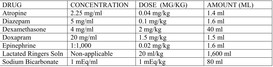
4. Administer 0.02 mg/kg of 1:1,000 (1.0mg/ml) epinephrine I.V. or intracardially and
continue with external cardiac massage. This dose is approximately 1.6 mg (1.6 ml) per
80 kg adult jaguar. Only veterinarians should attempt intracardial injections.
5. Administer 20 ml/kg cool Lactacted Ringer’s Solution as an IV bolus (i.e., a single
rapid infusion).
6. If no response, repeat 4 above at 5 minute intervals indefinately.
C. Hyperthermia is defined as an increase in body temperature to a point where oxygen demand exceeds supply due to increased metabolism.
Diagnosis of hyperthermia is easily determined by rectal thermometer. 1. Temperatures > 410 C (105.8 0 F) are true emergencies.
Causes of hyperthermia in field immobilization include 1) internal heat production due to
excessive physical exertion; 2) external heat absorption; 3) drug-induced compromise of
thermoregulation; and 4) inability to use behavioral thermoregulation.
Treatment of hyperthermia includes the following: 1. Make sure the jaguar is in the shade
2. Use portable “cold” packs that can be placed in the groin, axillae (armpit) and belly of
the jaguar.
3. Cool the jaguar by applying water over the body and/or alcohol to the extremities (legs
and feet).
5. Administer 20 ml/kg cool Lactacted Ringer’s Solution as an IV bolus (i.e., rapid fluid
infusion).
6. Take the temperature every 5-10 minutes to determine if the temperature is decreasing.
Continue to wet the animal if the temperature remains high.
7. Administer antagonist I.V. (I.M. if a vein is not readily identified). However, do this
cautiously as the antagonist will only reverse the drug it antagonizes and the jaguar may
be semi-anesthetized and difficult to handle after the antagonist is administered.
8. If it is believed that the hyperthermia is due to muscle rigidity and a light plane of
anesthesia, diazepam at a dose of 5 – 10 mg / jaguar TOTAL can be administered slowly
I.V. to decrease muscular activity.
Note - Hypothermia (<350 C = <95 0 F) - decreased body temperature to point of cellular death -
is much less likely under most field conditions in which jaguar will be immobilized. However,
this may occur (i.e., high altitude regions) and should be treated by warming the animal.
D. Vomiting and aspiration are defined as the ejection of stomach contents through the esophagus and mouth and inspiratory sucking into the airways of foreign material,
respectively.
Diagnosis of aspiration of vomit is not always straight forward. Clinical signs suggestive of aspiration are
1. Blue/gray mucous membranes (gums)
2. Choking and gasping
3. Gurgling sounds during respiration
4. Presence of material in the larynx and trachea
Causes of aspiration during field immobilizations include 1) drug-induced vomiting (i.e.,
xylazine) with subsequent aspiration; 2) stress; 3) excitement; and 4) positioning of the head
lower than the stomach.
Treatment of vomiting and aspiration include the following: 1. Do not administer any additional anesthetic agents.
2. Clear the airway.
3. If the jaguar is not breathing on its own, begin artificial ventilation (see respiratory
depression/arrest above).
4. If the laryngeal region is KNOWN to be irreversibly blocked with vomitus, a
tracheotomy may be attempted in the distal trachea to allow the passage of oxygen. (This
procedure should only be performed by veterinarians that are familiar with the surgical
procedure.)
5.Administer long-acting antibiotics (i.e., Penicillin G benzathine 40,000 IU/kg I.M.)
Aspiration of vomit can be an acute life-threatening situation due to the initial blockage
of the respiratory tract and asphyxiation. However, it should be noted that a more chronic
result of aspiration is pneumonia, which may also be life-threatening. Any jaguar known
to have aspirated vomit is susceptible to developing pneumonia. The use of a long-acting
antibiotic will help to decrease the likelihood of pneumonia developing but is often of
little help in those cases
in which a large amount of vomit has been aspirated.
E. Shock is defined as ineffective blood perfusion of tissues resulting in cellular hypoxia. Shock is typically classified into three categories: hypovolemic, cardiogenic, and
distributive. Shock associated with the capture and immobilization of free-ranging
jaguars can be any of these three categories but typically is distributive or cardiogenic.
2. Slow capillary refill time
3. Hyperventilation
4. Depressed mentation in those animals not anesthetized.
Causes include 1) prolonged physical exertion; 2) prolonged physiologic stress; 3)
prolonged psychological stress; and 4) severe blood loss.
Treatment should include the following:
1. Do not administer any additional anesthetic agents.
2 Administer 4 mg/kg dexamethasone I.V. (unless vein is not readily available; then use
I.M.)
3. Administer 30 ml/kg Lactated Ringer_s solution I.V.
4. If the jaguar is not breathing on its own, begin artificial ventilation (see respiratory
depression/arrest above).
F. Seizures are defined as disturbances of cerebral function characterized by a violent, involuntary contraction or series of contractions of the voluntary muscles.
Diagnosis is made based on clinical signs that include the following 1. Uncontrolled muscle and/or whole body spasms
2. Rigid extension of the limbs
Causes include 1) drug-induced (i.e., ketamine and tiletamine); 2) trauma; and 3)
hypoglycemia.
Treatment includes the following:
1. Administer 10 mg diazepam I.V. slowly over 10-15 seconds.
3. Monitor body temperature to determine if secondary hyperthermia results from the
seizure activity.
G. Wounds are often associated with the dart site as well as by trap or chase injuries. (Be especially cognizant of any oral lesions and/or broken teeth.)
Diagnosis is based on clinical signs. The severity of the wound will dictate the treatment modality chosen.
1. Physical examination to evaluate for traumatic lacerations and lesions
2. Oral examination to evaluate for oral lesions and broken teeth
Treatment should always include:
1. Clean the wound with a povidone-iodine or 2% chlorhexidine solution. If neither of
these are available, use soapy water.
2. If necrotic tissue is present and the field personnel are familiar with veterinary surgical
techniques, debride the dead tissue and repeat step 1.
3. Only suture those wounds that you KNOW are fresh (i.e., caused by the dart) and that
require sutures to minimize further tissue damage. Again, only field personnel who are
familiar with veterinary surgical techniques should attempt to suture any wounds.
4. Apply topical antibiotic and fly strike ointment to wound site.
5. Administer long-acting antibiotic I.M. (i.e., Penicillin G benzathine 40,000 IU/kg I.M.)
6. Administer Ivermectin 200 mcg/kg SQ (to prevent screw worm infestation at site of
broken skin)
Treatment of broken teeth: It is imperative that a fractured tooth (most commonly a canine is broken during jaguar captures and immobilizations) be repaired to minimize pain and infection
associated with the tooth. A calcium hydroxide product (i.e., Dycal R) can be used to cap the
H. Capture myopathy is defined as a complex alteration of metabolic processes that may cause peracute lethal acid-base and electrolyte imbalances or acutely produce necrosis of
cardiac and striated muscles. Although rarely ever encountered in felids (it is most
commonly a problem in ungulates), field researchers should be familiar with this
condition.
Diagnosis is based on clinical signs that include 1. Ataxia (abnormal walk) and weakness
2.Paresis and paralysis
3. Brownish urine
4. Death
Causes include 1) prolonged physical exertion; and 2) prolonged physiological and/or
psychological stress.
Treatment is often unrewarding and the backbone of treatment is PREVENTION. 1. Minimize the stress of any immobilization procedure.
2. Administer 5 meq/kg sodium bicarbonate IV
3. Administer 30 ml/kg Lactated Ringer’s solution IV
I. Dehydration defined as the reduction of the normal body fluid is often associated with immobilization of free-ranging wildlife.
Diagnosis is based on clinical signs that include 1. Weak pulse
2. Dry mucous membranes
3. Skin lacks pliability
4. Depressed mentation in those jaguar not anesthetized
Causes include 1) decreased water intake; 2) hyperthermia; and 3) chronic water loss (ie.,
Treatment should include the following:
1. Do not administer any additional immobilizing drugs.
2. Administer Lactated Ringers solution at a rate of 20 ml/kg, preferably I.V. but S.Q. can
be used as the second choice. Alternatively, it would be best to calculate fluid deficit
based on a scale of 5% (mucous membranes dry; skin tacky) to 8% (mucous membranes
dry and reddened; skin tacky and remains tented when pinched) and administer fluids at a
rate of - (% fluid deficit) X (body weight (kg)) / 100 = volume in liters
Example for 80 kg jaguar with 5% dehydration 5% X 80 kg / 100 = 4 liters
4. Medical kit for field immobilization
In addition to a strong knowledge base in immobilization and handling of jaguars, it is
imperative that researchers have the proper equipment with them in the field. Devices for
monitoring physiologic parameters will ensure that the researcher is alerted to possible anesthetic
problems / emergencies. Additionally, a few instruments and drugs are necessary for handling
emergencies should they occur. A standard medical kit for all researchers to take to the field is
presented in table 2.
IV. POST ANESTHETIC RECOVERY
The recovery period is just as important for proper handling and monitoring as the
induction and maintenance periods; in fact, most anesthetic complications occur during induction
and recovery. It is not uncommon for anesthetic related morbidity and mortality to occur during
recovery. Although there are reversal drugs for the zolazepam component of telazol (flumazenil),
xylazine (yohimbine), and medetomidine (atipamezole), jaguar recoveries can not be completely
reversed with one specific antidote as is available for narcotic (i.e., carfentanil, etorphine)
does not cause injury to itself or to people involved in the immobilization during the recovery
period.
During recovery, the jaguar should be positioned so that it can breathe easily and will not
harm itself on nearby objects. The animal should be placed in lateral recumbency with the head
and neck extended. Abrasive material should not be under the head due to possible head
movements that could lead to corneal abrasions. Additionally, the jaguar should be placed in a
shaded area to protect if from excessive heat and from the sun damaging the cat’s eyes. People in
the area must remain quiet and should NOT stimulate the jaguar. It should recover at its own
pace as it metabolizes the anesthetic agent(s).
If the jaguar was originally captured in a box trap, it may be beneficial to let the animal
recover in the cage where it is dark and quiet. However, it must be remembered that when the cat
is awake enough for release, the danger to field personnel may be significant when opening the
cage. While in the cage and recovering, the animal may also be aggressive and cause harm to
itself. Thus, if one is to use a box trap for recovery, it requires judgement to be sure the jaguar is
awake enough prior to release, but does not cause harm to itself while still in the cage.
Alternatively, when no trap is available (i.e., treed by dogs or darted from a blind), the
animal can be placed in a quiet, padded (i.e., with leaf litter), and protected (i.e., not near ledges,
hard structures) area to recover on its own. Risks are involved with both recovery methods.
People should move far away from the recovering cat with one or two observers remaining only
as close as needed to visually observe the animal.
V. ANIMAL SAMPLING
The number one priority for the JHP is related to the safe handling of all jaguars during
immobilization procedures. The second most important aspect of the JHP is that researchers
transported appropriately so that viable tissues are available for diagnostic testing. Samples can
be collected from immobilized live jaguars (i.e., blood, feces, ectoparasites, hair), field samples
(i.e., feces, urine, hair) and tissues salvaged from dead jaguars.
In this section we present the types of samples that should be collected, equipment
necessary to collect them, how to collect the samples, and what diagnostic tests can be performed
on these samples. Field researchers will have different equipment available, as well as different
levels of training and experience in the technical skills related to biological tissue sample
collection. For this reason, the type of samples collected will vary from one immobilization event
to another.
1. Sample collection, storage, and transport
A. Physical examinations - All jaguars that are immobilized, or observed free-ranging, should be clinically evaluated. A physical examination of the jaguar_s health will provide
valuable information about its health status. Visual observations are useful but more specific
physical examination techniques (i.e., thermometer, stethoscope, palpation, etc.) should be
performed when feasible. Morphometric measurements are also an important component of the
physical examination. An example of a standard physical examination form is provided in
appendix 4.
B. Blood samples - Blood should be collected from jaguars during immobilizations when field staff are familiar and comfortable with the procedure.
1. Collection - Vessels for collecting blood from jaguars include the medial and lateral saphenous veins, femoral vein, cephalic vein, lateral tail vein, and the jugular. We recommend
that biologist with no training in veterinary techniques should not attempt to collect blood. The
needle size should range from 18 - 22 gauge and 1 - 1 _ inches and syringes should be 6 - 25
ml. Ideally, a total of 25 ml whole blood per jaguar should be collected.
Additionally, a sample should be collected from a peripheral ear vein to prepare blood
slides for hemoparasite identification. This sample should be collected using a sterile 18 or 20
gauge needle to puncture the vessel. Microcapillary tubes should be used to draw blood from the
punctured site and slides prepared as described in Appendix 9.
2. Storage - Blood must be immediately transferred to red top tubes containing no anticoagulant and purple top tubes containing EDTA anticoagulant. Once blood is placed in
purple top tubes, these tubes must be gently inverted a few times to mix the blood with the
anticoagulant. This will help to prevent the blood from clotting. Red top tubes should be kept at
room temperature and purple top tubes are bested placed under refrigeration (i.e., cooler with
ice). Blood in both tubes should be centrifuged within 4 hours of collection to separate blood
fractions. A small amount of whole blood (blood in anticoagulant) should be put into a few
microcapillary tubes. This blood will be used for packed cell volume (PCV), total solids (TS),
white blood cell (WBC) counts, filter paper dot blots and blood smear slides (appendices 5 - 9).
The remainder of the blood should be spun at 3,500 rpm for 10 minutes and the separated serum
decanted into cryotubes for long-term storage. Slides should be air dried and fixed with slide
fixative. (Additionally, the quality of slides is improved if they are stained in the field.) Slides
should be placed in a protective holder (slide tray) and stored at room temperature. Cryotubes
should be stored frozen, preferably in liquid nitrogen but alternatively can be kept on ice.
3. Transport – Serum and plasma should be transported in liquid nitrogen or on dry ice to ensure that they remain frozen. Slides can be shipped at room temperature.
1. Collection - In live immobilized jaguars, feces should be collected directly from the animal’s rectum, using a gloved hand, and then placed in appropriate media in an airtight
container (see storage below). Five to 10 grams of feces is adequate to place in each type of
storage media.
2. Storage - Feces can be stored in a variety of media, depending on what diagnostic tests are to be performed. Feces should be placed in 5% formalin for parasite ova and larvae
identification.
Additional feces can be placed into 70 - 95% alcohol. This can be used for DNA analysis,
hormone analysis (i.e., cortisol, estrogens, progesterones, testosterone), and possibly polymerase
chain reaction (PCR) testing for bacteria and viruses.
Empty cryogenic container. This sample should be kept frozen either using dry ice or liquid
nitrogen. This may be used for testing reproduction and stress hormone levels, and virus
isolation.
3. Transport - Fecal samples should be transported in above mentioned containers as follows:
1) Feces in formalin and alcohol should be transported at room temperature.
2) Feces in cryogenic containers should be transported frozen either in liquid nitrogen or on dry
ice.
D. Ectoparasites samples should be collected from immobilized live jaguars, as well as fresh carcasses.
the tick to minimize post-removal lesions. Ticks commonly attach around the ears, groin region,
and axillae.
2. Storage - Place all ectoparasites in airtight containers with 70 - 95 % ethyl alcohol, and maintain at room temperature.
3. Transport - All ectoparasites can be transported in above containers at room temperature.
E. Hair samples should be collected from immobilized live jaguars, as well as from carcasses. (Please refer to the genetic handout.)
1. Collection - Hair samples can be plucked using forceps or fingers or cut using a knife or scissors. Plucked hair is preferable since it is more useful in genetic studies.
2. Storage - Hair samples should be placed in empty, dry paper envelopes and stored at room temperature.
3. Transport - Hair samples should be transported at room temperature in above mentioned containers.
2. Permits and laws for transporting samples internationally
All diagnostic laboratory testing will be performed at a few select laboratories in the USA
that are known for their specialty in non-domestic felids. Our ability to do this is contingent on
exporting samples from various countries and importing them into the US. Jaguars are listed on
CITES appendix I. Therefore, a CITES import and export permit are required for transporting
tissue samples into the US. The JCP is working to obtain blanket CITES permits for the work.
The importing country (USA) must issue an import permit and the exporting country
permit is presented to the office issuing the export. The U.S. Fish and Wildlife Service’s Office
of Management Authority is the agency which currently issues CITES import permits. In
general, they require written proof of permission to conduct the work in the host country to
accompany the application for an import permit. . For more information on permit requirements,
visit the technical pages on the FVP website at www.fieldvet.org.
3. Diagnostic tests that MUST be performed in the field
Blood tests that must be performed in the field include the use of whole blood, prior to
centrifugation, for PCV, TS, and WBC count. After the whole blood has been separated, using
the centrifuge, these test can no longer be performed on the resultant plasma. These tests require
a centrifuge, capillary tubes, PCV chart, and refractometer for the PCV and TS and a microscope
and WBC Unopette kit for a WBC count. Methods for performing these field based tests are
presented in appendices 5, 6 and 7.
4. Diagnostic tests to be performed in a laboratory
Most diagnostic testing will be performed in laboratories. The following is a list of the
tests we recommend for a complete health assessment of jaguars. These tests include an
assessment of parasitic and infectious disease agents to which they have been exposed.
A. Blood
1. White blood cell differential counts (in conjunction with blood test performed in the
field)
2. Chemistry profile
3. Serology for infectious and parasitic agents which will include -
Viral agents
FeLV - feline leukemia virus
FIP - feline infectious peritonitis (coronavirus)
FCV - calicivirus
FHV - feline rhinotracheitis (herpesvirus)
FIV - feline immunodeficiency virus
Puma lentivirus
CDV - canine distemper virus
Pseudorabies
Rabies
Bacterial agents
Leptospirosis (17 serovars)
Bartonella henselae
Hemobartonella felis
Parasitic infections
Toxoplasmosis gondii
Babesiosis
Dirofilaria immitis
Cytauxzoon felis
Hormone levels (i.e., cortisol, testosterone, estrogen, progesterone)
Toxins (i.e., mercury, organophosphates, carbamates, PCBs, hydrochlorinated carbons)
Vitamins / Minerals
B. Feces 1. Parasites
2. PCR test for bacteria and viruses
3. Feline coronavirus RNA antigen
Hormone levels (i.e., cortisol, testosterone, estrogen, progesterone)
C. Ectoparasites
All ticks should be identified to the species level by an acarologist. Other ectoparasites
D. Necropsy protocol
Appendix 10 contains a hard copy of the necropsy manual that can also be located on the internet
at http://www.vetmed.ucdavis.edu/whc/necropsy/toc.html.
The necropsy manual was written by Dr. Linda Munson of the University of California, Davis
and translated to Spanish by Dra. Marcela M. Uhart and to Portugese by Dra. Paulo Rogeria
Mangini. This manual provides practical information for performing necropsies in the field. It is
imperative that all researchers that collect samples from a dead jaguar record the following: age,
sex, GPS reading, date and time located, and condition of body. It is also extremely important
that the stomach contents be collected for studies on prey preference for jaguars across their
range. These contents may be dried and stored at room temperature or placed in 70 – 95%
alcohol.
E. Centralized laboratories - The Field Veterinary Program, WCS is establishing
agreements with a large number of experts for laboratory testing in order to standardize the data
from jaguar samples.
VI. DATA COLLECTION, ANALYSIS, AND DISTRIBUTION
One of the largest benefits of a collaborative health program throughout the jaguar’s
range is the ability to compile and correlate data from various habitats and populations of
jaguars. A standard format for data collection is invaluable for compiling the data in any
meaningful way. Appendix 2 is an example of the medarks form for collecting anesthesia data.
Additionally, a standard form to collect physical examination data would be beneficial. One
example is presented in appendix 4. This data will require statistical and non-statistical analysis
to allow us to achieve our primary objectives of assessing the overall health status of jaguars
across their range, determining the disease threats to jaguars, and using both these findings for
These data will be presented for the scientific community, governmental officials, and lay
VII. BIBLIOGRAPHY
Bauditz, R. 1972. Sedation, immobilization and anesthesia with Rompun in captive and
freeliving wild animals. Vet. Med. Rev. 3: 204-226.
Boever, W.J., J. Holden, K.K. Kane. 1977. Use of telazolTM (CI-744) for chemical restraint and
anesthesia in wild and exotic carnivores. Vet. Med./Sm. Ani. Clin., Exotic Species, 1722-1725
Bush, M. 1992. Remote drug delivery systems. J. Zoo Wildl. Med. 23: 159-180.
Crawshaw, P. G. 1992. Recommendations for study design on research projects on neotropical
felids. In: Felinos de Venezuela – Biología, Ecología y Conservacíon. Memorias del Simposio
Organizado por Fudeci del 01 al 04 de Septiembre de 1991. FUDECI, Caracus. Pp. 187-222.
de Wet, T. 1993. Physical capture of carnivores. In: McKenzie, A.A. (ed.).The Capture and Care
Manual. Wildlife Division Support Services CC and The South African Veterinary Foundation.
South Africa, Pp. 255-277.
Evans, A.T. 1996. Anesthetic emergencies and accidents. In: J.C. Thurmon, W.J. Tranquilli, and
G.J. Benson (eds.). Lumb and Jones_ Veterinary Anesthesia: third edition. Wilkins and Wilkins
Co., Philadelphia, PA. Pp. 849-860.
Fowler, M.E. 1995. Medical problems during restraint. In: Restraint and Handling of Wild and
Domestic Animals; second edition. Iowa State University Press. Ames, Iowa. Pp. 78-99.
Gray, C.W., M. Bush, and C.C. Beck. 1974. Clinical experience using CI-744 in chemical
restraint and anesthesia of exotic specimens. J. Zoo Ani Med. 5: 12-21.
Hoogesteijn, R., R. McBride, M. Sunquist, A. Hoogestiejn, and L. Farrell. 1996. Medetomidine
and rubber-padded leg-hold traps in Venezuelan cat studies. Cat News 25: 22-23.
Interational Wildlife Veterinary Services, Inc., 1991. Wildlife Restraint Series. Interational
Wildlife Veterinary Services, Inc. Salinas, Cali.
Jalanka, H.H., and B.O. Roeken. 1990. The use of medetomidine, medetomidine-ketamine
combinations, and atipamezole in nondomestic animals: a review. J. Zoo Wildl. Med. 21: 259-
282.
Kreeger, T.J. 1996. Handbook of Wildlife Chemical Immobilization. Pp. 175
Kreeger, T.J. 1996. Emergency treatment - Animal. In: Handbook of Wildlife Chemical
31 Immobilization. International Wildlife Veterinary Services, Inc., P.O., Laramie, WY, Pp.
79-96.
Lopez de Buen, L., J. M. Aranda Sanchez. 1986. Nota zoologica. Anestesia de mamiferos
silvestres con la combinacion ketamina-xilacina. Biotica, 67-71.
McLaughlin, R., and A. Kuzma. 1991. Surgical management of collapsed pelvis in a jaguar.
J.A.V.M.A. 198: 1789-1791.
Morato, R.G. 1997. Dissertacao Mestrado. 120 pp.
Morato, R.G., C.A. Moura, and P.G. Crawshaw. Chemical restraint of free ranging jaguars
(Panthera onca) with tiletamine-zolazepam combination. In: Jaguars in the New Millenium
Nielsen, L. 1999. Instrumentation. In: Chemical Immobilization of Wild and Exotic Animals.
Iowa State University Press. Ames, Iowa. Pp. 31-82.
Nielsen, L. 1999. Management of medical emergencies in the field. In: Chemical
Immobilization of Wild and Exotic Animals. Iowa State University Press. Ames, Iowa. Pp.
209-226.
Quigley, H. 1987. Ecology and conservation of the jaguar in the Pantanal Region, mato Grosso
do Sul, Brazil. Ph.D. Dissertation, University of Idaho, Moscow, Idaho, USA.
Quigley, K. Hornocker Wildlife Institute Immobilization and Biological Sampling Protocols.
Hornocker Wildlife Institute, Inc. Moscow, Idaho.
Rabinowitz, A.R., and B.G. Nottingham. 1986. Ecology and behaviour of the jaguar (Panthera
onca) in Belize, Central America. J. Zool. Lond. 210: 149-159.
Seal, U.S., and T.J. Keeger. 1987. Chemical immobilization of furbearers. In: Novak, M. et al.,
eds. Wild Furbearer Management and Conservation in North America. Toronto: Ontario
Ministry of Natural Resources, 191-215.
Shobert, E. 1987. Telazol use in wild and exotic animals. Vet. Med. 82: 1080-1088.
Spelman, L. Unpublished data, veterinarian for the National Zoological Park, Washington, D.C.
1997.
Wilson, D. E., Cole, F. R., Nichols, J.D. Rudran, R., and Foster, M. 1996. Measuring and
monitoring biological diversity. Standard Methods for Mammals. Smithsonian Institution Press,
Yates, R.A. Unpublished data, veterinarian for the National Zoological Park, Washington, D.C.
VIII. FIGURES
1. Dart placement sites for darting free-ranging jaguar.
.
Triceps - large muscle mass behind humerus and below scapula
Quadriceps - large muscle mass in front of femur
Semi-Memb. group - large muscle mass behind femur
Karesh and Deem, 2000