Effect of chronic oestrogen administration on androgen
receptor expression in reproductive organs and pituitary
of adult male rat
M. C. Kaushik1, M. M. Misro1, N. Sehgal2& D. Nandan1
1 Department of Reproductive Biomedicine, National Institute of Health and Family Welfare, Munirka, New Delhi, India; 2 Department of Zoology, University of Delhi, Delhi, India
Introduction
Chronic oestrogen exposure in foetal or neonatal life has been implicated for the apparent increase in male repro-ductive disorders like cryptorchidism, hypospodiasis, low sperm counts and testicular cancer in men (Sharpe, 2003). These alterations are considered as symptoms of a syndrome known as the testicular dysgenesis syndrome (Skakkebaek et al., 2001; Skakkebaek & Jorgensen, 2005) reportedly altered in areas close to industry and exposed to environmental and industrial chemicals possessing oestrogenic potency (Mackenzie et al., 2005). It is per-ceived that these abnormalities first arise during foetal
development (Delbes et al., 2006). Male rats treated neo-natally with diethylstilboestrol (DES) revealed suppression of androgen action and gross abnormalities in reproduc-tive tract (McKinnell et al., 2001). Furthermore, studies on the sons of women who were exposed to DES during pregnancy show a similar range of abnormalities (Still-man, 1982; Toppariet al., 1996).
On the other hand, oestrogen is also known to have adverse effects on spermatogenesis, which are reversible following withdrawal of treatment (Toyama et al., 2001). Similar findings are also reported in adult male rats in which oestradiol 3-benzoate (EB) affects not only sper-matogenesis but also induces apoptosis in the prevailing
Keywords
Androgen receptor—apoptosis— immunostaining—oestrogen treatment—pituitary—RT-PCR— testis—Western blotting
Correspondence
M. M. Misro, Department of Reproductive Biomedicine, National Institute of Health and Family Welfare, Baba Gang Nath Marg, Munirka, New Delhi, India.
Tel.: 91-11-26165959; Fax: 91-11-26101623; E-mail: [email protected]
The study was funded jointly by ICMR and NIHFW.
Accepted: June 18, 2009
Summary
Following chronic (15 or 30 days) treatment with oestradiol 3-benzoate (75lg rat)1day)1in 100ll of olive oil) to adult rats, androgen receptor (AR)
population among the dwindling number of germ cells (Chaki et al., 2006). While foetal and neonatal exposure with oestrogen induces irreversible abnormal development of reproductive organs, the effect in adult animals is com-pletely reversible with the stoppage of treatment. This may be explained by the fact that neonatal treatment with DES can cause life long suppression of plasma testoster-one levels (Atanassova et al., 1999) while post-pubertal DES administration clearly results in reduced androgen production during the course of treatment (Abeny & Keel, 1986).
The molecular mechanism whereby oestrogen mediates such divergent effects is not completely understood and there are conflicting reports on the way it is mediated in different reproductive organs. In the prostate, neona-tal exposure to oestrogen induced lobe-specific altera-tions in AR receptor expression (Prins, 1992). The intracellular mechanism of AR down-regulation is described to be mediated at the post-translational level characterised by AR degradation through proteosome pathway with no difference in the mRNA levels in response to oestrogen exposure (Woodham et al., 2003). By contrast, adult rats fed with dietary phytooestrogens for 3 days demonstrated an increased AR mRNA expres-sion in the initial segment of the epididymis (Glover &
Assinder, 2006). This study was therefore initiated in adult rats exposed to EB at a higher pharmacological dose to determine the AR expression both at the mRNA and protein levels in different reproductive organs (testis, epididymis, prostate and seminal vesicle) and simultaneously in the pituitary through immunostaining, Western blotting and RT-PCR.
Materials and methods
Animals, treatment and sample preparation
Adult male rats, Holtzman strain, weighing 200–220 g were maintained under controlled temperature (25 ± 2C) and constant photoperiodic (12 h light : 12 h dark) conditions. Food and water were provided ad libitum. Twenty-four animals were divided into four groups of six animals each receiving either olive oil or oestrogen (EB in olive oil) as described below.
Group I: Vehicle control 15 days (olive oil 100ll rat)1
day)1
)
Group II: Vehicle control 30 days (olive oil 100ll rat)1day)1)
Group III: Oestradiol 3-benzoate 15 days (EB, 75lg rat)1day)1in 100ll of olive oil)
C E15 E30
C E15 E30
570 bp
539 bp
110 kda
42 kda
(a) (b)
(f)
(d) (c)
Group IV: Oestradiol 3-benzoate 30 days (EB, 75lg rat)1day)1in 100ll of olive oil)
Oestradiol 3-benzoate (Sigma, St Louis, MO, USA) was dissolved first in isopropanol, resuspended in olive oil and administered intramuscularly. At the end of the treatment, the animals were bled and killed under aether anaesthesia with strict adherence to Standard Institutional Guidelines for Animal Care. Appropriate clearance from Animal Ethics Committee of the Institute was earlier obtained. Serum was separated and stored at)20C till assayed for testosterone. Testis, epididymis, prostate, seminal vesicle and pituitary gland were dissected, weighed and divided in two equal parts. One part was immediately fixed in buf-fered formalin while the rest was frozen in liquid nitrogen and stored at )70C till utilised for RT-PCR, Western blotting and intra-testicular testosterone.
Immunohistochemistry (IHC)
The protocol was strictly followed as per manufacturer’s (Lab Vision Corporation, Feremont, CA, USA) instruc-tions provided in the kit. Briefly, the paraffin-embedded sections were deparaffinised in xylene and rehydrated in alcohol series, immersed in dH2O and washed two times in PBS pH 7.4. The sections were heated in sodium
citrate buffer (10 mmol l)1, pH 6.0) for 10 min in a
microwave oven for antigen retrieval, washed in PBS and incubated with the blocking solution (provided in the kit) for 5 min at RT to block nonspecific binding sites. Sections were incubated with AR primary antibody (Lab Vision Corporation) (dilution 1 : 100 in PBS) overnight at 4C, washed four times in PBS and incubated with
biotinylated second antibody ready-to-use followed by streptavidin alkaline phosphatase solution for 20 min each. The colour was developed using fast red as sub-strate followed by light counterstaining with aqueous haematoxylin.
Western blot analysis
Frozen tissues (testis, epididymis, prostate gland, seminal vesicles and pituitary gland) in liquid nitrogen were grin-ded in ice-cold RIPA (radioimmuno-precipitation assay) buffer pH 7.5 containing 100 ll of protease inhibitor cocktail (Roche, Basel, Switzerland). Following centrifuga-tion at 15000 g for 20 min at 4C, the supernatant was
collected and stored at )20C. Aliquots of supernatant
were assayed for protein content (Bradford, 1976) and electrophoresed through 10% SDS–PAGE and transferred to polyvinyl difluoride membrane (0.45 lm). Membranes
C E15 E30
C E15 E30
110 kda
42 kda 570 bp
539 bp
(a)
(c) (d)
(b)
(e)
(f)
were blocked with 3% gelatin provided in the immuno-blot assay kit (Bio-Rad Laboratories, Philadelphia, PA, USA). Membranes were washed and incubated with AR primary antibody (1 : 2000, Lab Vision Corporation) for overnight at 4C. Equal loading of samples with b-actin antibody (Santa Cruz Biotechnology, Santa Cruz, CA, USA) was carried out. The membranes were finally treated with goat anti-rabbit second antibody conjugated with alkaline phosphatase supplied in the immunoblot kit and the colour was developed as per the manufacturer’s (Bio-Rad Laboratories) instructions.
Semiquantitative RT-PCR
Total RNA was isolated from testes, epididymis, prostate gland, seminal vesicle and pituitary using acid guan-idinium thiocyanate–phenol–chloroform extraction as described (Chomczynski & Sacchi, 1987). Complementary DNA was made using 2lg of total RNA in the presence of Avian myeloblastosis virus reverse transcriptase from kit (Promega Corporation, Madison, WI, USA). The pri-mer sets used as a template to obtain PCR products of AR were 5¢-TGC TGC CTT GTT ATC TAG TCT CA-3¢ and 5¢-ACC ATA TGG GAC TTG ATT AGC AG-3¢ and forb-actin were 5¢-CTG TGC CCA TCT ATG AGG GTT
AC -3¢and 5¢-AAT CCA CAC AGA GTA CTT GCG CT -3¢. For semiquantitative analysis,b-actin was used as an inter-nal control and co-amplified with AR mRNA by using
b-actin primer and AR primer, 50 pmol each. All PCR reactions were performed for 25 cycles with an annealing temperature of 60C in 2.5 mmMgCl2.
The band intensities of AR andb-actin as obtained for each organ were quantified using the densitometric soft-ware; Lab Works Image Acquisition and analysis Software version 4.0 (UVP, CA, USA) and the differences in ratios (value of AR/b-actin) between groups were compared.
In situend labelling (ISEL)
In situApoptosis Detection Kit (R & D Systems Inc., Min-neapolis, MN, USA) was used to identify apoptotic cells by detecting DNA fragmentation through a combination of enzymology and immunohistochemistry techniques. Paraf-fin embedded sections of testis, epididymis, prostate gland, seminal vesicle and pituitary gland were deparaffinised in xylene rehydrated in alcohol, immersed in dH2O and washed in PBS. To make the DNA accessible to the label-ling enzyme, the cell membranes were permeabilised with proteinase K. Endogenous peroxidase activity was quenched using hydrogen peroxide and biotinylated
C
(a)
(c) (d)
(b)
(f)
E15 E30
C E15 E30
570 bp
110 kda
42 kda 539 bp
nucleotides were incorporated into the 3¢-OH ends of the
DNA fragments by terminal deoxynucleotidyl transferase. The biotinylated nucleotides were detected by using strep-tavidin-horseradish peroxidase conjugate followed by the substrate, diaminobenzidine (DAB). The enzyme reaction generates an insoluble coloured precipitate where DNA fragmentation has occurred. DAB-stained tissues sections were examined using a Nikon microscope image analyser and photographed. ISEL positive cells were counted out of every 100 cells examined serially in each section.
Quantitation of spermatogenesis
Quantitation of the spermatogenesis in the testes of con-trol and treated rats (n= 6) was carried out as described (Russell & Clermont, 1977). Briefly, 20 tubules in each testicular section (one section each from five different regions of testis) were randomly picked up and included in the analysis. Numbers of different types of germ cells present in a tubule were recorded through microscopic examination. An ocular calibrated micrometre fitted to the eyepiece was used to measure diameters of seminifer-ous tubules and germ cell nuclei. All the nuclear count (crude counts) of the germ cells were corrected for differ-ences in nuclear diameter by the formula of Abercrombie
(1946), i.e. true count = (crude count·section
thick-ness)/(section thickness + nuclear diameter of germ cells) and tubular shrinkage by the Sertoli cell correction factor (Clermont & Morgantaler, 1955).
Radio immunoassay of testosterone
Radio immunoassay of testosterone was performed following methods of Abraham (1974) with modifications. The antibodies for the purpose were raised in the labora-tory. Aether extraction of serum samples was carried out prior to setting up the radioimmunoassay. The inter- and intra-assay variations were 4.3% and 2.3% respectively.
Statistical analysis
Quantitative data are expressed as mean ± SD. Statisti-cally significant differences between groups were determined by student’st-test. P-value <0.05 was consid-ered statistically significant.
Results
The AR protein expression found using immunostaining, Western blotting and the semiquantitative RT-PCR in
C E15 E30
C E15 E30
570 bp
539 bp
110 kda
42 kda
(a)
(c)
(b)
(d)
(e)
(f)
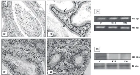
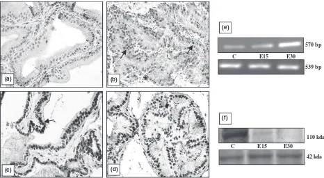
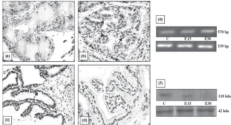
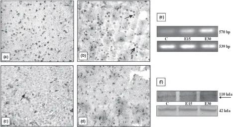
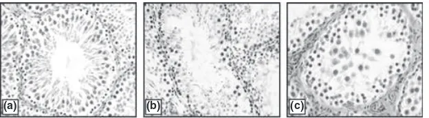
different organs is shown in Figs 1–5. All the organs (testis, epididymis, seminal vesicle, prostate and pitui-tary), showed a marked decline in the intensity of immu-nostaining at the end of 30 days of EB treatment compared with vehicle treated controls. In testis, conspic-uous staining was noted in the cells close to the basement membrane which included Sertoli cells, gonial cells and spermatocytes (Fig. 1b–d). Positive staining was also observed in peritubular myoid cells and Leydig cells but was absent in sections incubated with nonimmune serum (Fig. 1a). AR immunostaining in the epididymis was intensely observed both in epithelial as well in stromal cells. A pronounced reduction in the intensity of expres-sion was visible after 30 days compared with 15 days of EB treatment (Fig. 2b–d). Immunoexpression in the vehi-cle treated adult prostate was characterised by positive staining in all the luminal epithelial, stromal and peria-cinar smooth muscle cells which gradually declined with increasing duration of oestrogen treatment (Fig. 3b–d). The trend was similar in the seminal vesicle with intense AR staining in epithelial and stromal cells of control rats, which faded gradually after oestrogenisation (Fig. 4b–d). In the pituitary, it was difficult to differentiate the acid-ophils and basacid-ophils on the basis of staining. However, the intensity of immunostaining followed a similar trend,
gradually decreasing with the increase in the duration of EB treatment (Fig. 5b–d). No immunostaining was seen in tissue sections (negative control) incubated with non-immune serum (Figs 2–5a).
Identical to immunostaining, immunoblots of all the reproductive organs (testis, epididymis, seminal vesicle and prostate) demonstrated a marked attenuation in AR protein expression following oestrogen treatment. How-ever, AR protein in these organs failed to be probed in the immunoblots at the end of 30 days of EB treatment (Figs 1–4f). Only the pituitary presented a different pic-ture in which AR protein was immuno-precipitated into two different specific entities or bands (Fig. 5f). By con-trast, all the five organs demonstrated a consecutive rise in AR mRNA levels with respect to the increase in the duration of oestrogen treatment (Figs 1–5e). This was clearly resolved from the data obtained from the densito-metric analysis of band intensities of AR transcripts in between groups and in different organs (Table 1). A sum-mary observation of the rise and fall of AR expression following oestrogen treatment in different organs is presented in Table 2.
The spermatogenesis was severely affected both quanti-tatively and qualiquanti-tatively. There was a significant decrease in seminiferous tubule diameter and the number of
C E15 E30
C
(a)
(c)
(b)
(d)
(f)
E15 E30
110 kda
42 kda 570 bp
539 bp
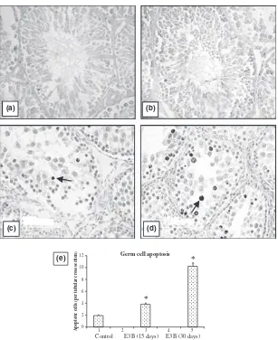
prevailing germ cells per tubule (Fig. 6). While the num-ber of elongated spermatids declined drastically by 15 days, most of the tubules were devoid of elongated spermatids and spermatozoa after 30 days of EB treat-ment (Fig. 6). Loss of germ cells in the seminiferous tubules was consistent with the rise in number of apopto-tic germ cells per tubule (Fig. 7). Germ cell apoptosis was not found specific to any particular germ cell type. There was decrease in weights of testis and epididymis with
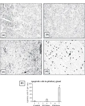
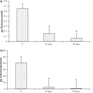
increasing duration of oestrogen treatment (Table 3). The reduction in the weight of accessory reproductive organs like prostate and seminal vesicle was significant (P< 0.001) after 15 days of EB treatment. However, no substantial decline was observed subsequently after 30 days treatment (Table 3). Cell apoptosis was also not found prevalent (data not shown) in all these organs. On the other hand, a significant rise in the number of apop-totic cells in the pituitary was observed at the end of 30 days of EB treatment (Fig. 8). Intra-testicular and serum testosterone levels declined significantly as a result of oestrogen treatment (Fig. 9).
Discussion
The present findings demonstrate that modulation of AR protein expression by oestrogen in all the five organs of adult rats (testis, epididymis, seminal vesicle, prostate and pituitary) was carried out through either translational or post-translational pathways as AR transcriptional activity was found up-regulated. Decrease in the weight of andro-gen-dependent organs (epididymis, seminal vesicle and prostate) was consistent with the decrease in serum levels of testosterone but independent of cell apoptosis found prevalent only in the testis and pituitary.
AR is a member of the steroid receptor super family with an apparent molecular mass of 110 kDa with 918 amino acids (Chang et al., 1988; Lubahn et al., 1988). Abnormalities in the AR signalling pathway have been linked to male infertility, Kennedy’s disease and prostate cancer. Regulation of the AR activity can be achieved by several different ways: modulation of AR gene expression,
Table 1Densitometry showing band intensities of AR andb-actin in different organs
Organs Control 15 days 30 days
Testis 1.035 ± 0.127 1.302 ± 0.159* 1.377 ± 0.180* Epididymis 0.906 ± 0.052 1.006 ± 0.068 1.063 ± 0.092* Prostate 0.853 ± 0.066 0.908 ± 0.080* 1.225 ± 0.100** Seminal
vesicle
0.721 ± 0.040 0.737 ± 0.040 0.918 ± 0.106*
Pituitary 0.633 ± 0.026 0.870 ± 0.011*** 0.982 ± 0.065**
*P< 0.05, **P< 0.01, ***P< 0.001 compared with control.
Table 2Summary of AR expression in different organs after EB treatment
Organs Immunostaining Western blotting RT-PCR
Testis fl fl ›
Epididymis fl fl ›
Prostate gland fl fl ›
Seminal vesicles fl fl ›
Pituitary gland fl fl ›
Seminiferous tubule diameter and maturing germ cells/tubule in control versus EB treatment rats.
Treatment
Vehicle
control 288 ± 12
255 ± 10 37 ± 3.2
androgen binding to AR, AR protein stability, AR nuclear translocation and AR transactivation (Lee & Chang, 2003). Both androgen and oestrogen regulate AR at the level of mRNA and protein. Neonatal exposure to oestro-gens resulted in an immediate and sustained decrease in AR protein levels in the developing and adult rat ventral prostate that in turn led to abnormal growth, decreased secretary capacity and a dampened activational response to exogenous androgen replacement (Prins, 1997).
It was also explained that this AR down-regulation in the rat prostate as result of neonatal oestrogen exposure
is not mediated at the transcriptional or translational level but rather through a post-translational mechanism at the level of AR protein degradation through ubiquitin-proteosome pathway (Woodham et al., 2003). In the adult model as followed in this study, the AR protein levels continued to decline from the control levels to post-15 days of EB treatment, and by 30 days there was hardly any protein resolved in the immunoblots of differ-ent organs except the pituitary (Figs 1–5f). AS decline in AR protein levels was observed by post-15 days of EB treatment in all the organs, the involvement of transla-2
Control E3B (15 days) *
*
E3B (30 days) Germ cell apoptosis
3 4 5
1 0 2
A
p
optotic cells (per tub
ular cr
oss section)
4 6 8 10 12 (a)
(c) (d)
(e)
(b)
Fig. 7In situend labelling of DNA in testis sections. Representative sections from (a) negative control, (b) vehicle treated control, (c) 15 days and (d) 30 days of EB treatment. Note the rise in ISEL positive cells ( ) in testis sections from treated rats, also shown in (e) bar chart. *P< 0.01, **P< 0.001, 400·.
Table 3Weight of reproductive organs from control and EB treated rats
Treatment Testis (g) Epididymis (g) Prostate gland (g) Seminal vesicle (g)
Vehicle control 1.4 ± 0.17 0.7 ± 0.11 0.416 ± 0.07 1.03 ± 0.235 EB (15 days) 0.85 ± 0.3* 0.339 ± 0.06* 0.141 ± 0.016* 0.3 ± 0.069* EB (30 days) 0.24 ± 0.03* 0.232 ± 0.04* 0.139 ± 0.093* 0.306 ± 0.064*
tional pathway in AR protein down-regulation cannot be ruled out. The complete disappearance of the AR protein probably supports the fact that it has undergone degrada-tion following long-term EB (30 days) exposure, by ubiquitin-proteosome or some other pathway in adult rats needs further investigation. However, the process of AR degradation probably provides sufficient input for additional AR transcriptional activity and quantitatively production of more AR mRNAs as presently observed (Figs 1–5e).
That the AR protein is degraded at the post-transla-tional level is further supported by the present findings in the pituitary gland. AR protein blot for the pituitary shows two specific bands instead of one, found closely allied to each other, possibly as a product of degradation (Fig. 5f). It is not clear at this stage whether AR protein degradation follows certain specific steps as seen in case of the pituitary or degrades without living any trace as shown in the immunoblots for reproductive organs. As the AR antibody recognises the two different specific
epitopes, it is quite possible that these bands represent the initial stages of degradation of AR protein in the pitu-itary following both 15 and 30 days of EB treatment. Fur-ther investigation of AR protein levels in the adult pituitary following extended oestrogen exposure than the present one is needed to confirm the sequences of events leading to total AR degradation.
Complete androgen withdrawal that elevated mRNA levels was also associated with a decrease in AR protein levels in the adult prostate. This autologous nature of regulation of AR expression was reported earlier in rats utilising a castrate model (Prins & Woodham, 1995; Pelletier, 2002). In the other androgen insufficiency model induced by ethane dimethane sulphonate treatment, AR protein expression in testis and prostate was suppressed independently of the up-regulation in mRNA levels (Turneret al., 2001; Tanet al., 2005). The possible expla-nation for such a discordant trend in AR protein and mRNA in the same organ was described as the inability or lack of availability of AR messages to be translated
0
1 2
Control E15 days E30 days
3 4
Apoptotic cells in pituitary gland
5 6 7
*
20 40 60 80 100
Apototic cells % per area
(a) (b)
(c) (d)
(e)
despite their cellular presence (Prins & Woodham, 1995).
The other possibility is that under limited androgen avail-ability as observed following chronic oestrogen treatment, androgens can regulate their own receptors both at tran-scription and translation levels completely independently of each other.
Some divergent trends on the effects of oestrogen on AR gene transcription rate and mRNA quantity in the same organ or different parts of the same organ are reported. In the rat ventral prostate, nuclear run-on-assays showed no alteration in AR gene transcription following neonatal exposure of EB on days 1–5 compared with the controls. In situ hybridisation and quantitative RT-PCR revealed no difference in mRNA levels in the stromal or epithelial cells of the prostate in response to oestrogen exposure, which supports the contention that oestrogen-induced down-regulation of AR is mediated at the post-transcriptional level (Woodham et al., 2003). In male rats fed with dietary phytooestrogens for 3 days, quantitative RT-PCR showed AR expression increasing in the initial but decreasing in the distal region of cauda epididymidis (Glover & Assinder, 2006). This is probably indicative of the fact that differential regulation of the steroid receptor (AR) does occur even in the same organ which could be associated with the specific differences in
physiological function in between the regions. Compared with the controls, up-regulation of AR transcripts was consistently observed following EB treatment in all the organs in this study and the increase was proportional to the duration of treatment; more in 30 days than 15 days of treatment (Figs 1–5, Table 1).
Animals, in this study were exposed to high pharma-cologic dose of oestrogen (75lg day)1rat)1) with
chronic interventions for longer periods (15 days and 30 days) and instead of a single reproductive organ, all the reproductive organs and the pituitary were simulta-neously investigated for AR expression through immu-nostaining as a tool. In earlier studies, in rats treated with DES, AR immno-expression was virtually absent from the affected tissues like testis, epididymis and vas deferens (McKinnell et al., 2001). Rat litters given EB on days 1, 3 and 5 and killed on day 90 demonstrated a significant reduction in AR immunoexpression in ventral and dorsal prostate. A near absence of prostate epithelial AR expression was reported due to impaired differentia-tion in these cells as a result of neonatal EB exposure (Prins, 1992). In our studies, not a single organ demon-strated a complete absence of AR expression on immu-nochemistry although a significant decline in the intensity of staining was resolved in all the organs from
0
0 10 20
pg T
estoster
one/testis
30 40 50 60
*
* * *
C 15 days 30 days
C 15 days 30 days
0.1 0.2 0.3 0.4 0.5
pg T
estoster
one/ml
(b)
treated rats compared with those of the controls. The suppression of immunoexpression of AR in the adult reproductive organs is similar to the effects seen in the reproductive organs (testis, epididymis and seminal vesi-cle) of pubertal rats (Williams et al., 2001) and also in organs (vas deferens, prostate and seminal vesicle) of adult animals exposed to oestrogen (Bianco et al., 2002) during the neonatal period (Atanassova et al., 2005; Prins et al., 2001). Androgen and oestrogen balance is believed to be critical for normal spermatogenesis to proceed and any impairment is always associated with sub-optimal androgen production and availability, which subsequently affects other reproductive tissues. Thus, the attenuation of AR immunoexpression in these tissues may be attributed partly to androgen insufficiency and partly to an anti-androgenic effect exerted by oestrogens. Oestrogen administration to adult rats reportedly reduced reproductive organ weights, epididymal sperm numbers and fertility (Goyal et al., 2001; Chaki et al., 2006). Present findings are very much identical as chronic EB treatment for 30 days down-regulated serum and intra-testicular testosterone, reduced testis and accessory sex organ weights and adversely affected spermatogenesis (Fig. 6, Table 3). Spermatogenesis is a hormonally regu-lated process and testosterone availability is essential for the survival and maintenance of germ cells (Sinha-Hikim & Swerdloff, 1999; Woolveridge et al., 1999). Although oestrogen is a survival factor (Pentikainenet al., 2000), a higher level of the hormone is apoptogenic for germ cells in vivo. Neonatal oestrogen administration was associated with germ cell apoptosis in the testis as a result of sup-pression of serum testosterone levels (Atanassova et al., 1999). In adult rats receiving oestrogen treatment, serum and intratesticular testosterone levels were severely depleted, leading to oxidative stress and germ cell apopto-sis in the testis (Chaki et al., 2006). Testosterone with-drawal was reportedly associated with the induction of apoptosis in the epididymis during the initial 5 days post-orchidectomy (Fan & Robaire, 1998). Similar find-ings on the induction of apoptosis in the prostate were described in organ cultures in Estrogen Receptor Knock Out (ERKO) mouse following high doses of oestradiol exposure which down-regulated prostate-specific AR pro-tein expression (Tayloret al., 2006). However, it is inter-esting to note that the decrease in the weight of reproductive organs in this study is associated with an increase in germ cell apoptosis only in the testis but not in the tissues that are entirely androgen-dependent (viz. epididymis, seminal vesicle or prostate). Unlike orchidec-tomy which drastically cuts down the source of androgen supply, oestrogen treatment in adults only reduces the same to sub-optimal levels (Fig. 7). In the present study, because of such sub-optimal but not complete withdrawal
of androgen availability, cell apoptosis was not observed in accessory reproductive organs although there was down-regulation of AR protein expression. Like testis, pituitary on the other hand, demonstrated a significant increase in cell apoptosis following 30 days of EB treat-ment (Fig. 8).
Apoptosis in pituitary examined through light and elec-tron microscopy has been reported in male rats in which hyperplasia of prolactin-secreting cells were induced by oestrogen implanted subcutaneously for 6 weeks (Drewett et al., 1993). More recently in female rats, utilising TUNEL, it was confirmed that chronic oestrogenisation induces apoptosis in the anterior pituitary gland and 75% of these apoptotic cells were identified as lactotropes using immunofluorescence (Pisera et al., 2004). The pres-ent findings in oestrogenised male rats confirmed the incidence of increased cell apoptosis in the pituitary and it is possible that the same cell types, lactotropes, are involved. However, its association with up-regulation of AR mRNA expression and protein degradation needs further investigation.
In conclusion, the above findings establish the fact that chronic oestrogen exposure to adult rats decreases AR immunolocalisation in testis, epididymis, seminal vesicle, prostate and pituitary which is associated with an increase in AR mRNA expression in these organs, probably initiat-ing AR protein degradation at translational/post-transla-tional level. It also induces cell apoptosis in the testis and pituitary but not in the accessory sex organs which needs to be investigated further in future studies. The mechanism of oestrogen action involves the suppression of androgen production and AR protein expression which may be common in the entire adult male reproductive tract.
Acknowledgement
Financial assistance (SRF) to Mahesh C. Kaushik from Indian Council of Medical Research (ICMR) is greatly acknowledged.
References
Abeny TO, Keel BA (1986) Temporal effects of diethylstilbes-trol administrationin vivoon testosterone production in Leydig cells.Arch Androl17:79–86.
Abercrombie M (1946) Estimation of nuclear population from microtome sections.Anat Rec94:239–247.
Abraham CE (1974) Radioimmunoassay of steroids in biological materials.Acta Endocrinol Suppl75:1–41. Atanassova N, Mckinnell C, Walker M, Turner KJ, Fisher JS,
induces stromal-epithelial abnormalities of the vas deferens and cauda epididymis in adulthood following delayed basal development.Reproduction129:589–601.
Bianco JJ, Handelsman DJ, Pedersen JS, Risbridger GP (2002) Direct response of the murine prostate gland and seminal vesicle to estradiol.Endocrinology143:4922–4933. Bradford MM (1976) A rapid and sensitive method for the
quantitation of microgram quantities of protein utilizing the principle of protein-dye binding.Anal Biochem72:248–254. Chaki SP, Misro MM, Gautam DK, Kaushik M, Ghosh D,
Chainy GB (2006) Estradiol treatment induces testicular oxidative stress and germ cell apoptosis in rats.Apoptosis 11:1427–1437.
Chang CS, Kokontis J, Liao ST (1988) Molecular cloning of the human and rat complementary DNA encoding androgen receptors.Science240:324–326.
Chomczynski P, Sacchi N (1987) Single–step method of RNA isolation by acid guanidinium thiocyanate–phenol-chloro-form extraction.Anal Biochem162:156–159.
Clermont Y, Morgantaler H (1955) Quantitative study of spermatogenesis in the hypophysectomized rat. Endocrinol-ogy57:369–382.
Delbes G, Levacher C, Habert R (2006) Estrogen effects on fetal and neonatal testicular development.Reproduction 132:527–538.
Drewett M, Jacobi JM, Willgoes LA, Lloyd MH (1993) Apop-tosis in the anterior pituitary gland of the rat: studies with estrogen and bromocriptine.Neuroendocrinology57:89–95. Fan X, Robaire B (1998) Orchidectomy induces a wave of
apoptotic cell death in the epididymis.Endocrinology 139:2138–2136.
Glover A, Assinder SJ (2006) Acute exposure of adult male rats to dietary phytoestrogen reduces fecundity and alters epidid-ymal steroid hormone receptor expression.J Endocrinol 189:565–573.
Goyal HO, Braden TD, Mansour M, Williams CS, Kamaleldin A, Srivastava KK (2001) Diethylstilbestrol-treated adult rats with altered epididymal sperm numbers and sperm motility parameters, but without alteration in sperm production and sperm morphology.Biol Reprod64:927–934.
Lee DK, Chang C (2003) Endocrine mechanisms of disease: expression and degradation of androgen receptor: mecha-nism and clinical implication.J Clin Endocrinol Metabol 88:4043–4054.
Lubahn DB, Joseph DR, Sullivan PM, Willard HF, French FS, Wilson EM (1988) Cloning of the human androgen receptor complementary DNA and localization to the X chromo-some.Science240:327–330.
Mackenzie CA, Lockridge A, Keith M (2005) Declining sex ratio in a first nation community.Environ Health Perspect 113:1295–1298.
diethylstilbestrol.J Androl22:323–338.
Pelletier G (2002) Effects of estradiol on prostate epithelial cells in the castrated rat.J. Histochem. Cytochem.50:1517– 1523.
Pentikainen V, Erkkila K, Suomalainen L, Parvinen M, Dunkel L (2000) Estradiol acts as a germ cell survival factor in the human testis in vitro. J Clin Endocrinol Metab 85:2057–2067.
Pisera D, Candolfi M, Navarra S, Ferraris J, Zaldivar V, Jaita G, Castro MG, Seilicovich A (2004) Estrogens sensitize ante-rior pituitary gland to apoptosis.Am J Physiol Endocrinol Metabol287:E767–E771.
Prins GS (1992) Neonatal estrogen exposure induced lobe-specific alterations in adult rat prostate androgen receptor expression.Endocrinology130:3703–3714.
Prins GS (1997) Developmental estrogenization of the prostate gland. In: Prostate: Basic and Clinical Aspects. Naz RK (ed). CRC Press, Florida, pp 247–265.
Prins GS, Woodham C (1995) Autologous regulation of androgen receptor messenger ribonucleic acid in the sepa-rate lobes of the rat prostate gland.Biol Reprod53:609–619. Prins GS, Birch L, Couse JF, Choi I, Katzenellenbogen B,
Korach KS (2001) Estrogen imprinting of the developing prostate gland is mediated through stromal estrogen recep-tora: studies withaERKO andbERKO mice.Cancer Res 61:6089–6097.
Russell LD, Clermont Y (1977) Degeneration of germ cells in normal, hypophysectomized and hormone treated hypo-physectomized rats.Anat Rec187:347–366.
Sharpe RM (2003) The ‘estrogen hypothesis’ – where do we stand now?Int J Androl26:2–15.
Sinha-Hikim AP, Swerdloff RS (1999) Hormonal and genetic control of germ cell apoptosis in the testis.Rev Reprod4:38– 47.
Skakkebaek NE, Jorgensen N (2005) Testicular dysgenesis and fertility.Andrologia37:217–218.
Skakkebaek NE, Rajpert-DE Meyts E, Main KM (2001) Testicular dysgenesis syndrome: an increasingly common developmental disorder with environmental aspects.Hum Reprod16:972–978.
Stillman RJ (1982)In uteroexposure to diethylstilbestrol: adverse effects on the reproductive tract and reproductive performance and male and female offspring.Am J Obstet Gynecol142:905–921.
Tan KAL, Turner KJ, Saunders PTK, Verhoeven G, De Gendt K, Atanassova N, Sharpe RM (2005) Androgen regulation of stage dependent cyclin D2 expression in Sertoli cells suggests a role in modulating androgen action on spermatogenesis. Biol Reprod72:1151–1160.
rodent prostate independently of ERaor ERb.Endocrinology 147:191–200.
Toppari J, Larsen JC, Christiansen P, Giwercman A, Grandjean P, Guillette LJ Jr, Je´gou B, Jensen TK, Jouannet P, Keiding N, Leffers H, McLachlan JA, Meyer O, Mu¨ller J, Rajpert-De Meyts E, Scheike T, Sharpe R, Sumpter J, Skakkebaek NE (1996) Male reproductive health and environmental xeno-estrogens.Environ Health Perspect104:741–803.
Toyama Y, Hosoi I, Ichikawa S, Maruoka M, Yashiro E, Ito H, Yuasa S (2001)b- estradiol 3-benzoate affects spermatogene-sis in adult mouse.Mol Cell Endocrinol178:161–168. Turner KJ, Morley M, MacPherson S, Millar MR, Wilson JA,
Sharpe RM, Saunders PTK (2001) Modulation of gene expression by androgen and estrogen in the testis and prostate of the adult rat following androgen withdrawal. Mol Cell Endocrinol178:73–78.
Williams K, Mckinnel C, Saunders PTK, Walker M, Fisher JS, Turner KJ, Atanassova N, Sharpe RM (2001) Neonatal exposure to potent and environmental estrogens and abnormalities of male reproductive system in the rat: evidence for the importance of the androgen-estrogen bal-ance and assessment of the relevbal-ance to man.Hum Reprod 7:236–247.
Woodham C, Birch l, Prins GS (2003) Neonatal estrogen down-regulates prostatic androgen receptor through a proteosome mediated protein degradation pathway. Endocrinology144:4841–4850.