Acne
Symposium at the World Congress of Dermatology
Paris, July, 2002
24 figures, 16 in color, and 18 tables, 2003
Editors
Ch.C. Zouboulis,
Berlin
M.I. Herane,
Santiago de Chile
D.Thiboutot,
Hershey, Pa.
S. Karger
Medical and Scientific Publishers Basel 폷Freiburg 폷Paris 폷London
New York 폷Bangalore 폷Bangkok
Singapore 폷Tokyo 폷Sydney
Drug Dosage
The authors and the publisher have exerted every effort to en-sure that drug selection and dosage set forth in this text are in accord with current recommendations and practice at the time of publication. However, in view of ongoing research, changes in government regulations, and the constant flow of informa-tion relating to drug therapy and drug reacinforma-tions, the reader is urged to check the package insert for each drug for any change in indications and dosage and for added warnings and precau-tions. This is particularly important when the recommended agent is a new and/or infrequently employed drug.
All rights reserved.
No part of this publication may be translated into other languages, reproduced or utilized in any form or by any means, electronic or mechanical, including photocopying, recording, microcopying, or by any information storage and retrieval system, without permission in writing from the publisher or, in the case of photocopying, direct payment of a specified fee to the Copyright Clearance Center (see ‘General Information’). © Copyright 2003 by S. Karger AG,
P.O. Box, CH–4009 Basel (Switzerland) Printed in Switzerland on acid-free paper by Reinhardt Druck, Basel
ISBN 3–8055–7548–3
Editors:
Christos C. Zouboulis
Department of Dermatology
University Medical Center Benjamin Franklin
The Free University of Berlin
Fabeckstrasse 60–62
14195 Berlin (Germany)
Tel.
49-30-84456910
Fax
49-30-84456908
E-mail [email protected]
Maria Isabel Herane
Department of Dermatology
West Unit
University of Chile
Hospital San Juan de Dios
Guardia Vieja 255 of. 901
Providencia, Santiago (Chile)
Tel.
56-2-3310449
Fax
56-2-3310450
E-mail [email protected]
Diane Thiboutot
Department of Dermatology
Milton Hershey Medical Center
Pennsylvania State University
P.O. Box 850
Vol. 206, No. 1, 2003
4 Editorial: Current and Future Aspects on Acne
Zouboulis, Ch.C. (Berlin); Herane, M.I. (Santiago); Thiboutot, D. (Hershey, Pa.)
5 Foreword and Critical Remarks Strauss, J.S. (Iowa City, Iowa)
7 Epidemiology of Acne Dreno, B. (Nantes); Poli, F. (Créteil)
11 Comedogenesis: Some Aetiological, Clinical and Therapeutic Strategies Cunliffe, W.J.; Holland, D.B.; Clark, S.M.; Stables, G.I. (Leeds)
17 New Aspects in Acne Inflammation Toyoda, M.; Morohashi, M. (Toyama)
24 Acne in Infancy and Acne Genetics Herane, M.I. (Santiago); Ando, I. (Kawasaki)
29 Topical Treatment in Acne. Current Status and Future Aspects Gollnick, H.P.M.; Krautheim, A. (Magdeburg)
37 Update and Future of Systemic Acne Treatment Zouboulis, Ch.C. (Berlin); Piquero-Martin, J. (Caracas)
54 Propionibacterium acnes Resistance: A Worldwide Problem Eady, E.A. (Leeds); Gloor, M. (Karlsruhe); Leyden, J.J. (Philadelphia, Pa.)
57 Update and Future of Hormonal Therapy in Acne Thiboutot, D. (Hershey, Pa.); Chen, W. (Tainan)
68 Less Common Methods to Treat Acne Kaminsky, A. (Buenos Aires)
74 Author Index
74 Subject Index
Contents
© 2003 S. Karger AG, Basel
Editorial: Current and Future Aspects on Acne
ABC
© 2003 S. Karger AG, BaselProf. Dr. Christos C. Zouboulis, Berlin
Prof. Dr. Maria Isabel Herane, Santiago de Chile
Prof. Dr. Diane Thiboutot, Hershey, Pa., USA
Dear colleagues and friends,
It is a great pleasure to present the Proceedings of the Symposium on Acne held at the 20th World Congress of Dermatology, July 1–5, 2002 in Paris. The topics dis-cussed have been selected to address current and future aspects of research, clinical entities and treatment of the most common human disease.
The manuscripts represent a cooperative effort of 20 experts on acne from literally all around the globe. They are state-of-the-art reports including data on the increas-ing evidence of acne occurrence in a considerable amount of adults, especially females, the cycling of normal folli-cles and of comedones that may explain the natural reso-lution of comedones and, in the longer term, of the disease itself, evidence that cutaneous neurogenic factors contrib-ute to the onset and/or exacerbation of acne inflamma-tion, first data that chromosomal abnormalities, HLA phenotypes, polymorphism of human cytochrome P-450 1A1 and the MUC1 gene may be involved in the patho-genesis of acne, new topical therapeutic regimens, sys-temic drugs, and concepts for their use, in association to the need of developing strategies to minimize use of anti-biotics in acne therapy, the endocrine aspects of acne and their selective treatment, and effective acne medication alternatives for countries which cannot afford modern treatments. In addition, Prof. John S. Strauss, Iowa City, wrote a comprehensive summary of the most important data and highlighted concepts for pathogenesis-tailored acne treatment.
This publication addresses equally clinicians and scientists interested in acne and determines the revolu-tion which occurred recently in acne research and will probably continue in the future.
We express our sincere thanks to Prof. Jean-Hilaire Saurat, Geneva, Switzerland, Editor-in-Chief of Derma-tology and Mr. Thomas Karger, Ms. Susanna Ludwig, and Ms. Elisabeth Anyawike from S. Karger AG for their help in the realization of this project as a peer-reviewed publi-cation under most favorable conditions.
Hoping that you will find this Dermatology thematic issue interesting, informative, and stimulating, we wish you a pleasant reading.
Prof. Dr. Christos C. Zouboulis, Berlin
Dermatology 2003;206:5–6 DOI: 10.1159/000067826
Foreword and Critical Remarks
John S. Strauss
Department of Dermatology, University of Iowa, Iowa City, Iowa, USA
John Strauss, MD 12 Brickwood Circle NE Iowa City, IA 52240 (USA)
Tel. +1 319 351 6655, Fax +1 319 356 6366
ABC
Fax + 41 61 306 12 34 E-Mail [email protected] www.karger.com
© 2003 S. Karger AG, Basel 1018–8665/03/2061–0005$19.50/0 Accessible online at:
www.karger.com/drm
The acne symposium held at the 20th World Congress in Paris in July 2002 was an opportunity for some of those working in the field to present their findings on a wide selection of topics related to the pathogenesis and treat-ment of acne. The presentations were indeed world-wide, including investigators from Argentina, Chile, France, Germany, Japan, Taiwan, United Kingdom, United States, and Venezuela. As is appropriate for the World Congress which is held every 5 years, these papers are a comprehensive review of the past, present, and future. The publication of these nine papers as a unit in this jour-nal covers varying points of view, and is an excellent refer-ence source for all those interested in acne. There is a need to focus our attention on acne, as it should not be forgot-ten that in developed countries, it is still responsible for more visits to the dermatologist than any other skin dis-ease.
A basic theme that runs throughout the nine papers is the importance of the four principles of treating acne, pro-posed many years ago by Kligman and myself. These include correcting the altered pattern of keratinization,
the inhibition of Propionibacterium acnes and the
produc-tion of extra-cellular pro-inflammatory products, the inhi-bition of sebum, and producing an anti-inflammatory effect. Almost all of the therapeutic approaches summa-rized in the presentations are related to these principles, and as is often mentioned, while we have made tremen-dous strides and are eminently successful in the ment of acne, we cannot rest on our laurels. The manage-ment of acne will change in the future, and indications of this are contained in the papers.
I will not comment on all the aspects of these papers, nor can I predict the future with any certainty. Nonethe-less, I want to emphasize three points. First of all, we must now reassess antibiotic care for acne. Antibiotics have been a cornerstone of our care, as pointed out by Eady and
co-authors. However, the development of P. acnes
6 Dermatology 2003;206:5–6 Strauss
inflammatory phase of acne, probably curb the use of sub-optimal doses of antibiotics, limit the use of oral erythro-mycin for acne to those in whom tetracyclines are con-traindicated (such as children under 8 years of age and pregnant or nursing mothers), and combine topical antibi-otics with benzoyl peroxide. The use of benzoyl peroxide should prevent the emergence of resistance strains of
P. acnes.
My second point relates to the report by Toyoda and Morohashi, who have found immunoreactive nerve fibers containing substance P in close apposition to the seba-ceous glands, and have also found the expression of neural endopeptidases in the germinative cells of the sebaceous glands of those with acne. These authors have also found an increase in the nerve fibers around the sebaceous glands in acne patients. These findings have great poten-tial importance in understanding the control of the seba-ceous gland stimulation, as well as inflammation. This may be the basis for a whole new group of therapeutic agents.
My last comment relates to future developments as mentioned throughout most of the papers. I want to emphasize in particular the concepts mentioned by Zou-boulis and Piquero-Martin, as well as Thiboutot and Chen. Their concepts of the control of the sebaceous glands are leading us to consider the roles of leukotrienes, transcription factors, insulin-sensitizing agents,
peroxi-some proliferator-activated receptors (PPAR), 5·
-reduc-tase, antisense oligonucleotides and Toll-like receptors, just to mention a few new substances that may be found to be the key regulating agents for the sebaceous glands. Within this group may be the future controlling mecha-nism for the sebaceous glands and acne.
Dermatology 2003;206:7–10 DOI: 10.1159/000067817
Epidemiology of Acne
Brigitte Dreno
aFlorence Poli
baDepartment of Dermatology, Hospital Hotel Dieu, Nantes, and bDepartment of Dermatology, Hospital Henri Mondor, Créteil, France
Dr. Brigitte Dreno
Clinique Dermatologique Hôtel Dieu, Centre Hospitalier Regional Universitaire de Nantes, Place Alexis Ricordieu
F–44093 Nantes Cedex 1 (France)
Tel. +33 2 400 83118, Fax +33 2 400 83117, E-Mail [email protected]
ABC
Fax + 41 61 306 12 34 E-Mail [email protected] www.karger.com
© 2003 S. Karger AG, Basel 1018–8665/03/2061–0007$19.50/0
Accessible online at: www.karger.com/drm
Acne vulgaris is a distressing condition related to the pilo sebaceous follicle and which is considered as an ‘ado-lescent’ disorder. It is characterized by spontaneous reso-lution in the late teens or early twenties in the majority of cases.
The first publication about the epidemiology of acne was in 1931 by Bloch [1]. Already at this time, the onset of acne was noted slightly earlier in girls (12.1 B 1.5) com-pared to boys (12.8 B 1.7 years), retentional lesions being the earliest lesions (13% at 6 years and 32% at 7 years of age).
Since this publication, no significant evolution has been noted concerning the age of onset of acne. According to different studies of the literature performed in different countries in the world, the mean onset of acne is 11 years in girls and 12 years in boys, remaining earlier in girls (1 or 2 years) with mainly retentional lesions (open and closed comedones). However, adult acne has also been described recently.
Adolescent Acne
Prevalence
The evaluation of the prevalence of adolescent acne is submitted to important variations directly related to the definition of ‘acne’ used in different studies, which is very variable. Indeed, in some studies one closed or opened
comedone is sufficient to consider the subject as a ‘patient with acne’ and in other studies such as the Daniel study [2], more than 20 inflammatory and retentional lesions were necessary to consider the subject as having acne. Thus, in Bloch’s study [1], realized among 4,191 subjects and in which one comedone was sufficient to classify the patient as having acne, the prevalence of acne was 68.5% in boys and 59.6% in girls. On the contrary, in Daniel’s study [2], performed in 914 patients, only 27.9% of the boys and 20.8% of the girls had acne lesions. Review of different studies in the literature shows a mean prevalence of between 70 and 87% without significant differences according to country.
Main Factors Influencing the Frequency of Adolescent Acne
Two main factors have to be considered:
Age
The frequency of acne in the population increases with age. Thus, among 409 patients (munroe-Ashman) only 22% of subjects had acne lesions at 13 years compared with 68% at 16 years of age.
Sex
8 Dermatology 2003;206:7–10 Dreno/Poli acne lesions at 12 years and 83% at 16 years with a
maxi-mum between 15 and 17 years. Among the boys, the prev-alence of acne was only 40% at 12 years but increased to 95% at 16 years with a maximum of frequency between 17 and 19 years.
Prognostic Factors in Adolescent Acne Two main factors have to be considered:
Genetic
Previous history of acne in the family and more specifi-cally in the father or mother increases the risk of acne in children. Thus, in an epidemiological study performed in French schools [2] among 913 adolescents between 11 and 18 years of age, in the group of acne patients, history of acne in the father was noted in 16 vs. 8% in the group without acne lesions. In a similar manner, a history of acne lesions in the mother was noted in 25% of subjects in the acne group vs. 14% in the group without acne lesions, and finally 68% of brothers or sisters had acne in the acne group vs. 57% in the group without acne lesions. More-over, family history of acne lesions in the father and mother is more often associated with severe acne or acne that responds less to acne treatment with agents such as cyclines [4].
Early Onset of Acne Lesions
Acne lesions beginning before puberty increases the risk of severe acne and often isotretinoin is necessary to obtain control of the acne lesions. At the beginning, reten-tional lesions are predominant [5].
Other Factors Known to Influence Acne Cigarette Smoking
A recent study indicates that acne is more frequent in smokers [6]. This work has been performed among 891 citizens in Hamburg (age 1–87 years; median: 42). The maximum frequency of acne lesions was noted between 14 and 29 years. 24.2% of the population were active smokers and among them 40.8% had acne lesions. 25% were ex-smokers and among them 23.5% had acne le-sions, and finally among the 50.8% of non-smokers acne lesions were identified in only 23.5%. The maximum risk of acne is obtained by the association of three factors: active smoker + male + young subject.
Skin Color
An evaluation of the difference in acne according to skin color has been performed at the Skin Color Center in New York. This study has been performed among 313
patients with acne vulgaris [7]. Thus, the mean age of acne onset appears lower in Hispanic (15.9 years old) com-pared to Black (20.3 years old) and Asian (18.9 years old) subjects. The frequency of acne at teenage is the highest in Hispanic (79.2%) and similar in Black (59.9%) and Asian (63.2%) groups. Scarring is clearly more frequent in His-panics (21.8%), remaining low in Blacks (5.9%) with an intermediate frequency in Asians (10.5%). The results are similar concerning severe acne with nodular and cystic lesions: Hispanic 25.5%, Black 18%, Asian 10.5%.
Oral Contraceptives
A recent study performed in Sweden [8] described the prevalence rate of acne among adolescents with allergic disease and studied the possible influence of oral contra-ceptives and tobacco smoking on disease prevalence. Among 186 subjects (15–22 years old) the prevalence of acne was 40.5% for males and 23.8% for females. The use of oral contraceptives was associated with a significantly lower prevalence of acne (yes 14.8%, no 32%; p = 0.038). However, in this study an increase of acne related to smoking is not found as in the previous study [6].
In summary, the frequency of adolescent acne in the population appears essentially dependent on age and to a minor degree on sex and skin color. An early onset of lesions and the notion of familial acne are two factors of bad prognosis.
Facial Acne in Adults
There are few studies about the prevalence and speci-ficities of facial acne in the adult population. Several stud-ies have been reported recently:
In England [9], 749 employees of a hospital, a universi-ty and a large manufacturing firm in Leeds, older than 25 years, were examined. Facial acne was recorded in 231 women and 130 men giving an overall prevalence of 54% in women and 40% in men. It was mainly ‘physiological acne’ but clinical acne (grade 10.75 on the Leeds scale) was recorded in 12% of the women and 3% of the men. Only 1% of the subjects with clinical acne had sought treatment. The majority believed that there was no effec-tive therapy for acne.
In Australia [10], 1,457 subjects from central Victoria
aged 620 years were examined. The prevalence of acne
Epidemiology of Acne Dermatology 2003;206:7–10 9 in 1.8%. Less than 20% were using a treatment on the
advice of a medical practitioner.
Two recent studies have demonstrated some specific features of acne in adult women:
– A postal survey was sent to 173 adult pre-menopausal women treated for acne between 1988 and 1996 in the USA [11]. 91 (52%) answered; all of them had received spironolactone at some point during the course of their treatment. The mean duration of acne was 20.4 years. Acne was reported to be persistent in 80% of the women and 58% of them had an ongoing need for treatment. In this selected population, acne in adult women was partic-ularly persistent and desperately recurring.
– Another survey investigated the effect of the menstrual cycle on acne [12] in 400 women aged 12–52 years: 44% had premenstrual flare. Women older than 33 years had a 53% rate of premenstrual flare. The above-mentioned study [11] noted a premenstrual flare in 83% of the adult women with acne.
We have conducted an epidemiological study of acne in adult females in France [13]. A self-administered ques-tionnaire was sent to 4,000 adult women aged 25–40 years representative of the French population. Three dermato-logists validated the questionnaire. A definition of acne severity, according to questionnaire answers was estab-lished before the questionnaire was sent out: ‘clinical acne’ was defined as 65 pustules or papulonodules on the face at the date of the questionnaire or during the pre-vious 3 months. ‘Physiological acne’ was defined as 1–4 papulonodules or pustules at the date of the questionnaire or during the previous 3 months.
A total of 3,394 women completed the questionnaire of which 3,305 were useable. Prevalence of acne was 41% in adult women. In 17% of the cases, it was ‘clinical acne’ – with 6.2 inflammatory lesions as a mean – and in 24% ‘physiological acne’ – with 1.3 inflammatory acne lesions as a mean. 97% and 94%, respectively, admitted that they used to scratch or squeeze their ‘pimples’. 49% of women with ‘clinical acne’ had acne sequelae, i.e. scars and/or pigmented macules. 34% of women with ‘clinical acne’ had not experienced acne during their adolescence. A pre-menstrual flare was recorded in 78% of women with ‘clin-ical acne’. The adult females with acne reported a signifi-cantly more oily or mixed type than the non-acne group, sensitive skin was slightly more prevalent in the acne (71%) and physiologic acne group (68%) than in the non-acne group (64%). The sensitivity of the skin to sun was no different among the 3 groups. Smoking, stressful life-style and professional occupation were not different among the three groups. Some differences were recorded
between the acne group and the non-acne group for poor sleep (35/32%), drug intake, especially benzodiazepine (10/8%), and daily skin make-up usage (16/13%). The quality of life assessed by a self-administered French translation of the DLQI was moderately impaired and more in the ‘clinical acne’ group.
Only 22% of women with ‘clinical acne’ were on medi-cal therapy at the date of the survey versus 11% of women with ‘physiological acne’.
This study confirms that acne in the adult female is more frequent than currently accepted. A high percentage starts during adulthood without any acne during adoles-cence. Scars are frequent. In all studies, few adult females had sought out medical treatment. The reasons varied: they were not bothered by their acne; they thought that their acne would clear spontaneously, or they believed that there was no effective therapy. In our study, among women in the acne group who received some form of medical treatment, one third were taking oral medication. Topical treatment is often irritating. Our study shows that women with acne had sensitive skin. The management of acne in the adult female is difficult. Oral therapies are not very effective and the acne is desperately recurring. Topi-cal therapy is not well tolerated.
10 Dermatology 2003;206:7–10 Dreno/Poli
References
1 Bloch B: Metabolism, endocrine glands and skin diseases, with special reference to acne vulgaris and xanthoma. Br J Dermatol 1931; 43:77–87.
2 Daniel D, Dréno B, Poli F, Auffret N, Beylot C, Bodokh I, Chivot M, Humbert P, Meynadier J, Clerson P, Humbert R, Berrou JP, Dropsy R: Epidémiologie descriptive de l’acné dans la population scolarisée en France métropolitaine pendant l’automne 1996. Ann Dermatol Ven-ereol 2000;127:273–278.
3 Rademaker M, Garioch JJ, Simpson NB: Acne in school children: No longer a concern for der-matologists. BMJ 1989;298:1217–1219. 4 Goulden V, McGeown CH, Cunliffe WJ: The
familial risk of adult acne: A comparison be-tween first-degree relatives of affected and unaffected individuals. Br J Dermatol 1999; 141:297–300.
5 Lucky AW, Barber BL, Girman CJ, Williams J, Tatterman J, Waldstreicher J: A multirater val-idation study to assess the reliability of acne lesion counting. J Am Acad Dermatol 1996;35: 559–565.
6 Schäfer T, Nienhaus A, Vieluf D, Berger J, Ring J: Epidemiology of acne in the general population: The risk of smoking. Br J Dermatol 2001;145:100–104.
7 Taylor SC, Cook-Bolden F, Rahman Z, Stra-chan D: Acne vulgaris in skin of color. J Am Acad Dermatol 2002;46:S98–S106.
8 Jemec GBE, Linneberg A, Nielsen NH, Fro-lund L, Madsen F, Jorgensen T: Have oral con-traceptives reduced the prevalence of acne? A population-based study of acne vulgaris, tobac-co smoking and oral tobac-contraceptives. Dermatol-ogy 2002;204:179–184.
9 Goulden V, Stables GI, Cunliffe WJ: Preva-lence of facial acne in adults. J Am Acad Der-matol 1999;4:577–580.
10 Plunkett A, et al: The frequency of common non-malignant skin conditions in adults in cen-tral Victoria, Auscen-tralia. J Dermatol 1999;38: 901–908.
11 Shaw JC, White LE: Persistent acne in adult women. Arch Dermatol 2001;137:1252–1253. 12 Stoll S, Shalita AR, Webster GF, Kaplan R,
Danesh S, Penstein A: The effect of the men-strual cycle on acne. J Am Acad Dermatol 2001;6:957–960.
Dermatology 2003;206:11–16 DOI: 10.1159/000067825
Comedogenesis: Some Aetiological,
Clinical and Therapeutic Strategies
W.J. Cunliffe D.B. Holland S.M. Clark G.I. Stables
Department of Dermatology, General Infirmary, Leeds, UK
Prof. William J. Cunliffe
Department of Dermatology, General Infirmary at Leeds Great George Street
Leeds LS1 3EX (UK)
Tel. +44 113 392 36 05, Fax +44 113 234 11 54, E-Mail [email protected]
ABC
Fax + 41 61 306 12 34 E-Mail [email protected] www.karger.com
© 2003 S. Karger AG, Basel 1018–8665/03/2061–0011$19.50/0 Accessible online at:
www.karger.com/drm
Key Words
ComedogenesisW HypercornificationW RetinoidsW Gentle cautery
Abstract
Hypercornification is an early feature of acne and usually precedes inflammation. It is associated with ductal hy-perproliferation, and there are many controlling factors such as androgens, retinoids, sebum composition and cytokines. Cycling of normal follicles and of comedones may explain the natural resolution of comedones and, in the longer term, resolution of the disease itself. There is a need to tailor treatment according to comedonal type. Suboptimal therapy can often result from inappropriate assessments of comedones, especially microcome-dones, sandpaper comemicrocome-dones, submarine comedones and macrocomedones. Macrocomedones can produce devastating acne flares, particularly if patients are inap-propriately prescribed oral isotretinoin. Gentle cautery under topical local anaesthesia is a useful therapy in the treatment of such lesions. The newer retinoids and new formulations of all-trans-retinoic acid show a better ben-efit/risk ratio.
Copyright © 2003 S. Karger AG, Basel
The purpose of this review is to discuss comedogenesis, which is one of the four major aetiological factors of acne [1]; the other three important aetiological factors are seborrhoea [2], colonization of the duct with Propionibac-terium acnes [3] and production of inflammation [4]. This review will discuss the aetiology of comedones, some new as well as the more commonly recognised clinical entities and their therapeutic modification.
Aetiology of Comedogenesis
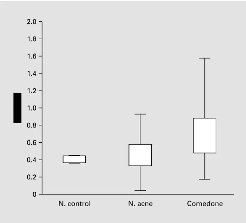
12 Dermatology 2003;206:11–16 Cunliffe/Holland/Clark/Stables Fig. 1. Ductal keratinocytes exhibit evidence of hyperproliferation
in contrast to control samples. The figure also shows that the so-called normal skin of acne patients – in an acne-prone area – evi-dences some ductal hyperproliferation.
expression partner of K16, are also found suprabasally in the follicle wall of microcomedones and comedones but not in control follicles [9]. In addition, our data also show that some of the so-called normal follicles of acne-prone skin may also show overexpression of Ki67 and K16. This suggests that topical therapy should be applied not just to the lesions, but also to the acne-prone areas. Limited data show no primary abnormality of ductal desmosomes [10].
Several factors may explain ductal hypercornification. There is evidence that abnormalities of the sebaceous lip-ids such as increased free fatty aclip-ids [11], squalene and squalene oxide [12] as well as a decrease in sebaceous lin-oleic acid [13] could all trigger hypercornification. The data incriminating fatty acids, squalene and squalene oxide emanate from studies on rabbits’ ears. The rele-vance of this to humans is questionable, particularly as the rabbit ear model is overpredictive for humans [14]. Sebaceous linoleic acid has been shown to be reduced in comedones. Linoleic acid is an essential fatty acid. Ani-mals deficient in linoleic acid become scaly. A comedo is due to the accumulation of much scale in the
piloseba-Fig. 2. The technique of in situ hybridization demonstrates (on the left) an increased expres-sion of K16 (a marker of hyperproliferation) in contrast to normal skin (on the right) which shows virtually no such expression.
Fig. 3. Comedone formation (in vitro) as a consequence of adding IL-1· to cultured ductal keratinocytes (with kind permission of Dr. T. Kealey).
2
Comedogenesis: Aetiological, Clinical and Therapeutic Strategies
Dermatology 2003;206:11–16 13
ceous duct. Androgens may have an important part in comedogenesis. 5·-Reductase (type 1) is present in the
infrainfundibulum part of the duct as well as in the seba-ceous gland [14]. The possible androgen-controlling effect is mirrored by a reduction in the number of comedones when a patient is prescribed anti-androgen therapy such as the oral contraceptive pill Dianette® [15].
Retinoids, both oral and topical, will suppress comedo-genesis [16–18]. After 2 months of therapy, many topical retinoids will suppress comedones by 30% whereas oral isotretinoin suppresses comedones by 52% and at 4 months of therapy, the suppression is about 80%. Cyto-kines are likely to be important [19, 20] (fig. 3). Kealey, like others, is able to maintain the pilosebaceous duct in culture. Comedones are produced in such a system under the influence of interleukin (IL) 1· (fig. 3), and this
pro-cess can be inhibited by adding IL-1 receptor antagonist to the growth medium.
From our own laboratories we have data to suggest that the comedo cycling could be important in comedogenesis and its resolution. As part of our research we realised that similar-looking pilosebaceous follicles and comedones showed different expressions of cycling cells (using Ki67) and proliferation markers (using K16). This led us to the concept that the duct may also undergo cycling just like the hair follicles [21]. Such cycling may explain why many blackheads and whiteheads disappear without treatment. If this were not to occur, then an adolescent developing acne, especially comedonal acne, early in his/her teens would, by the late teens, have no healthy skin on the acne-affected site: it would be a mass of comedones. A cycling phenomenon is probably not an unreasonable hypothesis given the close proximity of the hair follicle and piloseba-ceous duct.
Physicians are well aware that antimicrobial therapy which may also have a direct anti-inflammatory role sig-nificantly reduces comedones. One explanation for this has been its effect in reducing the ductal P. acnes, which in turn results in a reduction in free fatty acids – some of which may be comedogenic. More recently we have shown that biopsies of normal-looking skin from an area prone to acne exhibit, compared to control skin, a signifi-cant increase in a variety of inflammatory cells, in partic-ular lymphocytes [Jeremy, unpubl. observations 2002] Such biopsies show no evidence of comedone formation and no evidence of hyperproliferation using Ki67 as a marker for hyperproliferation. By analogy with psoriasis, could this lymphocytic infiltrate trigger comedone forma-tion?
Comedonal Types
The clinical type of comedo could, and perhaps should, influence the treatment prescribed.
Microcomedones
Biopsy sections of normal-looking skin in an acne-prone individual with comedonal acne will frequently (28%) show histological features of microcomedones. Biopsies of papules taken at up to 72 h of development will reveal a microcomedone in 52% of subjects, a white-head in 22% and a blackwhite-head in 10% [22], confirming even further the practical need to apply topical therapies to apparently non-involved skin.
Ordinary Comedones
Dermatologists recognise the typical pattern of come-dones seen in clinical practice, and so this requires no fur-ther explanation.
Missed Comedones
In all patients, it is essential to stretch the skin, using a good light, at a shallow angle, otherwise even ordinary comedones will not be recognised. Stretching of the skin will demonstrate, in about 20% of patients, comedones which would otherwise not be seen, and thus prevent the prescription of inappropriate topical therapy. Our treat-ment protocols for ordinary, missed and microcomedones are similar. The topical treatment must be applied not just to the lesions, but also to the adjacent subclinically ‘nor-mal’ skin. Physical methods of therapy such as blackhead removers are worthy of consideration in a small number of patients with obvious blackheads. Topical retinoids are the most effective topical therapy [16–18, 23–25].
Sandpaper Comedones
Patients with these comedones represent a difficult subgroup who present with predominantly very small, almost confluent closed comedones giving the feel of ‘sandpaper’ which may become inflamed. They are par-ticularly seen on the forehead and are difficult to treat. On the whole, they show little or varied response to oral anti-biotics and topical retinoids, and the optimum treatment is oral isotretinoin at a preferred dosage of 0.5 mg/kg/ day.
Submarine Comedones
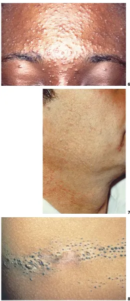
14 Dermatology 2003;206:11–16 Cunliffe/Holland/Clark/Stables Fig. 4. Submarine comedones.
Fig. 5. Macrocomedones.
surprisingly quite large and may reach a size of up to 1 cm. Treatment is difficult, and the optimum therapy is probably focal cautery using a technique described later in this review for the treatment of macrocomedones, which allows the drainage of retained corneocytes. Such a technique is successful in about 50% of submarine come-dones.
Macrocomedones
This term refers to blackheads and whiteheads which are 11 mm in size. Whiteheads are the most common. They need to be treated for two reasons. They are a cos-metic problem and may flare into inflamed lesions (fig. 5), especially in patients treated with oral isotretinoin. In such patients, they are the major reason for a severe flare of the acne and surprisingly are easily missed unless ade-quate lighting and examination techniques, i.e. stretching the skin, are used. The optimum therapy is gentle cautery [26–28]. This is performed under topical local anaesthesia using an anaesthetic cream such as EMLA® which is
applied for 60–75 min under an occlusive dressing such as Tegaderm®. The area is then lightly touched with a small
hot-wire cautery probe, the tip being grey in colour rather than vividly red and red-hot. The purpose is not to burn the skin significantly but to produce low-grade, localised thermal damage. This therapy is far superior to topical retinoids: at 2 weeks of treatment using light cautery there
is typically virtually 100% clearance compared with topi-cal retinoids which produce a reduction in the order of
!10% [28]. Not all patients respond perfectly. A test area
is always treated initially, and thereafter the remaining lesions are treated in further sessions. Five percent devel-op recurrent lesions requiring multiple treatments with gentle cautery. Scarring and pigmentary changes are un-common. If the patient has macrocomedones and is on oral isotretinoin and the acne flares, it is necessary to stop the oral isotretinoin, consider giving oral steroids and treat the macrocomedones. Macrocomedones are also a cause of a slow and poor response to oral isotretinoin ther-apy [29].
Drug-Induced Comedones
These may be due to corticosteroids [29, 30] or ana-bolic steroids [31, 32] and ‘blue comedones’ can occur, albeit very infrequently, due to minocycline-induced pig-mentation. Treatment of drug-induced comedones is by removal of the cause and by treating with either topical retinoids or gentle cautery.
Pomade Comedones
in-Comedogenesis: Aetiological, Clinical and Therapeutic Strategies
Dermatology 2003;206:11–16 15
cludes stopping the hair preparations, topical retinoids and possibly oral antibiotics.
Chloracne
This is also characterised by many comedones [33–36]. Indeed, comedonal acne is a hallmark of this disease (fig. 7), and inflammatory lesions are less frequent. In-flamed lesions may be treated with oral or topical benzoyl peroxide or antibiotics. Gentle cautery is very successful; there is usually a poor response to topical and oral reti-noids [27].
Naevoid Comedones
These are rare and may present before puberty but more often at and around puberty [37, 38]. The lesions may be typical confluent comedones (fig. 8) or white-heads, usually occurring asymmetrically. They may be localised or, in some unfortunate individuals, extremely extensive. Treatment is difficult. Response to oral and topical retinoids is unsatisfactory. Physical methods are also unsatisfactory, but gentle cautery, excision of locally affected areas and carbon dioxide laser therapy can be tried; however, as yet there seems to be no satisfactory solution for the majority of patients.
Conglobate Comedones
Patients with conglobate comedones are predominant-ly males with extensive truncal acne characterised by severe nodular inflammation and scarring. A hallmark of the disease is grouped comedones [40, 41], particularly on the posterior neck and upper trunk. The comedones may be blackheads, whiteheads or both. This is a really diffi-cult subgroup to treat. There are no satisfactory data to demonstrate which is the preferred way of treating such comedones.
New Topical Retinoids
New topical anti-acne therapies are required for sever-al reasons. There is no topicsever-al anti-acne therapy which reduces lesions by over 60% in contrast to, for example, oral isotretinoin which can suppress lesions by 100%. This may simply be a measure of penetration of the drug. Most topical therapies frequently produce an irritant der-matitis, and this will reduce compliance. Many antibiotics have been shown to produce resistant P. acnes, and this is associated in some patients with clinical failure. New reti-noid molecules such as adapalene [17, 18] have been developed, while old retinoids have been redeveloped
Fig. 6. Pomade acne. Fig. 7. Chloracne. Fig. 8. Naevoid acne.
6
7
16 Dermatology 2003;206:11–16 Cunliffe/Holland/Clark/Stables using new vehicle delivery systems [42, 43]. It is not the
intention of this review to discuss the pros and cons of such therapies, except to say that some newer drugs and new formulations of older therapies tend to show a better benefit/risk ratio.
Acknowledgements
This study was financially supported in part by the Leeds Foun-dation for Dermatological Research, Roche, Galderma and Dermik. This paper is extensively based on a paper published in the British Journal of Dermatology [2000;142:1084–1091]. With the permission of the British Journal of Dermatology to re-publish this paper in a shorter version.
References
1 Cunliffe WJ, Simpson NB: Disorders of the sebaceous gland; in Champion RH, Burton JL, Burns DA, Breathnach SM (eds): Textbook of Dermatology, ed 6. Oxford, Blackwell Science, 1998, pp 1927–1984.
2 Burton JL, Shuster S: The relationship between seborrhoea and acne vulgaris. Br J Dermatol 1971:84:600–601.
3 Leyden JL, McGinley KJ, Mills OH, et al: Pro-pionibacterium levels in patients with and without acne vulgaris. J Invest Dermatol 1975; 65:382–384.
4 Webster GF: Inflammation in acne vulgaris. J Am Acad Dermatol 1995;33:247–253. 5 Holmes RL, Williams M, Cunliffe WJ:
Pilose-baceous duct obstruction and acne. Br J Der-matol 1972;87:327–332.
6 Plewig G, Fulton JE, Kligman AM: Cellular dynamics of comedo formation in acne vulga-ris. Arch Dermatol Forsch 1971;242:12–29. 7 Knaggs HE, Holland DB, Morris C, et al:
Quantification of cellular proliferation in acne using the monoclonal antibody Ki-67. J Soc Invest Dermatol 1994;102:89–92.
8 Hughes BR, Morris C, Cunliffe WJ, Leigh IM: Keratin expression in pilosebaceous epithelia in truncal skin of acne patients. Br J Dermatol 1996;134:247–256.
9 Holland DB, Roberts SG, Cunliffe WJ: Local-isation of keratin 6 mRNA in acne (abstract). J Invest Dermatol 1994;103:443.
10 Knaggs HE, Hughes BR, Morris C, et al: Im-munohistochemical study of desmosomes in acne vulgaris. Br J Dermatol 1994;130:731– 737.
11 Kligman AM, Katz AC: Pathogenesis of acne vulgaris. I. Comedogenic properties of human sebum in external ear canal of the rabbit. Arch Dermatol 1968;98:53–57.
12 Motoyoshi K: Enhanced comedo formation in rabbit ear skin by squalene and oleic acid per-oxides. Br J Dermatol 1983;109:191–198. 13 Downing DT, Stewart ME, Wertz PW, et al:
Essential fatty acids and acne. J Am Acad Der-matol 1986;14:221–225.
14 Thiboutot DM, Knaggs H, Gilliland K, Hagari S: Activity of type 1 5·-reductase is greater in
the follicular infrainfundibulum compared with the epidermis. Br J Dermatol 1997;136: 166–167.
15 Stewart ME, Greenwood R, Cunliffe WJ, et al: Effect of cyproterone acetate-ethinyl oestradiol treatment on the proportion of linoleic and sebaceous acids in various skin surface lipid classes. Arch Dermatol Res 1986;278:481– 485.
16 Chalker DK, Lesher JL, Smith JG, et al: Effica-cy of topical isotretinoin 0.05% gel in acne vul-garis: Results of a multicenter, double-blind investigation. J Am Acad Dermatol 1987;17: 251–254.
17 Shalita A, Weiss JS, Chalker DK, et al: A com-parison of the efficacy and safety of adapalene gel 0.1% and tretinoin gel 0.025% in the treat-ment of acne vulgaris: A multicenter trial. J Am Acad Dermatol 1996;34:482–485.
18 Kligman AM: The treatment of acne with topi-cal retinoids: One man’s opinion. J Am Acad Dermatol 1997;36:S92–S95.
19 Guy R, Green MR, Kealey T: Modelling acne in vitro. J Invest Dermatol 1996;106:176–182. 20 Sanders DA, Philpott MP, Nicolle FV, Kealey T: The isolatioin and maintenance of the hu-man pilosebaceous unit. Br J Dermatol 1994; 131:166–176.
21 Aldana OL, Holland DB, Cunliffe WJ: Varia-tion in pilosebaceous duct keratinocyte prolif-eration in acne patients. Dermatology 1998; 196:98–99.
22 Norris JFB, Cunliffe WJ: A histological and immunocytochemical study of early acne le-sions. Br J Dermatol 1988;118:651–659. 23 Hughes BR, Norris JF, Cunliffe WJ: A
double-blind evaluation of topical isotretinoin 0.05% benzoyl peroxide gel 5% and placebo in pa-tients with acne. Clin Exp Dermatol 1992;17: 165–168.
24 Elbaum DJ: Comparison of the stability of top-ical isotretinoin and toptop-ical tretinoin and their efficacy in acne. J Am Acad Dermatol 1988;19: 486–491.
25 Verschoore M, Langner A, Wolska H, et al: Efficacy and safety of CD271 alcoholic gels in the topical treatment of acne vulgaris. Br J Der-matol 1991;124:368–371.
26 Pepall LM, Cosgrove MP, Cunliffe WJ: Abla-tion of whiteheads by cautery under topical anaesthesia. Br J Dermatol 1991;125:256– 259.
27 Yip J, Pepall LM, Gawkrodger DJ, Cunliffe WJ: Light cautery and EMLA® in the
treat-ment of chloracne lesions. Br J Dermatol 1993; 128:313–316.
28 Bottomley WW, Yip J, Knaggs H, Cunliffe WJ: Treatment of closed comedones – Compari-sons of fulguration with topical tretinoin and electrocautery with fulguration. Dermatology 1993;186:253–257.
29 Plewig G, Kligman AM: Induction of acne by topical steroids. Arch Dermatol Forsch 1973; 247:29–52.
30 Monk B, Cunliffe WJ, Layton AM, Rhodes DJ: Acne induced by inhaled corticosteroids. Clin Exp Dermatol 1993;18:148–150.
31 White Gl Jr, Tyler LS: Blackmarket steroids complicate acne therapy (letter). J Fam Pract 1987;25:214.
32 Fyrand O, Fiskdaadal HJ, Trygstad O: Acne in pubertal boys undergoing treatment with an-drogens. Acta Derm Venereol (Stockh) 1992; 72:148–149.
33 Crow KD: Chloracne and its potential clinical implications. Clin Exp Dermatol 1981;6:243– 257.
34 Rosas-Vasquez E, Campos-Macias P, Ochoa-Tirado JG, et al: Chloracne in the 1990s. Int J Dermatol 1996;35:643–645.
35 McConnell R, Anderson K, Russell W, et al: Angiosarcoma, porphyria cutanea tarda and probable chloracne in a worker exposed to waste oil contaminated with a 2,3,7,8-tetra-chlorodibenzo-p-dioxin. Br J Ind Med 1993;50: 699–703.
36 Coenraads PJ, Brouwer A, Olie K, Tang N: Chloracne: Some recent issues. Dermatol Clin 1994;12:569–576.
37 Beck MH, Dave VK: Extensive nevus come-donicus. Arch Dermatol 1980;116:1048–1050. 38 Barsky S, Doyle JA, Winkelmann RK: Nevus comedonicus with epidermolytic hyperkerato-sis: A report of our cases. Arch Dermatol 1981; 117:86–88.
39 Munro CS, Wilkie AOM: Epidermal mosai-cism producing localised acne: Somatic muta-tion in FGFR2. Lancet 1998;352:704–705. 40 Burns RE, Colville JM: Acne conglobata with
septicaemia. Arch Dermatol 1959;79:361– 363.
41 Williamson DM, Cunliffe WJ, Gatecliff M, Scott DG: Acute ulcerative acne conglobata (acne fulminans) with erythema nodosum. Clin Exp Dermatol 1977;2:351–354.
42 Quigley JW, Bucks DAW: Reduced skin irrita-tion with tretinoin containing polyolprepolym-er-2, a new topical tretinoin delivery system: A summary of preclinical and clinical investiga-tions. J Am Acad Dermatol 1998;38:S5–S10. 43 Embil K, Nacht S: The microsponge delivery
Dermatology 2003;206:17–23 DOI: 10.1159/000067818
New Aspects in Acne Inflammation
Masahiko Toyoda Masaaki Morohashi
Department of Dermatology, Faculty of Medicine, Toyama Medical and Pharmaceutical University, Toyama, Japan
M. Toyoda, MD
Department of Dermatology, Faculty of Medicine Toyama Medical and Pharmaceutical University 2630 Sugitani, Toyama 930-0194 (Japan)
Tel. +81 76 434 7305, Fax +81 76 434 5028, E-Mail [email protected]
ABC
Fax + 41 61 306 12 34 E-Mail [email protected] www.karger.com
© 2003 S. Karger AG, Basel 1018–8665/03/2061–0017$19.50/0
Accessible online at: www.karger.com/drm
Key Words
AcneW NeuropeptidesW Substance PW Neutral endopeptidaseW Nerve growth factorW Sebaceous glandsW Stem cell factorW Mast cellsW Nerves
Abstract
There is ample clinical evidence suggesting that the ner-vous system such as emotional stress can influence the course of acne. We examined possible participation of cutaneous neurogenic factors including neuropeptides, neuropeptide-degrading enzymes and neurotrophic fac-tors, in association with inflammation in the pathogene-sis of acne. Immunohistochemical studies revealed that substance P (SP)-immunoreactive nerve fibers were in close apposition to the sebaceous glands, and that neu-tral endopeptidase (NEP) was expressed in the germina-tive cells of the sebaceous glands in the skin from acne patients. Nerve growth factor showed immunoreactivity only within the germinative cells. In addition, an increase in the number of mast cells and a strong expression of endothelial leukocyte adhesion molecule-1 on the post-capillary venules were observed in adjacent areas to the sebaceous glands. In vitro, the levels and the expression of stem cell factor by fibroblasts were upregulated by SP. When organ-cultured normal skin specimens were ex-posed to SP, we observed significant increases in the sizes of the sebaceous glands and in the number of sebum vacuoles in sebaceous cells. Furthermore, sup-plementation of SP to organ-cultured skin induced ex-pression of NEP, and we demonstrated the subcellular
localization of NEP in the endoplasmic reticulum and the Golgi apparatus within the sebaceous germinative cells using preembedding immunoelectron microscopy. These findings suggest that SP may stimulate lipogene-sis of the sebaceous glands which may be followed by proliferation of Propionibacterium acnes, and may yield a potent influence on the sebaceous glands by provoca-tion of inflammatory reacprovoca-tions via mast cells. Thus, cuta-neous neurogenic factors should contribute to onset and/or exacerbation of acne inflammation.
Copyright © 2003 S. Karger AG, Basel
Acne vulgaris is a skin disorder of the sebaceous folli-cles that commonly occurs in adolescence and young adulthood. Many lines of clinical evidence suggest that components of the nervous system, such as psychological and neurogenic factors, can influence the course of acne [1–3]. The disease has been reported to be initiated and/or exacerbated as a result of emotional or psychosocial stress. However, the nature of the association between stress and acne remains unclear, due in part to a lack of substantial evidence regarding the participation of cuta-neous neurogenic factors in the pathogenesis of acne.
Cutaneous Innervation and Neuropeptides
18 Dermatology 2003;206:17–23 Toyoda/Morohashi
sympathetic nerves. The cutaneous sensory nervous sys-tem comprises a network of fine C fibers within the skin that innervate multiple cell types and play an important role of in inflammation [4, 5]. Various stimuli may direct-ly activate peripheral nerve endings of primary sensory neurons and impulses are conveyed centrally as well as, through antidromic axon reflexes, peripherally. Upon re-lease of neuropeptides (NPs) from sensory terminals, im-portant visceromotor inflammation and trophic effects occur in the peripheral tissues. This proinflammatory NPs release causes the set of changes collectively referred to as neurogenic inflammation [6–8].
Neuropeptides can manifest immunomodulatory ac-tivity, and they contribute to the cross-talk between the nervous system and the immune system in the skin [8– 10]. NPs are a heterogeneous group of several hundred biologically active peptides, present in neurons of both the central and the peripheral nervous system and in-volved the transmission of signals not only between nerve cells, but also with the immune system where they appear to be critical mediators of different processes. Normal human skin expresses a variety of NPs that are either directly derived from sensory neurons or from skin cells such as keratinocytes. In addition, immune cells that ei-ther constitutively resides in the skin such as mast cells (MCs) or infiltrating cells into the skin under inflammato-ry conditions have been reported to produce NPs [8]. Clinical evidence in support of a connection between neu-ropeptide secretion and the development of inflammation is found in various skin diseases such as atopic dermatitis, psoriasis and alopecia areata, which are commonly exac-erbated during periods of emotional stress [9–15]. Indeed, stress has been shown to elicit the release of substance P (SP) [8], a neuropeptide belonging to the tachykinin fami-ly, which can induce neurogenic inflammation. Clinical evidence in support of a connection between neuropep-tide secretion and the development of inflammation is found in various skin diseases such as atopic dermatitis, psoriasis and alopecia areata which are commonly exacer-bated during periods of emotional stress [10–16]. Indeed, stress has been shown to elicit the release of SP [17], a neuropeptide belonging to the tachykinins family, which can induce neurogenic inflammation. SP is associated with multiple cellular responses, including vasodilatation, increased blood flow, plasma extravasations, mast cell degranulation, the wheal and flare reaction via axon reflex, neutrophil and macrophage activation, modula-tion of the release of proinflammatory cytokines and che-mokines, and the upregulation of adhesion molecule ex-pression required for trafficking of leukocytes [11, 12, 18].
Nevertheless, none of those previous studies addressed the effects of SP on the sebaceous glands or on the disease process of acne.
SP-Containing Nerves in Acne
Nerve fibers showing immunoreactivity for SP were rarely observed in skin specimens from the face devoid of acne lesions in healthy subjects. On the other hand, speci-mens from acne patients showed a strong immunoreactiv-ity for SP with many fine nerve fibers around the ceous glands. Some of them were invading into the seba-ceous glands and were located in close apposition to the sebocytes [19].
Effects of SP on the Sebaceous Glands
New Aspects in Acne Inflammation Dermatology 2003;206:17–23 19
testosterone and lipogenesis were examined in sebaceous glands of Syrian hamsters, and demonstrated that immo-bilization-induced stress lowered the levels of testosterone in plasma as well as in the skin, which resulted in decreased lipogenesis in the skin [22]. Although these data suggest that psychological or physiological stress can in-fluence sebaceous gland function by inducing changes in the neuroendocrine system, they provide no appropriate explanation for the effects of stress-induced exacerbation of acne. Taking into account that stress can elicit SP release from peripheral nerves [17], it is tempting to spec-ulate that SP should be partially involved in stress-induced exacerbation of the disease.
Neutral Endopeptidase in the Sebaceous Glands in Acne
Tissue responsiveness to NPs depends on the presence of specific receptors and on the distribution of neuropep-tide-degrading enzymes which play essential roles in the removal of NPs from the extracellular environment and are thus important regulators in neurogenic inflamma-tion. Recent studies indicate that neutral endopeptidase (NEP; EC 3.4.24.11; enkephalinase), a zinc metallo-pro-tease, is a cell surface enzyme and has the potential to degrade several NPs such as SP, and thereby terminates their biologic actions [5]. NEP is localized in keratino-cytes, vascular endothelial cells, fibroblasts, the outer root sheath of hair follicles and mast cells in the skin [23, 24]. Administration of NEP inhibitors magnifies the proin-flammatory effects of SP and other tachykinins in several tissues [25]. Thus, upregulation of NEP is a potential mechanism of limiting proinflammatory effects of NEP-degradable NPs, notably SP, by reducing the amounts of bioactive NPs.
Immunohistochemical staining for NEP in normal fa-cial skin was negative within the sebaceous glands. On the other hand, NEP was highly expressed in the sebaceous glands of acne patients in which immunoreactivity for NEP in the sebaceous glands were restricted to the germi-native cells. There was a statistically significant difference in the percentage of NEP-positive sebaceous acini to all acini between acne patients and controls [26]. We next examined effects of SP on NEP expression in the seba-ceous glands using organ-cultured skin in vitro. Although normal facial skin specimens supplemented with medium alone showed no expression of NEP in sebaceous cells, skin specimens stimulated with SP revealed prominent NEP staining in the germinative cells of the sebaceous
acini, which appeared to be analogous to the staining pat-tern of the sebaceous glands in acne patients. In addition, SP induced NEP expression in sebaceous glands in a dose-dependent manner [26]. Taking into account the lack of NEP expression in tissue not stimulated with SP, seba-ceous germinative cells may begin to synthesize NEP fol-lowing stimulation by SP. To examine the subcellular localization of NEP in sebaceous cells more precisely, we performed ultrastructural immunocytochemistry using an indirect immunoperoxidase technique. NEP expression was restricted to the Golgi apparatus and the endoplasmic reticulum within sebaceous germinative cells [26], which indicates that NEP is synthesized through the pathway of protein synthesis in the usual fashion.
Innervation and Nerve Growth Factor in the Sebaceous Glands in Acne
20 Dermatology 2003;206:17–23 Toyoda/Morohashi
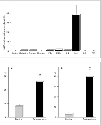
Fig. 1. Induction of NGF in the sebaceous glands by mast cell-derived mediators.
Fig. 2. IL-6 within MCs. There are statisti-cally significant increases in the percentages of IL-6-immunoreactive MCs (a) and of IL-6-containing specific granules of MCs in fa-cial skin of acne patients (n = 32) versus healthy volunteers (n = 35) as controls (b). * p ! 0.01 compared with control (unpaired Student’s t test).
that some MC-derived mediators may exert inducible activity of NGF in the sebaceous glands of acne patients. When organ-cultured normal skin was stimulated with various MC-derived mediators and cytokines including histamine, tryptase, chymase, leukotriene D4, prostaglan-din E2, IL-4, IL-6, IL-8, TNF-·, IFN-Á and
platelet-acti-vating factor, IL-6 specifically induced expression of NGF in the sebaceous glands (fig. 1). Preincubation of explants with anti-IL-6 receptor, followed by exposure to IL-6, resulted in abrogation of NGF induction in the seba-ceous glands. Immunohistochemical and immunoelec-tron microscopic studies revealed the presence of IL-6 within specific granules of MCs around the sebaceous glands in the skin of acne patients. The numbers of
IL-6-positive MCs and IL-6-containing MC granules were significantly increased in acne patients compared with the control (fig. 2). These findings suggest that MC-derived IL-6 has potential to induce NGF in sebaceous cells, which may result in promoting innervation within and around the sebaceous glands in acne patients.
New Aspects in Acne Inflammation Dermatology 2003;206:17–23 21
tempting to speculate that NGF plays an important role in spontaneous inflammatory dermatoses, such as acne, by modulating NPs. There is increasing evidence that NGF, in addition to its actions within the nervous system, elicits a number of biologic effects on local and systemic cells of the immune-inflammatory compartment. In vivo, admin-istration of NGF to neonatal rats increases the size and the number of mast cells in several peripheral tissues, and, in vitro, NGF induces mast cell degranulation and media-tor release. NGF enhances survival, phagocytosis, and superoxide production of mature murine neutrophils, causes mediator release from basophils, stimulates T and B lymphocyte proliferation, and stimulates B-cell differ-entiation into immunoglobulin-secreting plasma cells [10, 20]. These data imply possible participation of NGF in the inflammatory process in the pathogenesis of acne.
Mast Cells in Acne Inflammation
Increasing attention has been directed towards interac-tions between components of the nervous system and multiple target cells of the immune system. Communica-tion between nerves and MCs is a prototypic demonstra-tion of such neuroimmune interacdemonstra-tions. Several studies have demonstrated that MCs are often found in close con-tact with nerves and that there may be a functional inter-action between mast cells and the nervous system [28]. In addition, recent evidence suggests that SP is an important mediator in intimate nerve-mast cell cross talk [29]. When organ-cultured normal facial skins were exposed to SP uniformly degranulated MCs adjacent the sebaceous glands were observed at the electron microscopic level. Venules around the sebaceous glands of specimens stimu-lated with SP showed expression of ELAM-1 on the endo-thelia after subsequent culture. Furthermore, preincuba-tion of explants with the SP analogue or with cromolyn sodium, one of the MC inhibitors, abrogated the ability of SP to induce ELAM-1. These findings suggest that SP endogenously released by dermal nerve fibers may be important in the regulation of endothelial-leukocyte inter-action via MCs. It has been demonstrated that the proin-flammatory effect of ELAM-1 induction by MC degranu-lation products is inhibited by blocking antiserum to TNF-·. Thus, SP, contained within dermal nerve fibers,
may represent a crucial initial mediator of a cascade of cellular events involving MC degranulation and release of proinflammatory cytokines such as TNF-·, with
subse-quent induction of adhesion molecules such as E-selectin on adjacent venular endothelia [30]. This would then
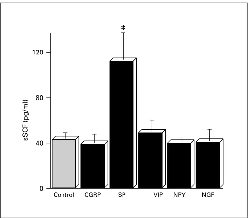
Fig. 3. Effects of neuropeptides and NGF on the levels of soluble SCF from cultured human fibroblasts. Cultured medium was col-lected 72 h after exposure to 100 ng/ml of each substance and then levels of soluble SCF were examined by ELISA. Means were obtained from triplicate cultures of four independent experiments. CGRP = Calcitonin gene-related peptide; VIP = vasoactive intestinal polypep-tide; NPY = neuropeptides Y. * p ! 0.01 compared with control (un-paired Student’s t test).
facilitate the local accumulation of blood leukocytes dur-ing the inflammatory response. Immunohistochemical study demonstrated that most of venules around the seba-ceous glands not in normal subjects but in acne patients expressed E-selectin (data not shown). We have recently found using immunoelectron-microscopic method that SP is localized within specific granules of human skin MCs [31]. In addition to cutaneous sensory nerves, MC-derived SP may also affect the morphologic and immuno-logic alterations associated with the sebaceous glands and may contribute to the development of the inflammatory events in acne.
pre-22 Dermatology 2003;206:17–23 Toyoda/Morohashi
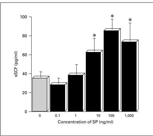
Fig. 4. A dose-dependent response of soluble SCF from cultured human fibroblasts stimulated with SP. Cultured medium were col-lected after 48 h exposure to a series of concentration of SP and then levels of soluble SCF were examined by ELISA. Means were obtained from triplicate cultures of four independent experiments. * p ! 0.01 compared with medium alone (0) (unpaired Student’s t test).
dicted 414-bp cDNA product was produced. When the PCR bands were quantified and the results were ex-pressed as ratios of densitometric scores for SCF and GAPDH for each sample, SCF message after treatment with 102 to 104 ng/ml of SP was relatively more intense than that platelet-derived growth factor, a well-known SCF enhancer [32] (data not shown). These findings
sug-gest that SP may be able to enhance MC proliferation through upregulation of SCF secretion and expression by fibroblasts.
On the basis of all the data mentioned above, the fol-lowing seven findings were found in association with acne inflammation from our in vivo and in vitro studies: (1) Many SP-containing nerve fibers were in close apposition to the sebaceous glands of acne patients (in vivo). (2) SP promoted both the proliferation and the differentiation of the sebaceous glands (in vitro). (3) NEP was expressed in the germinative cells of the sebaceous glands in acne patients (in vivo). SP-induced expression of NEP in seba-ceous glands which was localized in the endoplasmic reticulum and the Golgi apparatus (in vitro). (4) There was an increase in the number of nerve fibers around the sebaceous glands in acne patients, which were sometimes invading into the sebaceous glands (in vivo). (5) Immuno-reactivity of NGF was seen in the sebaceous glands only in acne patients (in vivo) and mast cell-derived IL-6 induced expression of sebaceous glands (in vitro). (6) An increase in the number of activated mast cells and a strong expression of E-selectin in postcapillary venules were observed in adjacent areas to the sebaceous glands in acne (in vivo). Mast cell-derived TNF-· induced expression of
E-selectin on venules (in vitro). (7) The levels of soluble form of and the expression of membrane-bound form of SCF by fibroblasts were upregulated by SP (in vitro).
Taken together, these findings suggest involvement of neurogenic factors including innervation, NPs, neuropep-tides-degrading enzymes and neurotrophic factors in the inflammatory process of acne and provide new insight into the possible mechanism of exacerbation of acne from the neurological point of view.
References
1 Koo JY, Smith LL: Psychologic aspects of acne. Pediatr Dermatol 1991;8:185–188.
2 Koblenzer CS: Psychotherapy for intractable inflammatory dermatoses. J Am Acad Derma-tol 1995;32:609–612.
3 Panconesi E, Hautmann G: Psychotherapeutic approach in acne treatment. Dermatology 1998;196:116–118.
4 Ansel JC, Kaynard AH, Armstrong CA, Olerud J, Bunnett N, Payan D: Skin-nervous system interactions. J Invest Dermatol 1996;106:198– 204
5 Misery L: Skin, immunity and the nervous sys-tem. Br J Dermatol 1997;137:843–850.
6 Bowden JJ, Baluk P, Lefevre PM, Vigna SR, McDonald DM: Substance P (NK1) receptor immunoreactivity on endothelial cells of the rat tracheal mucosa. Am J Physiol 1996;270: L404–L414.
7 Bozic CR, Lu B, Hopken UE, Gerard C, Ge-rard NP: Neurogenic amplification of immune complex inflammation. Science 1996;273: 1722–1725.
8 Scholzen T, Armstrong CA, Bunnett NW, Lu-ger TA, Olerud JE, Ansel JC: Neuropeptides in the skin: Interactions between the neuroendo-crine and the skin immune system. Exp Der-matol 1998:7:81–96.
9 Foreman J, Jordan C: Neurogenic inflamma-tion. Trends Pharmacol Sci 1984;5:116–119.
10Pincelli C, Fantini F, Gianetti A: Neuropep-tides and skin inflammation. Dermatology 1993;187:153–158.
11 Farber EM, Nickoloff BJ, Recht B, Fraki JE: Stress, symmetry and psoriasis: Possible role of neuropeptides. J Am Acad Dermatol 1986;14: 305–311.
New Aspects in Acne Inflammation Dermatology 2003;206:17–23 23
13 Pincelli C, Fantini F, Romualdi P, Sevignani C, Lesa G, Benassi L, Giannetti A: Substance P is diminished and vasoactive intestinal peptide is augmented in psoriatic lesions and these pep-tides exert disparate effects on the proliferation of cultured human keratinocytes. J Invest Der-matol 1992;98:421–427.
14 Naukkarinen AN, Nickoloff BJ, Farber EM: Quantification of cutaneous sensory nerves and their substance P content in psoriasis. J Invest Dermatol 1989;92:126–129.
15 Toyoda M, Morimatsu S, Morohashi M: The alterations of cutaneous innervation and neu-ropeptide expression by cyclosporin A treat-ment in atopic dermatitis: An immunohisto-chemical study. Jpn J Dermatol 1997;107: 1275–1279.
16 Toyoda M, Makino T, Kagoura M, Morohashi M: Characteristic expression of neuropeptide-degrading enzymes in alopecia areata: An im-munohistochemical study. Br J Dermatol 2001;144:46–54.
17 Singh LK, Pang X, Alexacos N, Letourneau R, Theoharides TC: Acute immobilization stress triggers skin mast cell degranulation via corti-cotropin releasing hormone, neurotensin, and substance P: A link to neurogenic skin disor-ders. Brain Behav Immun 1999;13:225–239. 18 Manske JM, Sullivan EL, Anderson SM:
Sub-stance P mediated stimulation of cytokine lev-els in cultured murine bone marrow stromal cells. Adv Exp Med Biol 1995;383:53–64.
19 Toyoda M, Nakamura M, Morohashi M: Neu-ropeptides and sebaceous glands. Eur J Derma-tol 2002;12:422–427.
20Pincelli C: Nerve growth factor and keratino-cytes: A role in psoriasis. Eur J Dermatol 2000; 10:85–90.
21 Toyoda M, Morohashi M: Pathogenesis of acne. Med Electron Microsc 2001;34:29–40. 22 Tsuchiya T, Horii I: Immobilization-induced
stress decreases lipogenesis in sebaceous glands as well as plasma testosterone levels in male Syrian hamsters. Psychoneuroendocrinology 1995:20:221–230.
23 Olerud JE, Usui ML, Seckin D, Chiu DS, Hay-cox CL, Song I-S, Ansel JC, Bunnett NW: Neu-tral endopeptidase expression and distribution in human skin and wounds. J Invest Dermatol 1999;112:873–881.
24 Toyoda M, Makino T, Kagoura M, Morohashi M: Expression of neuropeptide-degrading en-zymes in alopecia areata: An immunohisto-chemical study. Br J Dermatol 2001:144:46– 54.
25 Nadel JA, Borson DB: Modulation of neuro-genic inflammation by neutral endopeptidase. Am Rev Respir Dis 1991:143:33–36. 26 Toyoda M, Nakamura M, Makino T,
Moroha-shi M: Sebaceous glands in acne patients ex-press high levels of neutral endopeptidase. Exp Dermatol 2002;11:241–247.
27 Donnerer J, Schuligoi R, Stein C: Increased content and transport of substance P and calci-tonin gene-related peptide in sensory nerves innervating inflamed tissue: Evidence for a reg-ulatory function of nerve growth factor in vivo. Neuroscience 1992;49:693–698.
28 Toyoda, M, Morohashi M: Morphological as-sessment of the effects of cyclosporin A on mast cell – nerve relationship in atopic dermatitis. Acta Derm Venereol (Stockh) 1998;78:321– 325.
29 Suzuki R, Furuno T, McKay DM, Wolvers D, Teshima E, Nakanishi M, Bienenstock J: Di-rect neurite-mast cell communication in vitro occurs via the neuropeptide substance P. J Im-munol 1999;163:2410–2415.
30Matis WL, Lavker RM, Murphy GF: Sub-stance P induces the expression of an endothe-lial-leukocyte adhesion molecule by microvas-cular endothelium. J Invest Dermatol 1990;94: 492–495.
31 Toyoda M, Makino T, Kagoura M, Morohashi M: Immunolocalization of substance P in hu-man skin mast cells. Arch Dermatol Res 2000; 292:418–421.
Dermatology 2003;206:24–28 DOI: 10.1159/000067819
Acne in Infancy and Acne Genetics
Maria I. Herane
aIwao Ando
baDepartment of Dermatology, West Unit Faculty of Medicine, Hospital San Juan de Dios, University of Chile, Santiago, Chile; bDepartment of Dermatolgy, Teikyo University, Mizonokuchi Hospital, Kawasaki, Japan
Prof. Maria Isabel Herane
Department of Dermatology, West Unit, Hospital San Juan de Dios
ABC
© 2003 S. Karger AG, Basel1018–8665/03/2061–0024$19.50/0
Key Words
AcneW InfancyW GeneticsW Hereditary factors
Abstract
Acne is a disease that can be seen in the first year of age, early childhood, prepubertal age and puberty. Neonatal acne is due mainly to considerable sebum excretion rate, and infantile acne because of high androgens of adrenal origin in girls and of adrenal and testes in boys. These pathogenic mechanisms are characteristic in these ages. Important factors like early onset of comedones and high serum levels of dehydroepiandrosterone sulfate are pre-dictors of severe or long-standing acne in prepubertal age. Hereditary factors play an important role in acne. Neonatal, nodulocystic acne and conglobate acne has proven genetic influences. Postadolescent acne is relat-ed with a first-degree relative with the condition in 50% of the cases. Chromosomal abnormalities, HLA pheno-types, polymorphism of human cytochrome P-450 1A1 and MUC1 gene are involved in the pathogenesis of acne. Several other genes are being studied.
Copyright © 2003 S. Karger AG, Basel
Neonatal Acne
Neonatal acne is present at birth or appears shortly after. It is more common than fully appreciated; if the diagnosis is based in a few comedones more than 20% of
newborns are affected [1]. The most common lesions are comedones, papules and pustules. They are few in num-ber and usually localized on the face, more often cheeks and forehead. Involvement of the chest, back or groins has been reported. Most cases are mild and transient. Lesions appear mainly at 2–4 weeks healing spontaneously, with-out scarring, in 4 weeks to 3–6 months. Neonatal acne has been suggested to be more frequent in male infants [2, 3].