MOLECULAR AND STRUCTURAL BASIS OF THE EVOLUTION
OF PARVOVIRUS TROPISM
P. TIJSSEN*
Laboratory of Structural and Molecular Virology, Centre de Microbiologie et Biotechnologie, INRS-Institut Armand-Frappier, Building 12, Université du Québec,
531, boul. des Prairies, Laval, QC, Canada H7N 4Z3 (Received March 12, 1999; accepted April 8, 1999)
Parvoviruses have small genomes and, consequently, are highly dependent on their host for various functions in their reproduction. Since these viruses gener-ally use ubiquitous receptors, restrictions are usugener-ally intracellularly regulated. A lack of mitosis, and hence absence of enzymes required for DNA replication, is a powerful block of virus infection. Allotropic determinants have been identified for several parvoviruses: porcine parvovirus, canine parvovirus (CPV), feline parvo-virus (feline panleukopenia parvo-virus), minute parvo-virus of mice, Aleutian disease parvo-virus, and GmDNV (an insect parvovirus). Invariably, these identifications involved the use of infectious clones of these viruses and the exchange of restriction fragments to create chimeric viruses, of which the resulting phenotype was then established by transfection in appropriate cell lines. The tropism of these viruses was found to be governed by minimal changes in the sequence of the capsid proteins and, often, only 2 or 3 critical amino acids are responsible for a given tropism. These amino acids are usually located on the outside of the capsid near or on the spike of the threefold axis for the vertebrate parvoviruses and on loops 2 or 3 for the insect parvoviruses. This tropism is not mediated via specific cellular receptors but by interactions with intracellular factors. The nature of these factors is unknown but most data point to a stage beyond the conversion of the single-stranded DNA genome by host cell DNA polymerase into monomeric duplex intermediates of the replicative form. The sudden and devastating emergence of mink enteritis virus (MEV) and CPV in the last 50 years, and the possibility of more future outbreaks, demonstrates the importance of understanding parvovirus tropism.
Key words: Parvovirus, virus structure, virus tropism, viral evolution, host range
Parvoviruses, among the smallest viruses known, have a relatively simple, isometric capsid with a diameter of about 25 nm and contain a linear, single-stranded DNA genome of about 46 kb (Siegl, 1976; Tijssen, 1990; Berns, 1996; Tijssen et al., 1999). Some parvoviruses are autonomous, whereas others require
a helper virus (usually an adenovirus or a herpesvirus) for their replication. The limited coding capacity of the parvovirus genome implies a heavy dependence on cellular functions, especially those required for DNA replication and supplied by cells during the S-phase. As a result, autonomous parvoviruses infect dividing cells [e.g., in mitosis-active tissues, fetuses or tumour cells (oncolytic)]. Patho-genesis is highly tissue specific, at least for the vertebrate parvoviruses, and pro-ductive parvoviral infections lead to cell lysis. Targeting of dividing cells, either mitotropism or oncotropism, is therefore a sine qua non as the first level of tropism of parvoviruses.
The tropism of parvoviruses that require a helper virus, the adeno-associated viruses or AAVs, seems to be determined primarily by the tropism of their helper viruses. This virotropism is usually much wider than tropism of autonomous parvoviruses. When AAV viruses enter a cell and are uncoated in the absence of a helper virus, their genomes can be integrated in a specific chromosome site to establish a latent infection (Kotin et al., 1992). These latent genomes can be activated by a subsequent helper virus infection. As a conse-quence, AAVs prevent superinfecting adenoviruses from transforming cells (Berns, 1996). Epidemiological studies have shown that, while 90% of the gen-eral population is seropositive for AAVs, patients suffering from cervical carci-noma are much less AAV-seropositive (Mayor et al., 1976). AAVs, in contrast to autonomous parvoviruses, have as yet to be associated with any pathogenicity and have received a considerable interest for potential use as vectors in gene therapy. Since goose and muscovy duck parvoviruses are closely related to these viruses (Zádori et al., 1995), it will be interesting to establish whether they are integration-competent and whether goose and duck adenoviruses could rescue these parvoviruses from non-susceptible cells.
Not all replicating cells are sensitive to autonomous parvoviruses and there are relatively few cell cultures that support parvoviruses. Cellular restric-tions may act at different levels: (i) receptors; (ii) intracellular transport and de-capsidation; (iii) virus-replication; and (iv) encapsidation and cell lysis. Allo-tropic determinants have been identified in the capsids of several animal and in-sect parvoviruses and often consist of just a few amino acids, but their cellular partners, that would interact with them, are hitherto elusive. In principle, these allotropic determinants could act at any of the four stages mentioned above but, as will be discussed in this review, most data point to the second and third stage.
Here, the biology and molecular biology of parvoviruses will be briefly re-viewed, followed by a discussion of their three-dimensional structure and struc-ture-function relationships, and finally the nature and evolution of viral tropism.
General properties and classification of parvoviruses
Parvoviruses have been isolated from tissues from both vertebrates and invertebrates. Although other DNA virus families exist with members that infect either vertebrates or invertebrates (i.e., pox and iridoviruses), these replicate in the cytoplasm and contain considerable genetic information to code for proteins required for viral replication and, consequently, depend considerably less on host functions than parvoviruses. The observations that parvoviruses replicate in the nucleus, closely in tune with the physiology of the host cell, and are able to do so with both vertebrate and invertebrate cells, suggest that vertebrate and in-vertebrate parvoviruses may be more different than usually accepted. Indeed, when recently the first 3D structure of an invertebrate parvovirus was solved by X-ray crystallography (Simpson et al., 1998), major differences were observed between the two virus groups. Despite many differences in the molecular biol-ogy of these virus groups, it is surprising how many aspects of their genome or-ganization and molecular biology are shared, justifying their classification as one family. For example, we discovered recently (Zádori et al., unpublished re-sults) that the capsids of both vertebrate and invertebrate parvoviruses carry an enzymatic site with multiple activities (phospholipase A2 involved in infection process and signal transduction pathway). Similar activities have not yet been shown in other DNA viruses. The parvovirus family (Parvoviridae) is thus di-vided into two subfamilies, the Parvovirinae (vertebrates, i.e. human and other mammals) and the Densovirinae (invertebrates, i.e. insects, shrimps, crab, etc.).
The most recent classification of parvoviruses (Berns et al., 1995) needs to be revised. Classifications tend to evolve with the shift of research emphasis; for example, in early days, viruses were classified according to the symptoms of the diseases they caused (respiratory, hepatitis, etc.). Later, in the heyday of the biochemical and physicochemical characterization of viruses in the mid-sixties, Lwoff (1967) and Gibbs and Harrison (1968) each suggested a classification system based on these physicochemical properties. Currently, the classification hierarchy is governed by sequence homologies and molecular-biology functions and strategies. Conveniently, this did not change the general outline of the univer-sal taxonomic system that evolved in the 1970s but merely fine-tuned it, particu-larly for the DNA viruses.
Although unofficial, the classification presented in Table 1 reflects, to our knowledge, best the hierarchy according to clades of similar sequences among par-voviruses. Compared to the official (ICTV) classification, many parvoviruses have been moved to other genera, some have been assigned a separate genus and, where necessary, names have been corrected. A comprehensive review of this classifica-tion is in progress (Zádori et al., unpublished results). Due to its unofficial nature, no attempt has been made at establishing a nomenclature for the genera.
Table 1
Classification of parvoviruses according to sequence similarities (clades) Subfamily Clade (Genus) Viruses and distinguishing features Parvovirinae 1 (Parvovirus) Porcine parvovirus (PPV), canine parvovirus, feline
parvovirus (= feline panleukopenia virus; FPV), mink enteritis virus, raccoon parvovirus, minute virus of mice, murine parvovirus, LuIII, Kilham rat virus, H-1. Genome 5 kb, unique terminal hairpins, similar splicing mechanisms; similar patterns of VPs
2 Aleutian Disease virus. Different genome organization
and VP pattern
3 Bovine parvovirus. Genome 5.5 kb
4 (Erythrovirus) B19, simian parvovirus, chipmunk parvovirus. Genome 5.6 kb, ITRs, One promoter
5 (Dependovirus) AAV (many serotypes), goose parvovirus, muscovy duck parvovirus. Genome 4.7 kb. The genome termini of the goose and duck parvoviruses resemble those of B19 Densovirinae 1-a (Densovirus) Densoviruses from Galleria mellonella (GmDNV),
Pseudoplusia includens (PiDNV), Diatraea saccharalis (DsDNV), Junonia coenia (JcDNV), Mythimna loreyi (MlDNV). Ambisense genome of 6 kb (one strand codes for NS proteins, one strand codes for VP pro-teins), no splicing; large ITRs
1-b (Densovirus) Densovirus from Culex pipiens (CpDNV); 5.5 kb genome, ambisense; splicing in NS proteins
1-c (Densovirus) Densovirus from Acheta domesticus (AdDNV); 5.5 kb genome, ambisense; splicing in VP; small ITRs 1-d (Densovirus) Densovirus from Periplaneta fuliginosa (PfDNV); 5.5 kb
genome, ambisense; splicing in VP
2 (Brevidensovirus) Genus was earlier named Contravirus by the ICTV, but these viruses do not have an ambisense organization, therefore the genus was renamed Brevidensovirus (Tijssen and Bergoin, 1995). Almost all densoviruses tentatively classified in this genus by ICTV belong to other genera. Large number of densoviruses from mos-quitoes [e.g., from Aedes aegypti (AaeDNV) and from Aedes albopictus (AalDNV)]. Monosense genome or-ganization. Genome 4 kb with unique terminal hairpins 3 (Iteravirus) Densonucleosis viruses from Bombyx mori (BmDNV-1)
and Casphalia extranea (CeDNV). Genome 5 kb; ITRs of about 225 nucleotides; genome organization monosense but we observed that the published organi-zation is incorrect (Zádori et al., unpublished results) There are a large number of unclassified viruses (e.g., those from crab and shrimps) that cannot be classified due to a lack of information about their genome.
There are only a limited number of striking physicochemical differences among the parvoviruses. All parvoviruses have a genome of 46 kb with large palindromic sequences (hairpins) at both ends. The complete virion has a buoy-ant density of about 1.40 g/ml and all have 60 protein subunits. These subunits each consist of one protein from a nested set (co-C-terminal, but different N-termini) of a few (up to 4) proteins with molecular masses between 40 and 90 kDa. The virion is also resistant to various enzymes (proteases, nucleases) and adverse environmental conditions (stable in a wide pH range, high thermo-stability). Some parvoviruses encapsidate only the strand (complementary to the mRNA) whereas others encapsidate, into separate virions, also the + strand (up to 50%, depending on virus and cell). Some viruses, for which both strands are coding, encapsidate both strands equally into separate particles.
Parvovirus infection and replication
It has been shown for several parvoviruses that nonproductive infections are often due to intracellular factors. These viruses enter both permissive and non-permissive cells, apparently because they often use ubiquitous receptors, but continue only in the permissive cells with a productive infection. This resembles infection by AAVs in the absence of adenovirus. Although integration for autonomous parvoviruses in the absence of replication has not received much attention, densoviruses have been shown to be able to do so (e.g., when an es-sential gene is knocked out; Bossin, 1998). Minute virus of mice (MVM) di-rected integration into episomes has also been observed in an experimental sys-tem (Corsini et al., 1997). Intracellular factors may thus be important for tropism and interact with the allotropic determinant on the capsid.
The parvoviral genome contains two sets of genes, the nonstructural (NS) or Rep genes and the viral protein (VP) or Cap genes. The NS proteins are cen-tral in the replication and expression of the viral genome, while the VP proteins form the capsid to protect the viral genome and play roles in assembly, viral maturation and infection. These VPs are, therefore, far from static proteins and our recent finding of an enzyme activity in the capsid attests to that fact.
Parvoviruses have only a few (23) NS proteins to mobilize cellular func-tions for replication and expression of the viral genome and to execute funcfunc-tions not available in the cell. Sometimes several variants of the NS proteins are gen-erated by minute variations. The largest NS protein, NS-1, has multiple activi-ties, such as ATPase/helicase, nuclear targeting, homo- and hetero-dimerization, transcription-activation domains, DNA-binding and site-specific nickase, and is involved in transcription and replication regulation (reviewed by Vanacker and Rommelaere, 1995). NS-2-negative mutants of MVM grow poorly in mice due
to inefficient translation of viral mRNAs (Brownstein et al., 1992; Naeger et al., 1993). NS-2 has also been reported to be required for the formation of capsids (Cotmore et al., 1997). However, NS-2-knock-out mutants of canine parvovirus (CPV) replicate in dogs and appeared to assemble their capsids in the same way as wild-type virus (Wang et al., 1998). Brockhaus et al. (1996) observed that NS-2 associates with 14-3-3 protein family members that are involved in signal transduction and/or cell cycle regulation pathways.
The role of NS-1 in oncolysis is quite interesting. Cells, even if they pro-liferate actively, become more sensitive to parvovirus infection upon oncogenic transformation (Rommelaere and Cornelis, 1991). The function inactivation of the tumour suppressor gene product p53 was found to be paralleled with a sensi-tization of rat cells with H-1 parvovirus (Telerman et al., 1993). AAV Rep prod-ucts can also suppress transformation without killing the cells (Schlehofer, 1994).
The linear parvovirus genome has palindromic terminal sequences that can fold into hairpin duplexes (often Y- or T-shaped structure) that serve as primers for replication by host cell DNA polymerase d (Cossons et al., 1996) and contain most of the cis-acting elements, both for replication and transcrip-tion (Cotmore and Tattersall, 1995). These telomers can vary in size from about 100 to over 500 nucleotides for the different parvoviruses. Some of the parvovi-ruses (Parvovirus and Brevidensovirus genera) have unique sequences at each end whereas others have inverted terminal repeats (ITRs). The viral DNA is rep-licated through double-stranded concatemeric intermediates by a unidirectional, quasi-circular rolling-hairpin mechanism (Tattersall and Ward, 1976). The hairpin transfer in the cross-linked ends of the duplex molecules results in a flip/flop se-quence inversion (flip is the reverse complementary sese-quence of flop) and the creation of new replication origins by a terminal resolution reaction (Snyder et al., 1990). For parvoviruses with unique ends, there is a bias toward right-end initia-tion (Willwand et al., 1998). In addiinitia-tion to nuclear components, NS-1, a plei-otropic viral phosphoprotein of about 80 kDa, is critical in this event, by virtue of its site-specific nickase activity (Nuesch et al., 1995), but also for the generation of dimer duplex intermediates. The replicative functions of NS-1 are regulated in turn by phosphorylation through protein kinase C (Nuesch et al., 1998). The left-end origin of some parvoviruses (MVM, PPV) occurs only in the flip orientation and a junction resolution model has been proposed (Liu et al., 1994; Cotmore and Tat-tersall, 1995). However, many of its elements remain speculative.
The strategies of expression may differ significantly among the various gen-era. For instance, they may differ with respect to the number of promoters in the genome (13), transactivation of promoters, absence or presence of (alternative) splicing, and the use of translational frameshifts. The compact genomes of parvo-viruses, except for the largest densoparvo-viruses, increase their coding capacity from overlapping reading frames by using alternative splicing. Both elements in introns
and exons govern these processes in a cis-acting fashion (Pintel et al., 1995). Regulated splicing is also one of the critical factors that control the steady-state levels of the alternative transcripts. In particular, a bipartite enhancer consisting of CA- and purine-rich elements in the NS-2 specific exon mediate proper levels of this exon into the mRNA (Gersappe and Pintel, 1999). Although parvovirus alter-native splicing does not seem to be regulated in a temporal fashion during infec-tion, it may be regulated in a cell-type specific way and so have an impact on the tropism of parvoviruses. Liu et al. (1992) observed a block in full-length transcript maturation in cells non-permissive for B19. Also, COS cells, which do not fully support B19, demonstrated a variability in splice boundaries of exon 2 of the 500/600 nucleotide class of RNA when compared to B19-infected human leukemic cells (St Amand et al., 1991).
The necessity for additional chaperone proteins in the formation of empty capsids is not clear and any role of the allotropic determinant at this level re-mains to be established. Encapsidation of single-stranded DNA requires ongoing DNA synthesis. Defective genomes always contain sequences from the 5-end of the strand which could, therefore, contain the cis-acting packaging signal (Faust and Ward, 1979). Since NS-1 proteins are attached to the 5-ends of all newly synthesized single-stranded DNA (and remain so during packaging; Cot-more and Tattersall, 1989), they may have a role in this process.
Three-dimensional structure of parvoviruses
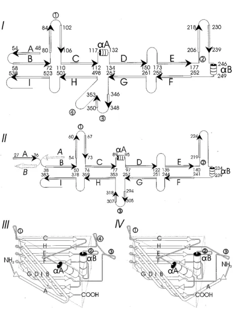
The 3D structures of several vertebrate parvoviruses (CPV, FPV, MVM) and, recently, of an invertebrate parvovirus (GmDNV) have been solved to near-atomic resolution using X-ray crystallography (Simpson et al., 1998). All have a common structure arranged with T = 1 icosahedral symmetry with each subunit having the same eight-stranded anti-parallel b-barrel motif (Fig. 1). This motif contains less than 1/3 of the protein mass of each subunit, is mostly below the cap-sid surface and consists of two b-sheets (b-strands BIDG and CHEF, respectively). Loops (large insertions between these b-strands) form much of the capsid surface and interactions with neighbouring subunits. There are three major insertions: be-tween b-strands B and C (loop 1), bebe-tween b-strands E and F (loop 2) and bebe-tween b-strands H and G (loop 3 for invertebrate parvovirus and loops 3 and 4 for verte-brate parvoviruses). Loops 1 and 2 are centrally located in the asymmetric unit and loops 3 or 3+4 are found around the 3-fold axis. In the case of CPV, FPV and MVM, loops 3 and 4 form a 22 Å-long spike, whereas for GmDNV, where loop 4 is absent, no spike but a b-annulus is found at the 3-fold axis. Although the struc-ture of the human parvovirus, B19, has not been solved to near-atomic resolution, it is clear (Agbandje et al., 1994) that it also lacks the 3-fold spike.
Fig. 1. Diagram of secondary structures of vertebrate and invertebrate parvoviruses. The b-strands are represented by arrows and the a-helices by cylinders. These drawings are not to scale. The loops that interconnect the b-strands are much longer than the a- and b-structures and form most of the surface and monomer connecting proteins. The backbone of the capsid structure is the pro-tein sequence in VP2 (for CPV, MVM, PPV) or VP4 (for GmDNV). Secondary structures are
rep-resented in diagrams I (canine parvovirus with numbering of VP2 amino acids) and II (GmDNV with numbering of VP4 amino acids). The most striking differences are (i) the length of the loops;
(ii) the presence or absence of loop 4; and, (iii), the orientation of b-strand A. The b-barrels (III, IV) are also highly schematic and show how the BIDG and CHEF b-sheets interact and are quite similar for the vertebrate (III) and invertebrate parvovirus (IV). These triangular subunits corre-spond to the isometric units represented in Fig. 2. The b-barrel in GmDNV must be rotated by 7.5°
Other differences between the vertebrate and invertebrate parvoviruses in-clude the orientation of the b-strand A which folds back to its own fivefold axis in the case of vertebrate parvoviruses, but interacts with the neighbouring subunit (via the twofold axis) in the case of GmDNV, an invertebrate parvovirus. Also the loops of GmDNV capsid protein are much shorter, as the mass of the structural proteins of this and related viruses is about 35% less than that of the vertebrate parvoviruses, and the surface of the capsid is generally much smoother (Fig. 2). It is not clear whether this is a result of the differences in the immune systems in vertebrates and invertebrates (who do not produce immuno-globulins). According to the canyon hypothesis, postulated by Rossmann (1989), vertebrate viruses would have evolved structures to protect crucial sites from antibody recognition (i.e., critical sites would be at the bottom of canyons and inaccessible to antibodies).
Fig. 2. Computer-generated reconstructions of invertebrate (GmDNV) and vertebrate (CPV) parvovi-ruses viewed along the twofold axis. Two isometric units (triangles defined by 3-fold and 5-fold axis of symmetry; 60 per particle) are represented on each particle with the positions of the allotropic de-terminants. The circles in GmDNV correspond to loop 2 sequences, whereas the circles in the
verte-brate parvovirus indicate amino acid differences in the allotropic determinant of PPV Sequence comparisons demonstrate that b-strand sequences are more con-served than those of the loops and, as a consequence, the greatest variation oc-curs at the capsid surface. Closely related viruses (CPV vs. FPV or among bio-logically different strains of PPV) with very few amino acid differences show that these are located in the loops and often at the surface of the capsid. For GmDNV, most differences are located at the surface and almost exclusively in two domains (not necessarily from the same loop). Since biological differences among these viruses must be accounted for by genetic differences, it was postu-lated that these few structural differences in the capsid are responsible.
Allotropic determinants of parvoviruses and their function
Allotropic determinants have been determined for a few parvoviruses and were found in the capsid (Gardiner and Tattersall, 1988; Chang et al., 1992; Bergeron et al., 1996). Generally, infectious clones (plasmids containing the complete viral genome) were generated that, upon transfection of cells, would excise the viral genome through replication and thus generate virus. This allows the convenient construction of chimeric viruses or mutants and their production in bacteria in order to establish, subsequently, the phenotype of the resultant vi-rus after transfection into host cells. A second prerequisite is the availability of a practical system that allows the recognition of the different phenotypes. For ex-ample, in the case of PPV it is known that Kresse and NADL-2 strains have a different phenotype in pigs. NADL-2 virus is hardly pathogenic (low viraemia) whereas the Kresse strain of PPV is highly pathogenic, even to immunocompe-tent fetuses. It is difficult and costly to find enough PPV-free pigs to study the biological differences between these two virus strains and in vitro systems are needed. However, at this level these virus strains usually do not show recogniz-able differences and it required a significant effort to find cells that would dis-tinguish these PPV strains. The primary cells (bovine testis) that were able to do so (Bergeron et al., 1996) were then immortalized with SV40 T-antigen and the clones obtained (Laakel et al., unpublished results) demonstrated different phe-notypes to these PPV strains (susceptibility vs. non-susceptibility, absence or presence of cytopathic effect in infected cells).
An alternative approach to elucidate parvovirus host range determinants is to pseudotype a genome of a given parvovirus with capsids of closely-related vi-ruses and to study the resultant tropism (Spitzer et al., 1996). It was shown with these transducing particles that the LuIII genome in MVM or H-1 capsids has the tropism of the capsids, not that of the genome.
Only very few genomic differences exist among the strains of PPV (Tijssen et al., 1995; Bergeron et al., 1996) that can be segregated into four, phenotypically distinct, groups. Chimeric constructs generated by exchanging restriction fragments among the infectious clones of these strains pinpointed three amino acid changes in the external loop (3/4) of the capsid protein. In par-ticular one of these (S436P) is prominent as it is located on the top of the three-fold spike (Fig. 2). The VP2 differences in yet another strain (P2; Vasudevach-arya and Compans, 1992) map structurally closely to the differences we ob-served between the non-pathogenic NADL-2 and Kresse strains (Bergeron et al., 1996). Oraveerakul et al. (1992) demonstrated that PPV strains may enter non-permissive cells but that they are restricted at some unknown level.
The sudden emergence of CPV in the late seventies and its close rela-tionship to FPV suggested that CPV may have arisen as an FPV variant.
Se-quence analysis of early CPV strains do not support this hypothesis (Truyen et al., 1998). The initial CPV-2 strains had a strict canine tropism, whereas the strains that replaced these strains in nature in the early eighties, i.e. CPV-2a and 2b, had acquired the ability to infect cats as well (10% of parvovirus isolates from feline diagnostic samples is in fact CPV) (Truyen et al., 1996). CPV VP2 residue changes K93N, A103V, and D323N determined that replication could occur in both dogs and canine cells (Chang et al., 1992; Horiuchi et al., 1994). Chimera between FPV and CPV demonstrated that two regions of the genome are important (VP2 residues K80R, N564S, or A568G). These residues are close together in the 3D-structure of the capsid on the top edge of the twofold dimple-like depression in a region where loops 1, 3, and 4 of three different monomers interact. Additional changes in this area may revert the CPV strain to a feline host range (Truyen and Parrish, 1995). A single mutation in VP2, A300D, causes a loss in canine host range (Llamas-Saiz et al., 1996). This D300 forms a salt bridge with R81 inducing also local changes within the antigenic site. In ad-dition, the loop between residues 359 and 374 (in loop 4) adopts a structure similar to that of FPV. Horiuchi et al. (1992) observed that the stage in FPV vi-rus replication cycle at which the host specificity of this subgroup is regulated in canine cells is found after cell entry of the virus.
It was observed for MVM that the MVMp strain infects fibroblast cells (A9) and the MVMi strain infects lymphoid cells (EL4 T-lymphocytes). The fi-brotropic determinant in MVMp was mapped to residues 317 and 321 and MVMi could become fibrotropic by mutations at these positions (Ball-Goodrich et al., 1991). In contrast, two segments of MVMi (one in the NS and one in the VP region) were required to confer its phenotype to MVMp (Colomar et al., 1998). The NS segment influences apparently the virus-strain-specific differ-ences in the regulation of splicing, whereas the VP segment impacts on the virion structure. Mutations in NS genes that affected the expression of the repli-cative form of the viral DNA restricted the viral reproduction more in murine cells than in other cells (Naeger et al., 1993). Spalholz and Tattersall (1983) also demonstrated that the strain-specific target cell specificity is mediated by intra-cellular factors.
MVM and PPV resemble each other in several aspects: (i) the nonpatho-genic PPV-NADL-2 and MVMp strains are much less viraemic in vivo than the pathogenic strains; (ii) the fetal infection by PPV finds its equivalent in the gen-eralized infection for MVM of hematopoietic cells and the capillary endothe-lium; and (iii) the position and function of the allotropic determinants. Unfortu-nately, we do not have yet the sequence of the NADL-8 strain (only pathogenic to non-immunocompetent fetuses), which has been shown to have a very differ-ent tropism towards the various organs when compared to the pathogenic Kresse strain (Oraveerakul et al., 1993).
There is a considerable variability among the different strains of ADV, particularly in a hypervariable region of about 25 nucleotides in the capsid gene. Some ADV strains grow only in the animal (e.g. ADV-Utah) whereas others grow in tissue culture (e.g. the nonpathogenic ADV-G). Chimeric constructs of an infectious clone of ADV-G with wild-type sequences indicated that se-quences within 5565 m.u. of ADV-Utah inhibited replication in vitro (Bloom et al., 1993). Construction of pathogenic molecular clones of ADV that replicate both in vivo and in vitro revealed two supplementary regions, at 6569 and 73 88 m.u., that, in tandem, can abolish growth in vitro (Bloom et al., 1998). A novel ADV strain (ADV-TR) that is closely related to ADV-G, but is pathogenic for Aleutian mink, was found to be able to infect raccoons and could be respon-sible for transmission of ADV infections (Oie et al., 1996). Twelve amino acids in the VP region were identified that could be at the basis of this phenotype.
The identification of the allotropic determinant of GmDNV is also under-way. This virus group offers the advantage that the host animals can be manipu-lated easily. Infectious clones have been generated and exchange of restriction fragments have been shown that two regions on the capsid are implicated (Fig. 2), one is around loop 2 in the middle of the isometric subunit and one straddles loop 3 and is continuous in two subunits along the 3-fold to 3-fold axis. Further investi-gations should allow us to reduce the number of amino acids that are essential for the species-specific tropism.
Conclusion
The various parvoviruses for which tropism has been studied all show that minimal changes in the sequence of the structural protein can determine cell tro-pism or host range. The location of these critical amino acids, usually in loops 3 and 4 and at the C-terminus of the capsid protein, are at or near the capsid sur-face, suggesting strongly that they interact with a cellular molecule. Ample evi-dence indicates that this cellular counterpart is not the receptor. Two possibili-ties exist: the cellular factor enables the virus infection in permissive cells or in-hibits virus infections in non-permissive cells. Although the decapsidation stage seems to be a logical candidate, the limited replication and expression may indi-cate that later steps in the viral cycle are critical.
The sudden and devastating emergence of MEV and CPV in the last 50 years demonstrates the importance of parvovirus tropism. Whether they arose by a few mutations from FPV or have another origin is academic. Despite its im-portance and considerable efforts, we know still precious little about the mecha-nisms of tropism changes of this virus group.
Acknowledgements
I wish to acknowledge the help from my collaborators at Purdue (M. Rossmann and A. Simpson), Cornell (C. Parrish), VIDO (L. Babiuk) and University of Montpel-lier/INRA (M. Bergoin, F.-X. Jousset) and from everybody in my laboratory working on this project, especially the post-doctoral fellows (M. Laakel, Y. Li, and Z. Zádori), stu-dents (M. David, S. Forest, S. Gariépy, B. Hébert, M.-C. Lacoste), research assistant (M. Letarte) and technical staff (J. Reid, L. Forget, L. Paris-Nadon). Furthermore, I want to express my gratitude to the Natural Science and Engineering Research Council of Canada for their financial support.
References
Agbandje, M., Kajigaya, S., McKenna, R., Young, N. S. and Rossmann, M. G. (1994): The struc-ture of human parvovirus B19 at 8 Å resolution. Virology 203, 106115.
Ball-Goodrich, L. J., Moir, R. D. and Tattersall, P. (1991): Parvoviral target cell specificity acqui-sition of fibrotropism by a mutant of the lymphotropic strain of minute virus mice involves multiple amino acid substitutions within the capsid. Virology 184, 175186.
Bergeron, J., Hébert, B. and Tijssen, P. (1996): Genome organization of the Kresse strain of por-cine parvovirus. Identification of the allotropic determinant and its comparison with those of NADL-2 and field strains. J. Virol. 70, 25082515.
Berns, K. I. (1996): Parvoviridae: the viruses and their replication. In: Fields, B. N., Knipe, D. M. and Howley, P. M. (eds) Fundamental Virology, 3rdedition. Lippencott-Raven Publishers,
Philadelphia. pp. 10171041.
Berns, K. I., Bergoin, M., Bloom, M., Lederman, M., Muzyczka, N., Siegl, G., Tal, J. and Tatter-sall, P. (1995): Parvoviridae. VIth report of International Committee on Taxonomy of Vi-ruses. Arch. Virol. Suppl. 10, 169178.
Bloom, M. E., Berry, B. D. Wei, W., Perryman, S. and Wolfinbarger, J. B. (1993): Characteriza-tion of chimeric full-length molecular clones of Aleutian mink disease parvovirus (ADV): identification of a determinant governing replication in cell culture. J. Virol. 67, 59765988. Bloom, M. E., Fox, J. M., Berry, B. D., Oie, K. L. and Wolfinbarger, J. B. (1998): Construction of
pathogenic molecular clones of Aleutian mink disease parvovirus that replicate both in vivo and in vitro. Virology 251, 288296.
Bossin, H. (1998): Development of stable expression vectors derived from JcDNV: application to constitutive expression of heterologous proteins in Lepidopteran cell lines and as a marker during Drosophila development [in French]. Ph. D. Thesis, University of Montpellier II, France. 226 pp.
Brockhaus, K., Plaza, S., Pintel, D. J., Rommelaere, J. and Salomé, N. (1996): Nonstructural pro-teins NS2 of minute virus of mice associate in vivo with 14-3-3 protein family members. J. Virol. 70, 75277534.
Brownstein, D. G., Smith, A. L., Johnson, E. A., Pintel, D. J., Naeger, L. K. and Tattersall, P. (1992): The pathogenesis of infection with minute virus of mice depends on expression of the small nonstructural protein NS2 and on the genotype of the allotropic determinants VP1 and VP2. J. Virol. 66, 31183124.
Chang, S.-F., Sgro, J.-Y. and Parrish, C. R. (1992): Multiple amino acids in the capsid structure of canine parvovirus coordinately determine the canine host range and specific antigenic and hemagglutinating properties. J. Virol. 66, 68586867.
Colomar, M. C., Hirt, B. and Beard, P. (1998): Two segments in the genome of the immunosup-pressive minute virus of mice determine the host-cell specificity, control viral DNA repli-cation and affect viral RNA metabolism. J. Gen. Virol. 79, 581586.
Corsini, J., Tal, J. and Winocour, E. (1997): Directed integration of minute virus of mice DNA into episomes. J. Virol. 71, 90089015.
Cossons, N., Faust, E. A. and Zannis-Hadjopoulos, M. (1996): DNA polymerase delta-dependent formation of a hairpin structure at the 5 terminal palindrome of the minute virus of mice genome. Virology 216, 258264.
Cotmore, S. F. and Tattersall, P. (1989): A genome-linked copy of the NS-1 polypeptide is located on the outside of infectious parvovirus particles. J. Virol. 63, 39023911.
Cotmore, S. F. and Tattersall, P. (1995): DNA replication in the autonomous parvoviruses. Sem. Virol. 6, 271281.
Cotmore, S. F., DAbramo, A. M., Jr., Carbonell, L. F., Bratton, J. and Tattersall, P. (1997): The NS2 polypeptide of parvovirus MVM is required for capsid assembly in murine cells. Vi-rology 231, 267280.
Faust, E. A. and Ward, D. C. (1979): Incomplete genomes of the parvovirus minute virus of mice: selective conservation of genomic termini. J. Virol. 32, 276292.
Gardiner, E. M. and Tattersall, P. (1988): Mapping of the fibrotropic and lymphotropic host range determinants of the parvovirus minute virus of mice. J. Virol. 62, 26052613.
Gersappe, A. and Pintel, D. J. (1999): CA- and purine-rich elements form a novel bipartite exon enhancer which governs inclusion of the minute virus of mice NS2-specific exon in both singly and doubly spliced mRNAs. Mol. Cell. Biol. 19, 364375.
Gibbs, A. I. and Harrison, B. O. (1968): Realistic approach to virus classification and nomencla-ture. Nature 218, 927929.
Horiuchi, M., Ishiguro, N., Goto, H. and Shinagawa, M. (1992): Characterization of the stage(s) in the virus replication cycle at which the host-cell specificity of the feline parvovirus sub-group is regulated in canine cells. Virology 189, 600608.
Horiuchi, M., Goto, H., Ishiguro, N. and Shinagawa, M. (1994): Mapping of the determinants of the host range for canine cells in the genome of canine parvovirus using canine parvovi-rus/mink enteritis chimeric viruses. J. Gen. Virol. 75, 13191328.
Kotin, R. M., Linden, R. M. and Berns, K. I. (1992): Characterization of a preferred site on human chromosome 19q for integration of adeno-associated virus DNA by non-homologous re-combination. EMBO J. 11, 50715078.
Liu, J. M., Green, S. W., Shimada, T. and Young, N. S. (1992): A block in full-length transcript maturation in cells nonpermissive for B19 parvovirus. J. Virol. 66, 46864692.
Liu, Q., Yong, C. B. and Astell, C. R. (1994): In vitro resolution of the dimer bridge of the minute virus of mice (MVM) genome supports the modified rolling hairpin model for MVM repli-cation. Virology 201, 251262.
Llamas-Saiz, A. L., Agbandje-McKenna, M., Parker, J. S., Wahid, A. T., Parrish, C. R. and Ross-mann, M. G. (1996): Structural analysis of a mutation in canine parvovirus which controls antigenicity and host range. Virology 225, 6571.
Lwoff, A. (1967): Principles of classification and nomenclature of viruses. Nature 215, 1314. Mayor, H. D., Drake, S., Stahmann, J. and Mumford, D. M. (1976): Antibodies to
adeno-associated satellite virus and herpes simplex in sera from cancer patients and normal adults. Am. J. Obstet. Gynecol. 126, 100104.
Naeger, L. K., Salomé, N. and Pintel, D. J. (1993): NS2 is required for efficient translation of viral mRNA in minute virus of mice-infected murine cells. J. Virol. 67, 10341043.
Nuesch, J. P. F., Cotmore, S. F. and Tattersall, P. (1995): Sequence motifs in the replicator protein of parvovirus MVM essential for nicking and covalent attachment to the viral origin: iden-tification of the linking tyrosine. Virology 209, 122135.
Nuesch, J. P., Dettwiler, S., Corbau, R. and Rommelaere, J. (1998): Replicative functions of min-ute virus of mice NS1 protein are regulated in vitro by phosphorylation through protein kinase C. J. Virol. 72, 99669977.
Oie, K. L., Durrant, G., Wolfinbarger, J. B., Martin, D., Costello, F., Perryman, S., Hogan, D., Hadlow, W. J. and Bloom, M. E. (1996): The relationship between capsid protein (VP2) sequence and pathogenicity of Aleutian mink disease parvovirus (ADV): a possible role for raccoons in the transmission of ADV infections. J. Virol. 70, 852861.
Oraveerakul, K., Choi, C. S. and Molitor, T. W. (1992): Restriction of porcine parvovirus replica-tion in nonpermissive cells. J. Virol. 66, 715722.
Oraveerakul, K., Choi, C. S. and Molitor, T. W. (1993): Tissue tropisms of porcine parvovirus in swine. Arch. Virol. 130, 377389.
Pintel, D. J., Gersappe, A., Haut, D. and Pearson, P. (1995): Determinants that govern alternative splicing of parvovirus pre-mRNAs. Sem. Virol. 6, 283290.
Rommelaere, J. and Cornelis, J. J. (1991): Antineoplastic activity of parvoviruses. J. Virol. Meth. 33, 233251.
Rossmann, M. G. (1989): The canyon hypothesis. Hiding the host cell receptor attachment site on a viral surface from immune surveillance. J. Biol. Chem. 264, 1458714590.
Schlehofer, J. R. (1994): The tumor suppressive properties of adeno-associated viruses. Mutation Res. 305, 303313.
Siegl, G. (1976): The parvoviruses. Virol. Monogr. 15, 1109.
Simpson, A. A., Chipman, P. R., Baker, T. S., Tijssen, P. and Rossmann, M. G. (1998): The structure of an insect parvovirus (Galleria mellonella densovirus) at 3.7 Å resolution. Structure 6, 13551367.
Snyder, R. O., Samulski, R. J. and Muzyczka, N. (1990): In vitro resolution of covalently joined AAV chromosome ends. Cell 60, 105113.
Spalholz, B. A. and Tattersall, P. (1983): Interaction of minute virus of mice with differentiated cells: strain-dependent target cell specificity is mediated by intracellular factors. J. Virol. 46, 937943.
Spitzer, A. L., Maxwell, F., Corsini, J. and Maxwell, I. H. (1996): Species specificity for transduc-tion of cultured cells by a recombinant LuIII rodent parvovirus genome encapsidated by canine parvovirus or feline panleukopenia virus. J. Gen. Virol. 77, 17871792.
St Amand, J., Beard, C., Humphries, K. and Astell, C. R. (1991): Analysis of splice junctions and in vitro and in vivo translation potential of the small, abundant B19 parvovirus RNAs. Vi-rology 183, 133142.
Tattersall, P. and Ward, D. C. (1976): Rolling hairpin model for replication for replication of par-vovirus and linear chromosomal DNA. Nature 263, 106109.
Telerman, A., Tuynder, M., Dupressoir, T., Robaye, B., Sigaux, F., Shaulian, E., Oren, M., Rom-melaere, J. and Amson, R. (1993): A model for tumor suppression using parvovirus H-1. Proc. Natl. Acad. Sci. USA 90, 87028706.
Tijssen, P. (Editor) (1990): Handbook of Parvoviruses vol. I (309 pp) and vol. II (312 pp). CRC Press, Boca Raton, Florida.
Tijssen, P. and Bergoin, M. (1995): Densonucleosis viruses constitute an increasingly diversified family among parvoviruses. Sem. Virol. 6, 347355.
Tijssen, P., Bergeron, J., Dubuc, R. and Hébert, B. (1995): Minor genetic changes among porcine par-vovirus groups are responsible for major distinguishing properties. Sem. Virol. 6, 319328. Tijssen, P., Laakel, M., Zádori, Z. and Hébert, B. (1999): Parvoviruses: rodents, pigs, cattle and
waterfowl. In: Webster, R. G. and Granoff, A. (eds) Encyclopedia in Virology. 2ndedition.
Academic Press, San Diego, CA (in press).
Truyen, U. and Parrish, C. R. (1995): The evolution and control of parvovirus host ranges. Sem. Virol. 6, 311317.
Truyen, U., Evermann, J. F., Vieler, E. and Parrish, C. R. (1996): Evolution of canine parvovirus involved loss and gain of feline host range. Virology 215, 186189.
Truyen, U., Geissler, K., Parrish, C. R., Hermanns, W. and Siegl, G. (1998): No evidence for a role of modified live virus vaccines in the emergence of canine parvovirus. J. Gen. Virol. 79, 11531158.
Vasudevacharya, J. and Compans, R. W. (1992): The NS and capsid genes determine the host range of porcine parvovirus. Virology 187, 515524.
Vanacker, J.-M. and Rommelaere, J. (1995): Non-structural proteins of autonomous parvovirus: from cellular effects to molecular mechanisms. Sem. Virol. 6, 291297.
Wang, D., Yuan, W., Davis, I. and Parrish, C. R. (1998): Nonstructural protein-2 and the replica-tion of canine parvovirus. Virology 240, 273281.
Willwand, K., Mumtsidu, E., Kuntz-Simon, G. and Rommelaere, J. (1998): Initiation of DNA rep-lication at palindromic telomeres is mediated by a duplex-to-hairpin transition induced by the minute virus of mice nonstructural protein NS1. J. Biol. Chem. 273, 11651174. Zádori, Z., Stefancsik, R., Rauch, T. and Kisary, J. (1995): Analysis of the complete sequences of
goose and muscovy duck parvoviruses indicates common ancestral origin with adeno-associated virus 2. Virology 212, 562573.