RENAL WATER AND ION TRANSPORT SY STEMS
James A. Schafer
Depa rtm ents of Physiology a nd Biophysics, Nephrology Tra ining a nd Resea rch Center, University of Ala ba m a a t Birm ingha m , Birm ingha m , Ala ba m a 35294
T
he teaching of renal physiology is an ever-evolving and fascinating opportunity to learn as well as to teach. Regardless of the particular format of our teaching, a constantly waxing tide of detailed information forces us to be selective. We cannot teach it all. In my own lectures and discussions of renal ion and water transport systems, I try to1) avoid complex and detailed areas that are not essential to a basic understanding of renal function,2) highlight the commonalities of transport processes among segments, 3) relate the details of transporters to clinical conditions that are especially interesting,4) make connections between physiological details and clinical therapy, and5) stress quantitative problems. Although generally simple, the quantitative understanding is essential to appreciate the role of the kidney in homeostasis and derangements thereof. When I review what I have taught, I try to integrate multiple basic mechanisms in individual segments to produce a picture of ‘‘whole kidney’’ function.AM. J. PHYSIOL. 275 (ADV. PHYSIOL. EDUC. 20): S119–S131, 1998.
Key words: isosmotic volume reabsorption; glucose reabsorption; aquaporins; renal diseases; diuretics; homeostasis; steady state; creatinine balance
The teaching of renal physiology is an ever-evolving and fascinating opportunity to learn as well as to teach. Whatever the format in which we teach this subject, be it traditional lectures, problem-based learn-ing, or some combination, an ever-growing body of information forces us to select those details that are most relevant for the medical student and put them in useful and interesting contexts. This focus on the medical student is also valid for our PhD graduate students. There is simply too much information for us to expect them to learn the whole body of informa-tion about each organ system, as might have been possible 20 or 30 years ago. On the other hand, our graduate students still need the breadth of a general physiology course to fulfill their roles as both scien-tists and teachers. Hopefully, the future physiologist will have this breadth of knowledge that will facilitate the integration of molecular and genetic information with physiological and pathophysiological processes; i.e., to be the link from molecule to clinic.
WHY TEACH NEPHRON FUNCTION?
Many of our nephrology and pathology colleagues might disagree with the detail with which we present the transepithelial transport processes involved in the function of the nephron. After all, they might argue, the most important and common renal diseases are not disorders of tubular transport. Glomerular immu-nopathology, specifically glomerulonephritis, is by far the most prevalent cause of chronic and end-stage renal disease. It might seem, therefore, that we should concentrate our teaching on glomerular function and immunology to the exclusion of renal transport pro-cesses. The counterpoint to this argument is that an understanding of how the kidney maintains salt and water balance through regulation of its multiple trans-port processes is essential for understanding fluid and electrolyte disorders and for the rational choice of therapy for those disorders. Fluid and electrolyte disorders are much more common than glomerular disease, and most physicians deal with these disorders daily. As just one of many examples, the most com-mon fluid and electrolyte disorder in the hospital setting is hyponatremia, and 50% of hyponatremic patients die. Also, the concepts learned in understand-ing renal epithelial transport processes are closely related to similar processes occurring in many other systems, for example, intestinal absorption and secre-tion, and electrolyte balance in the respiratory system and in the brain.
AVOID EXPLAINING COMPLEX CONCEPTS WHEN THEY ARE NOT ESSENTIAL
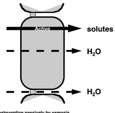
In addressing the more complex topics in renal salt and water transport, the lecturer has to make the decision of whether a full discussion is warranted. All of the topics that we teach involve details and com-plexities that could require hours of self-study for the student to begin to appreciate. A primary job of the teacher is to decide whether a full discussion of the topic is worth the time that it would take from more important, more clinically applicable, subjects. An example dear to my own heart is the process by which fluid is reabsorbed isosmotically from the proximal tubule. Despite the large volume of information on this subject that my colleagues and I, and many others, have laboriously gathered (2, 4, 18, 28), I feel that the student can appreciate the essence of isosmotic vol-ume absorption according to the simple principles
outlined in Fig. 1. The important points to stress are as follows (18). 1) Water movement across the epithe-lium and all regions of the nephron occurs in response to an osmotic gradient, whether that gradient is localized to a small region such as the intracellular spaces or between two compartments such as the tubular lumen and the interstitial fluid.2) The essen-tial ingredient for (nearly) isosmotic volume absorp-tion is the extremely high osmotic water permeability of the proximal tubule. Because of this high water permeability, only a very small osmolality difference, one that was not easily measured in early studies, is needed to drive the observed rates of volume reabsorp-tion. 3) The active transport of solutes makes the lumen slightly dilute and the interstitial fluid slightly hypertonic.4) The actively reabsorbed solutes such as glucose, amino acids, and bicarbonate have higher reflection coefficients than NaCl; thus the preferential reabsorption of these solutes results in the
develop-FIG. 1.
ment of a larger ‘‘effective’’ osmolality difference across the epithelium. Students should already be familiar with the concept of a reflection coefficient, but even if they are not, they should be able to intuitively grasp the fact that solutes of lower permeability (and thus higher reflection coefficient) will make a greater contribution to the true osmolality of interstitium com-pared with the fluid remaining in the tubule lumen, which becomes increasingly composed of NaCl, which replaces the preferentially reabsorbed solutes.
Another area that I find too demanding in time for the student to understand in detail is the mechanism of countercurrent multiplication, especially passive coun-tercurrent multiplication in the inner medulla. Cer-tainly, the postulation and experimental verification of the countercurrent multiplication system was one of the most intellectually gratifying achievements of renal physiology research. For that reason, I believe many of us have given it undue emphasis in our practical teaching. If the students want to know these details, I refer them to an appropriate text. However, at least in the lecture setting, I stress the essential ingredients for the development of medullary hyperto-nicity and the clinically important events that would interfere with the urinary concentrating and diluting ability. Obviously, the crucial element is active NaCl reabsorption by the thick ascending limb of the loop of Henle via the Na1-K1-2Cl cotransporter (BSC1). The
students can well appreciate that this active solute reabsorption, which occurs in a segment that is water impermeable, should concentrate the medullary inter-stitium. They can also readily appreciate that the countercurrent arrangement and low blood flow rate through the vasa recta curtail the ‘‘washout’’ of the hypertonicity that would occur in most other tissues. The second component is the addition of urea to the tubular fluid in the thin limbs of the loop of Henle, which increases the amount of urea that is delivered to the thick ascending limb of the loop of Henle. Urea is then trapped in the tubular lumen, because all nephron segments from the thick ascending limb through the outer medullary collecting duct are imper-meable to urea. However, because of the presence of a urea transporter (UT1) in the most distal portion of the inner medullary collecting duct, urea can diffuse passively across this epithelium. When the urine urea concentration is very high, as in antidiuresis, urea diffuses from the inner medullary collecting duct to
the medullary interstitium, where it contributes to the medullary hypertonicity and enhances water reabsorp-tion from the descending limb of the loop of Henle. These points encompass the essential ingredients of the countercurrent multiplication system, and using these points, the teacher can examine mechanisms by which the medullary osmolality gradient can be dis-turbed. One can discuss, for example, the effects of changes in medullary blood flow, saline loading, loop diuretics, low-protein diets, and water diuresis on the medullary concentration gradient.
SIMPLIFY THE DETAILS OF EPITHELIAL TRANSPORT AND HIGHLIGHT THE COMMONALITIES AMONG SEGMENTS
One of the most important characteristics of the nephron is its ability to dissociate salt and water transport in various segments. Some nephron seg-ments, including the thin and thick ascending limbs of the loop of Henle and the collecting duct in the absence of vasopressin (AVP), are very water imperme-able. In these water-impermeable segments, solute reabsorption results in dilution of the tubular fluid. In contrast, most cell membranes, including those of the proximal tubule and thin descending limb of the loop of Henle, are very water permeable under all circum-stances. This high water permeability is a conse-quence of the presence of a water channel that has come to be referred to as aquaporin-1 (AQP1). I find the students interested to know that this water chan-nel is found in many cells, including red blood cells, and that it contributes to the very high water perme-ability of the proximal tubule and the descending limb of the loop of Henle, where it is present in both luminal and basolateral membranes (Fig. 2).
two other aquaporins: AQP3 and AQP4. At this point, it is instructive to point out that mutations in human AQP2 that result in nephrogenic diabetes insipidus have been identified and that there may be as yet unidentified abnormalities in AQP3 or AQP4 that could also interfere with the ability to concentrate the urine. (For references to aquaporins, see Refs. 1, 7, 12, 14, and 23.)
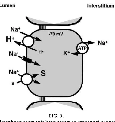
In discussing the details of transport processes, it is also useful to generalize about transepithelial trans-port before dealing with the details of individual systems. For example, when I introduce transport in the proximal tubule, as shown in Fig. 3, I first point out the aspects that are common to all regions of the nephron.1) The epithelium is a single cell layer, and cells are joined by junctional complexes near their apical border. This topology provides two pathways by which solutes can traverse the epithelium, either through the cell or through the paracellular pathway, i.e., the junctional complexes and lateral intercellular spaces.2) All epithelial cells in the nephron have the Na1-K1-ATPase that is common to virtually every cell
in the body. This Na1-K1-ATPase is responsible for
maintaining a low intracellular Na1concentration and
for developing the ion gradients that lead to a negative
intracellular voltage. However, most epithelia, includ-ing all the nephron segments, are unique because this transporter is localized only to the basolateral mem-brane.3) In all nephron segments, the Na1-K1-ATPase
develops an Na1 electrochemical potential gradient
across the opposite membrane, i.e., the luminal mem-brane; thus there is a driving force for the passive entry of Na1from the lumen into the epithelial cell.
This entry may occur through an electrogenic Na1
channel, such as the amiloride-sensitive Na1channel
in the collecting duct, in which case the potential energy embodied in the Na1electrochemical
poten-tial gradient is dissipated. However, if the movement of Na1is coupled to the movement of another solute,
for example, by an antiporter, such as the Na1/H1
exchanger (NHE3) in the proximal tubule, or by a cotransporter, such as the Na1-glucose symporter,
then the energy available in the Na1electrochemical
potential gradient can be used to perform useful work. That useful work is the active transport of the coupled solutes against their own electrochemical potential gradients. It should be emphasized to the student that this general plan for the transport of Na1
holds true throughout the nephron. That is, the Na1
electrochemical potential gradient is maintained by
FIG. 2.
Characteristics of primary aquaporin water channels found in kidney. CHIP28 and WCH-CD ar e alter nate names that wer e pr eviously used for AQP1 and AQP2, r espectively. AVP, vasopr essin; DLH, descending limb of the loop of Henle; IMCD, inner medullary collecting duct; DI, diabetes insipidus. Right arr ow indicates pr oduction. See tex t for fuller ex planations.
FIG. 3.
All nephron segments have common transport proper-ties: a single layer of cells connected by junctional complex es near their luminal aspects, basolateral Na1
-K1-ATPase, and passive Na1entry acr oss the luminal
the basolateral Na1-K1-ATPase, and this potential
energy gradient provides the driving force for passive entry of Na1 into the cell across the luminal
mem-brane either alone or in combination with secondary active transport of other solutes. It is only the details of the transporters, usually those on the luminal membrane, that vary from one nephron segment to another. I try constantly to refer back to this overall scheme to avoid the students becoming belabored by the details of specific transporters in each nephron segment.
RELATE TRANSPORTER DETAILS TO CLINICAL CONDITIONS THAT ARE ESPECIALLY INTERESTING
In my experience, medical students are more willing to address the details of an epithelial transport process that are associated with clinically relevant defects. Graduate students are now acutely aware of the direct associations between gene mutations and functional defects and are also receptive to these practical applications of basic science. Thus, whenever pos-sible, I try to relate the details of the operation of a membrane transporter to a particular clinical situa-tion. A perfect example is a comparison of the two Na1-glucose cotransporters that are found in the
kidney: SGLT1 and SGLT2 (Fig. 4). (The number associated with the acronyms of these two transport-ers presents the order in which they were cloned and sequenced. This can be somewhat confusing, because the SGLT1 protein cotransports two Na1 in
associa-tion with one glucose, whereas the SGLT2 has a 1:1 stoichiometry.) In the kidney, SGLT2 is the primary transporter and is found in the proximal convoluted tubule, whereas the SGLT1 cotransporter is found in the proximal straight tubule. Because of the higher stoichiometry of SGLT1, the transporter can develop a concentration gradient (lumen to cell) of up to 1:10,000, whereas SGLT2 develops only a 1:100 gradi-ent. Thus it appears that SGLT2 serves to reabsorb the bulk of the glucose filtered in the kidney, whereas SGLT1 is able to reabsorb the last bit of sugar against a high transepithelial concentration gradient (3, 19, 22).
Where is the clinical interest here? The answer is that genetic defects occur in both transporters in humans that result in completely different phenotypes as illustrated in Fig. 4. A single base pair mutation in SGLT1, which causes a loss of function, produces the
glucose-galactose malabsorption syndrome that is char-acterized by severe diarrhea and dehydration, but it has little effect on the renal reabsorption of glucose. There are multiple mutant forms of SGLT2 in the human population that are associated with different forms of familial renal glucosuria. However, this condition is relatively benign, and its effects are restricted to the kidney because SGLT2 is not found in the intestine. This example provides students with an excellent example of how relatively small differences in quite similar transporters can lead to quite different disease phenotypes (for details, see Refs. 9, 10, 15, and 29).
Another condition that integrates genetics, molecular biology, physiology, and disease in a most elegant collage is Liddle’s syndrome. Liddle’s syndrome is a rare autosomal dominant disease that was first de-scribed by Grant Liddle et al. (13) in 1963. It is characterized by severe hypertension, often leading to stroke even at a young age. It is also frequently associated with hypokalemia and metabolic alkalosis. Thus, in most aspects, Liddle’s syndrome looks very
FIG. 4.
Secondary active transport of glucose by Na1-glucose
cotransporters SGLT1 and SGLT2. Although these 2 transporters ar e quite similar, and both ar e ex pr essed in pr ox imal tubule, subtle differ ences in molecular details of their operation ex plain pr ofound pheno-typic differ ences pr oduced by loss-of-function muta-tions. G, glucose; Tm, max imum r enal r eabsorption
similar to primary aldosteronism, i.e., the overproduc-tion of aldosterone by an adrenal carcinoma or diffuse renal hyperplasia. However, as determined by bioas-says, which at the time of Liddle’s report were available in his laboratory and a few others, it was determined that plasma renin and aldosterone levels were both immeasurable, and the condition was not ameliorated by the aldosterone receptor antagonist spironolactone. Liddle and his co-workers could find no hormonal explanation for the abnormality, and, on the basis of the physiological information of that time, they correctly concluded in 1963 that the defect probably represented an unregulated absorption of Na1by the distal nephron. Their suggested treatment,
which is the administration of triamterene or amilo-ride in combination with a low-Na1diet, remains the
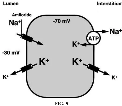
treatment of choice. As shown in Fig. 5, since 1963, we have learned that the target of amiloride and triamterene action is the electrogenic Na1 channel
found in the luminal membrane of the principal cell of the collecting duct. This channel, referred to as the epithelial Na1channel (ENaC), consists of three
sub-units labeleda,b, andg, which were originally cloned and sequenced by Cecilia Canessa in Bernard Rossier’s laboratory just four years ago (5). Once the genetic sequence for these channel subunits was known and their position on human chromosomes ascertained, it was possible to examine whether there was a muta-tion associated with one of the subunits that results in Liddle’s syndrome. Fortunately, the Liddle’s kindred,
which resides in Southern Alabama, is relatively exten-sive, and immortalized lymphocytes obtained from family members were used for DNA analysis. It was found that Liddle’s syndrome was associated with a mutation in theb-ENaC sequence that resulted in an early truncation in the COOH-terminal region (20). This truncation results in an overactive Na1 channel
that is not regulated normally and can explain the excessive Na1reabsorption seen in Liddle’s patients.
Since these original findings, several similar kindreds have been identified with other mutations in the COOH-terminal regions of theb- andg-subunits (24). It is also interesting that the congenital form of pseudohypoaldosteronism is associated with a muta-tion in the internal region of the a-ENaC subunit that results in a loss-of-function mutation rather than the gain-of-function observed in Liddle’s syndrome (6).
Students are fascinated with this kind of information because it so beautifully transfers information from the basic physiology of the kidney, which allowed Grant Liddle to interpret the syndrome he observed, to the final resolution of the clinical problem by the application of molecular biology and physiology. This example can be used to demonstrate the reasons for the other manifestations of the Liddle’s syndrome. As shown in Fig. 5, if the Na1channel is unregulated and
‘‘wide open,’’ the high Na1 conductance of the
luminal membrane would depolarize it, just as the activation of Na1channels in nerve or muscle
mem-brane results in depolarization of those cells. The result of this depolarization is a more favorable K1
electrochemical potential gradient that drives the passive movement of K1out of the principal cells into
the lumen, thus enhancing K1excretion and leading
to the hypokalemia that is observed in at least some of the patients. The associated alkalosis may also be a result of an increased lumen-negative voltage that favors H1secretion.
It is also important to point out that these syndromes illustrate the importance of Na1balance in
maintain-ing normal blood pressure and volumes of body fluid compartments. About 25,000 meq of Na1are filtered
per day by the glomeruli in two adult human kidneys, but less than 2,000 meq/day are reabsorbed in the distal tubule and collecting duct, with the vast major-ity being reabsorbed in the proximal tubule and the ascending limb of the loop of Henle. Aldosterone
FIG. 5.
regulates the reabsorption of,500 meq of the 2,000
meq reabsorbed by the distal tubule and collecting duct. Thus, in the total absence of aldosterone, only
,500 meq/day are excreted, and at very high
aldoste-rone levels, Na1 excretion can be reduced nearly to
zero. The important point to stress is that aldosterone is operating on only a very small fraction of the filtered Na1load, but this regulation of Na1excretion in the
range of 0–1% of the filtered load is extremely critical. The retention of only 150 meq of Na1(with
accompa-nying monovalent anions) requires the retention of 1 liter of water to make it isotonic. Given the fine nature of this regulation, it is remarkable that disturbances, such as those produced by defects in ENaC, can lead to either salt wasting in the case of the a-subunit mutation or Na1retention with severe hypertension
in the case of theb- andg-subunit mutations.
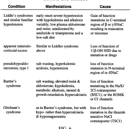
There are additional excellent examples of transporter or enzyme mutations that have interesting clinical correlations, some of which are described in Fig. 6. In the case of apparent mineralocorticoid excess, the underlying facts are that glucocorticoids such as cortisol
have reasonable affinity for both the mineralocorticoid and glucocorticoid (type I and type II) receptors in aldosterone responsive tissues. However, because of the presence of an enzyme, 11b-hydroxysteroid dehy-drogenase (11b-OH HSD), cortisol is degraded to the inactive cortisone. This allows aldosterone, which is usually present in much lower concentration in the plasma than glucocorticoids, to react with mineralocor-ticoid and, to some extent, glucocormineralocor-ticoid receptors in aldosterone target tissues such as the principal cell of the cortical collecting duct. The 11b-HSD can be inhibited by certain compounds and drugs such as glycyrrhetinic acid (in licorice) and gossypol, which leads to a syndrome that is very similar to Liddle’s syndrome or apparent hyperaldosteronism (11, 25, 26). The syndrome is also caused by mutations in the human 11b-HSD gene (16, 27).
Bartter’s syndrome (Fig. 6) illustrates the importance of specific transporters in the thick ascending limb of the loop of Henle that result in the development of a lumen-positive voltage and thus favor Ca21
reabsorp-tion. Mutations in BSC1 as well as mutations in the luminal K1channel (ROMK) or in the basolateral Cl2
channel have been linked to Bartter’s syndrome. All these defects lead to decreased salt reabsorption in the thick ascending limb of the loop of Henle, and thus to a concentrating defect with increased Na1
delivery to the collecting duct. The latter leads to salt wasting with an elevation of renin and aldosterone concentrations. Na1 reabsorption by the collecting
duct is increased by the aldosterone. In addition, the increased Na1 delivery as well as the aldosterone
produce inappropriate levels of K1and H1secretion,
leading to hypokalemia and metabolic alkalosis. These effects are accompanied by a relative failure to absorb Ca21 and hypercalciuria. Gitelman’s syndrome is a
similar condition with all of the characteristics of Bartter’s syndrome but withhypo-rather than hyper-calciuria and hypomagnesemia. This syndrome results from a loss-of-function mutation in the thiazide-sensitive NaCl cotransporter (TSC1), which is found in the distal convoluted tubule. By examining abnor-malities in transporters or enzymes associated with syndromes such as these, the teacher can provide superb examples of the importance of physiological detail in the context of clinical understanding (for references to Bartter’s and Gitelman’s syndromes, see Ref. 8, 17, and 21).
FIG. 6.
Associations between defects at molecular level and pathophysiology of disease states they pr oduce. 11b-OH HSD, 11b hydr ox yster oid dehydr ogenase; ENaC, epithelial Na1channel; ROMK, inwar dly r
ectify-ing K1channel found in luminal membrane of thick
RELATING PHYSIOLOGICAL DETAILS TO THERAPY
As shown in Fig. 7, the mechanisms of action of drugs such as diuretics can also be used to illustrate physi-ological processes. The action of acetazolamide (Dia-mox) can be used to illustrate the mechanism of bicarbonate reabsorption in the proximal tubule. The diuretic and natriuretic effects of a failure to reabsorb bicarbonate can also be related to the similar effects of mannitol operating as an osmotic diuretic. It is also known that furosemide and similar drugs such as bumetanide interfere with the chloride recognition site on BSC1, which results in inhibition of Na1and
Ca21reabsorption by the thick ascending limb of the
loop of Henle, leading to natriuresis and diuresis. Again, the teacher should stress the effects on in-creased salt and water delivery to the distal nephron in promoting K1secretion, resulting in hypokalemia as
well as hypercalciuria.
In the distal convoluted tubule, the thiazide diuretics interfere with the chloride recognition site on TSC1. Thus the diuretic inhibits NaCl reabsorption in the distal convoluted tubule and increases its delivery to the collecting duct with the same consequences on K1secretion. However, because the thiazides act only
in the cortex, where the distal convoluted tubule is located, they interfere primarily with the ability of the kidney to dilute the urine. In contrast, furosemide, by inhibiting NaCl reabsorption in the medulla, interferes with both the concentrating and diluting abilities of the kidney. I have already discussed the fourth panel in Fig. 7, which is the action of amiloride and triamterene to block the Na1channel in the principal
cell of the collecting duct.
QUANTITATIVE PROBLEMS ARE ESSENTIAL
Despite the fact that our students generally have a good background in the physical sciences, they are
FIG. 7.
Unique transporters and enzymes ar e tar gets for diur etic therapy. In pr ox imal tubule, acetazolamide inhibits carbonic anyhdrase, decr easing HCO3
2 and Na1 r eabsorption;
mannitol acts as an osmotic diur etic. In thick ascending limb, fur osemide and bumetanide inhibit Na1-K1-2Cl cotransporter (BSC1) by competition for Cl2r eceptor. Cl2r eceptor is
often slow to transfer basic concepts from the physi-cal sciences to biology. For example, students fre-quently confuse the concentration of a solute with the amount of the solute in a fluid compartment. Students should be led to recognize that a concentration is defined by an amount of the solute in a given volume. Similarly, they need to grasp the concept that mass flow, which is the amount of material flowing per unit of time at some point along the nephron (for example, the rate of filtration or excretion, or the rate of Na1
flow at the end of the proximal tubule), is equal to the product of the concentration and the flow rate, shown with the use of some sample units as
Mass flow (mmol/min)5concentration (mmol/l)
3flow (l/min)
Students lacking this understanding often do not appreciate why, in mannitol diuresis, more Na1 is
excreted (a natriuresis) despite the fact that the Na1
concentration of the tubular fluid in the late proximal tubule is less than that in the plasma and thus less than normal. As simple as the concept may seem, the teacher must be sure that the students appreciate that the mass flow of Na1at this point in the nephron is
greater because of the highly increased flow rate despite the lower Na1concentration.
Similarly, the determinants of concentration need to be appreciated by the student to understand why the plasma Na1 concentration is not an indicator of
extracellular fluid (ECF) volume. Because Na1and its
associated anions make up most of the osmolality of the plasma, the plasma Na1 concentration is a good
indicator of the plasma osmolality, which can be approximated as
Posm<2?PNa
or yet more exactly as
Posm<2?PNa1 Pglucose
18 1
BUN
2.4
where Posm, PNa, and Pglucose are, respectively, the plasma osmolality, Na1 (in mmol/l), and glucose (in
mg/dl) concentrations, and BUN is the blood urea nitrogen (in mg/dl). Plasma osmolality is normally
maintained constant by the AVP feedback system, and as a consequence, plasma Na1 concentration is also
normally kept quite constant. If the plasma Na1
concentration is constant, one can then immediately see that ECF volume is proportional to the total body Na1and is not reflected by the plasma Na1
concentra-tion. In other words, if PNa . constant, then ECF
volume is proportional to the amount of Na1 in the
body by rearrangement of the equation
PNa<
total body amount of Na1
ECF volume <constant
thus
ECF volume~total body amount of Na1
In summary, changes in plasma osmolality are usually indicative of problems in water balance, whereas changes in ECF volume are related to body Na1
balance.
Another difficult quantitative concept is that of a ‘‘steady state.’’ Students can intuitively appreciate homeostasis in terms of the maintenance of a constant volume and composition of body fluid compartments, which can be attributed to the fact that daily intake and production of substances are matched by an equivalent daily output of those substances. This matching of output to input and production is a steady state, and when that steady state is poised with ideal (i.e., normal) body fluid volumes and composition, the condition is referred to as homeostasis. Whenever there are disturbances of body solute, water, or acid-base balance, a new steady state is generally achieved fairly quickly, but that steady state repre-sents a nonideal steady state, one not equivalent to homeostasis. I have found it very useful to present these concepts to the student in terms of a very important clinical parameter that is used to assess the progress of renal disease—the plasma creatinine con-centration or creatinine clearance.
As shown in Fig. 8, for a steady state of creatinine to exist in the body, the rate of creatinine excretion must be equal to the rate of creatinine production by metabolism. This rate of production is ,1.8 g for a
approximate that the creatinine excretion rate is equal to the creatinine filtration rate, which, in turn, is equal to the product of the glomerular filtration rate (GFR) and the plasma creatinine concentration. This rate of filtration must be equal to the rate of excretion or the rate of production of ,1.8 grams/day. Thus the
plasma creatinine concentration is inversely propor-tional to the GFR as shown by the last equation in Fig. 8. We are all familiar with the graphical form of the last equation in Fig. 8, which shows an asymptotically increasing plasma creatinine concentration with pro-gressive reductions in GFR.
This analysis certainly helps the student to understand why the plasma creatinine concentration is a fairly good indicator of the glomerular filtration rate, pro-vided there are no large fluctuations in diet or meta-bolic rate. However, I find that most students still have a preconception that, in chronic renal failure, plasma creatinine concentration and BUN are elevated be-cause the kidney is excreting lesser amounts of these by-products of metabolism than are being produced. This assumption seems to be intuitively connected with the idea of kidney failure. However, the student needs to appreciate that a patient with chronic renal failure is actually in creatinine balance, i.e., a steady state with respect to creatinine, but only when the plasma creatinine concentration has increased so that the rate of creatinine filtration is equal to the rate of production.
The question that I challenge the students with is, ‘‘How quickly is such a new steady state achieved?’’ I choose the hypothetical example of removing one kidney and assuming no compensation by the other kidney and no other complications. Most students can quickly determine that the patient will come back into balance when the plasma creatinine concentration has doubled so that at one-half the GFR and double the plasma creatinine concentration, the excretion rate is equal to the normal production rate. How long would it take to attain this new steady state after removal of the one kidney? A rigorous solution of this problem requires compartmental analysis with the solution of a differential equation, the result of which is shown by the plot in Fig. 9. However, I would never attempt to explain it to any student (other than an engineer) in
FIG. 8.
Steady-state balance of cr eatinine in the body persists even in patients with chr onic r enal failur e, but only because the plasma cr eatinine concentration (PCr)
rises to make ex cr etion equal pr oduction. This ex -ample assumes a daily cr eatinine pr oduction of 1.8 g/ day, which is typical for a 70- to 80-kg adult. GFR, glomerular filtration rate.
FIG. 9.
this way. Instead, I attempt to get the student to appreciate the ‘‘ball park’’ rate at which the plasma creatinine concentration would change based on the total body content of creatinine and the rate of its production. As outlined at the bottom of Fig. 9, if one assumes that creatinine is distributed at a concentra-tion of,1 mg/dl uniformly throughout the total body
water volume of ,40 liters, then the total body
content of creatinine would be,400 mg. If the daily
rate of creatinine production is 1,800 mg/day, the student can then readily appreciate that it will prob-ably take less than one day for the patient to come into a new steady state of creatinine balance, even with this very abrupt decrease in GFR. The important point to be made here is that until end-stage renal disease is reached, the individual with chronic renal failure is really in a steady state with regard to the balance of water and solutes by the kidney. However, this steady state can be achieved only with nonideal or nonhomeo-static fluid volumes and solute concentrations. Again, I find these quantitative details to be of the utmost importance for the student to appreciate what is really meant by salt (or solute) and water balance.
REVIEW: PUTTING MULTIPLE BASIC
MECHANISMS INTO AN INTEGRATED PICTURE OF ‘‘WHOLE KIDNEY’’ FUNCTION
When I review salt and water transport systems in the kidney, I try to provide the student with examples that integrate multiple mechanisms of nephron function and the daily balance of solutes and water by the body. One such example is the effect of the diuretic furose-mide, as shown in Fig. 10. Of course, the primary action of furosemide as denoted bystep 1in Fig. 10 is inhibition of BSC1, which results in decreased reab-sorption of NaCl, K1, and divalent cations and also
results in a decrease in the medullary hypertonicity. Second, the reduction in medullary hypertonicity decreases osmotic reabsorption of water from the descending limb of the loop of Henle. Third, this decrease in water extraction and salt reabsorption in the loop of Henle results in increased water and salt delivery to the distal convoluted tubule and collecting duct. The presence of an increased Na1concentration
enhances the driving force for Na1 reabsorption via
the amiloride-sensitive Na1 channel in the principal
FIG. 10.
cells of the collecting duct and also depolarizes the luminal membrane so that there is an increase driving force for K1 secretion. The overall result, shown as
step 5 of Fig. 10, is an increased excretion of NaCl, water, K1, and divalent cations, and the urine
osmolal-ity approaches isotonicosmolal-ity. The loss of salt and water in turn serves as a stimulus to renin production, leading to increased aldosterone secretion by the adrenal glands, which in turn, enhancesstep 4, i.e., increased reabsorption of Na1 and secretion of K1 in the
principal cells of the collecting duct. Obviously, exactly the same sequence of events would occur if there were a loss-of-function mutation in BSC1, as occurs in Bartter’s syndrome (see above). One can also discuss the changes in reabsorption of Ca21and
Mg21and how the balance of these divalent cations is
affected. Using schema such as these, the teacher can review and reinforce many of the important details of the transport processes involved in salt and water reabsorption, while at the same time integrating those processes to develop a comprehensive picture of ‘‘whole kidney’’ physiology and pathophysiology.
I acknowledge my own teachers, particularly Drs. Horace Daven-port, John Jacquez, Richard Malvin, and Arthur Vander at the University of Michigan, and Dr. Thomas Andreoli, who was my postdoctoral mentor. All of these individuals held the teaching art in high esteem and were recognized by students for the excellence of their teaching. My first-hand exposure to such masters of the art were essential to my own development. What examples! I am also grateful for the patience of my students, past and present, and for the questions they constantly ask that made me reevaluate what and how I am teaching. I very much appreciate the helpful comments on the manuscript by Dr. Teresa Wilborn and Ryan Morris.
Research support for some of the studies mentioned from this lab came from National Institute of Diabetes and Digestive and Kidney Diseases Grants DK-25519 and DK-45768.
Address for reprint requests: J. A. Schafer, Dept. of Physiology and Biophysics, 958 MCLM Bldg., 1918 Univ. Blvd., Birmingham, AL 35294-0005.
Refer ences
1. Agr e, P., G. M. Pr eston, B. L. Smith, J. S. Jung, S. Raina, C. Moon, W. B. Guggino, and S. Nielsen.Aquaporin CHIP: the archetypal molecular water channel.Am . J. Physiol. 265 (Rena l Fluid Electrolyte Physiol. 34): F463–F476, 1993.
2. Andr eoli, T. E., and J. A. Schafer.External solution driving forces for isotonic fluid absorption in proximal tubules.Federa -tion Proc. 38: 154–160, 1979.
3. Bar fuss, D. W., and J. A. Schafer.Differences in active and passive glucose transport along the proximal nephron.Am . J. Physiol. 241 (Rena l Fluid Electrolyte Physiol. 10): F322–F332, 1981.
4. Bar fuss, D. W., and J. A. Schafer. Rate of formation and composition of absorbate from proximal nephron segments.
Am . J. Physiol. 247 (Rena l Fluid Electrolyte Physiol. 16): F117–F129, 1984.
5. Canessa, C. M., L. Shild, G. Buell, B. Thor ens, I. Gautschi, J.-D. Horisber ger, and B. C. Rossier. Amiloride-sensitive epithelial Na1channel is made of three homologous subunits.
Na ture367: 463–467, 1994.
6. Chang, S. S., S. Grunder, A. Hanukoglu, A. Rosler, P. M. Mathew, I. Hanukoglu, L. Schild, Y. Lu, R. A. Shimkets, C. Nelson-Williams, B. C. Rossier, and R. P. Lifton.Mutations in subunits of the epithelial sodium channel cause salt wasting with hyperkalaemic acidosis, pseudohypoaldosteronism type 1.
Na t. Genet. 12: 248–253, 1996.
7. Chou, C. L., T. Ma, B. Yang, M. A. Knepper, and A. S. Verkman. Fourfold reduction of water permeability in inner medullary collecting duct of aquaporin-4 knockout mice.Am . J. Physiol. 274 (Cell Physiol. 43): C549–C554, 1998.
8. Hebert, S. C., and S. C. Gullans.Editorial comment on ‘‘The molecular basis of inherited alkalosis: Bartter’s and Gitelman’s syndrome.’’ Am . J. Physiol. 271 (Rena l Fluid Electrolyte Physiol. 40): F957–F959, 1996.
9. Hediger, M. A., M. J. Coady, T. S. Ikeda, and E. M. Wright.
Expression cloning and cDNA sequencing of the Na1/glucose co-transporter.Na ture330: 379–381, 1987.
10. Hediger, M. A., and D. B. Rhoads.Molecular physiology of sodium-glucose cotransporters. Physiol. Rev. 74: 993–1026, 1994.
11. Kamel, K. S., M. L. Halperin, M. D. Faber, S. P. Steigerwalt, C. Heilig, and R. G. Narins.Disorders of potassium balance. In:The Kidney(5th ed.), edited by B. M. Brenner. Philadelphia, PA: Saunders, 1996, p. 999–1037.
12. Knepper, M. A.Molecular physiology of urinary concentrating mechanism: regulation of aquaporin water channels by vasopres-sin.Am . J. Physiol. 272 (Rena l Physiol. 41): F3–F12, 1997. 13. Liddle, G. W., T. Bledsoe, and W. S. Coppage.A familial renal
disorder simulating primary aldosteronism but with negligible aldosterone secretion.Tra ns. Am . Assoc. Physicia ns. 76: 199– 213, 1963.
14. Ma, T., B. Yang, A. Gillespie, E. J. Carlson, C. J. Epstein, and A. S. Verkman.Severely impaired concentrating ability in transgenic mice lacking aquaporin-1 water channels (Abstract).
FASEB J. 12: A331, 1998.
15. Martı´n, M. G., E. Turk, M. P. Lostao, C. Ker ner, and E. M. Wright.Defects in Na1glucose cotransporter (SGLT1) traffick-ing and function cause glucose-galactose malabsorption.Na t. Genet. 12: 216–220, 1996.
16. Mune, T., F. M. Rogerson, H. Nikkila¨, A. K. Agarwal, and P. C. White.Human hypertension caused by mutations in the kidney isozyme of 11b-hydroxysteroid dehydrogenase. Na t. Genet. 10: 394–399, 1995.
17. Pollak, M. R., V. B. Delaney, R. M. Graham, and S. C. Hebert.Gitelman’s Syndrome (Bartter’s variant) maps to the thiazide-sensitive cotransporter gene locus on chromosome 16q13 in a large kindred.J. Am . Soc. Nephrol. 7: 2244–2248, 1996.
18. Schafer, J. A.Transepithelial osmolality differences, hydraulic conductivities, and volume absorption in the proximal tubule.
19. Schafer, J. A., and J. C. Williams, Jr.Transport of metabolic substrates by the proximal nephron.Annu. Rev. Physiol. 47: 103–125, 1985.
20. Shimkets, R. A., D. G. War nock, C. M. Bositis, C. Nelson-Williams, J. H. Hansson, M. Schambelan, J. Gill, Jr., S. Ulick, R. V. Milora, J. W. Findling, C. M. Canessa, B. C. Rossier, and R. P. Lifton.Liddle’s syndrome: heritable human hypertension caused by mutations in the beta subunit of the epithelial sodium channel.Cell79: 407–414, 1994.
21. Simon, D. B., and R. P. Lifton. The molecular basis of inherited hypokalemic alkalosis: Bartter’s and Gitelman’s syn-dromes.Am . J. Physiol. 271 (Rena l Fluid Electrolyte Physiol. 40): F961–F966, 1996.
22. Tur ner, R. J., and A. Moran. Heterogeneity of sodium-dependentD-glucose transport sites along the proximal tubule: evidence from vesicle studies.Am . J. Physiol. 242 (Rena l Fluid Electrolyte Physiol. 11): F406–F414, 1982.
23. Verkman, A. S., A. N. Van Hoek, T. H. Ma, A. Frigeri, W. R. Skach, A. Mitra, B. K. Tamarappoo, and J. Farinas.Water transport across mammalian cell membranes.Am . J. Physiol. 270 (Cell Physiol. 39): C12–C30, 1996.
24. War nock, D. G.Liddle syndrome: an autosomal dominant form of human hypertension.Kidney Int. 53: 18–24, 1998. 25. Whorwood, C. B., J. I. Mason, M. L. Ricketts, A. J. Howie,
and P. M. Stewart.Detection of human 11b-hydroxysteroid dehydrogenase isoforms using reverse-transcriptase-polymer-ase chain reaction and localization of the type 2 isoform to renal collecting ducts.Mol. Cell. Endocrinol. 110: R7–R12, 1995. 26. Whorwood, C. B., M. C. Sheppar d, and P. M. Stewar d.
Lic oric e inhibits 11b-hydroxysteroid dehydrogenase ribo-nucleic acid level and potentiates glucocorticoid action. Endo-crinology132: 2287–2296, 1993.
27. Whorwood, C. B., and P. M. Stewart.Human hypertension caused by mutations in the 11b-hydroxysteroid dehydrogenase gene: a molecular analysis of apparent mineralocorticoid ex-cess.J. Hypertens. Suppl.14: S19–S24, 1996.
28. Williams, J. C., Jr., and J. A. Schafer.Cortical interstitium as a site for solute polarization during tubular absorption.Am . J. Physiol. 254 (Rena l Fluid Electrolyte Physiol. 23): F813–F823, 1988.
29. Wright, E. M.The intestinal Na1/glucose cotransporter.Annu.