The Difference of Gastric pH in Dyspepsia Patients
:LWKRU:LWKRXW7\SH'LDEHWHV0HOOLWXV
Ilum Anam*, Ari Fahrial Syam**, Dante Saksono Harbuwono***
*Department of Internal Medicine, Cut Nyak Dien Meulaboh Hospital, Aceh **Division of Gastroenterology, Department of Internal Medicine, Faculty of Medicine University of Indonesia/Dr. Cipto Mangunkusumo General National Hospital, Jakarta ***Division of Metabolic and Endocrine, Department of Internal Medicine, Faculty of Medicine
University of Indonesia/Dr. Cipto Mangunkusumo General National Hospital, Jakarta
Corresponding author:
Ilum Anam. Department of Internal Medicine, Cut Nyak Dhien Meulaboh Hospital. Jl. Gajah Mada Aceh Indonesia. Phone: +62-55-21137; Facsimile: +62-55-21352. E-mail: [email protected]
ABSTRACT
Background: Dyspepsia syndrome often experienced by patients with diabetes mellitus (DM). Gastric acid is
RQHRIWKHDJJUHVVLYHIDFWRUVRIV\QGURPHG\VSHSVLDDQGSHSWLFXOFHUV7KLVVWXG\DLPHGWR¿QGWKHGLIIHUHQFHRI
gastric pH in dyspepsia patients with DM and without DM, and also to determine whether there is any correlation between gastric pH with proteinuria and hemoglobin A1c (HbA1c).
Method:Two groups of patients consisted of 30 patients with DM and 30 patients without DM. Basal gastric pH of each group counted. Basal gastric pH was measured by inserting electrode catheter into the stomach for 30 minutes and then recorded on PH Metri brand Digitrapper pH-Z. Complication of DM was measured by microalbuminuria, while blood sugar control was measured by HbA1c. Chi-square test was done to look for
GLIIHUHQFHRIJDVWULFS+EHWZHHQWKHGLDEHWLFSDWLHQWVJURXSDQGQRQGLDEHWLFSDWLHQWVJURXSE\¿UVWGHWHUPLQLQJ
the point of intersection with receiver operating characteristic (ROC) analysis. Correlation test between basal gastric pH with microalbuminuria and HbA1c were done.
Results: Basal gastric pH in dyspepsia patients with DM vs. dyspepsia patients without DM was 2.30 ± 0.83
YV:LWKWKH&KLVTXDUHWHVWWKHUHLVDVLJQL¿FDQWGLIIHUHQFHEHWZHHQWKHGLDEHWLFSDWLHQWVJURXSDQG
non-diabetic patients group. With the correlation test between gastric pH and microalbuminuria was found r = 0.47 and p < 0.05, whereas the correlation test between gastric pH and HbA1c was found r = 0.59 and p > 0.05.
Conclusion: 7KHUHLVDVLJQL¿FDQWGLIIHUHQFHEHWZHHQEDVDOJDVWULFS+LQGLDEHWLFG\VSHSVLDSDWLHQWVDQG non-diabetic dyspepsia patients. There is a correlation between basal gastric pH and microalbuminuria, whereas there is no correlation between basal gastric pH and HbA1c.
Keywords: basal gastric pH, dyspepsia, diabetes, microalbuminuria, HbA1c
ABSTRAK
Latar belakang:Sindroma dispepsia sering dialami oleh penderita diabetes mellitus (DM). Asam lambung salah satu faktor agresif terjadinya sindroma dispepsia dan tukak lambung. Penelitian ini bertujuan untuk mencari perbedaan pH lambung pada pasien dispepsia DM dengan yang bukan DM dan untuk mengetahui apakah ada korelasi antara pH lambung dengan proteinuria dan juga dengan hemoglobin A1c (HbA1c).
30 menit kemudian direkam dengan alat PH metri merk Digitrapper pH-Z. Komplikasi DM diukur dengan mikroalbuminuria, sedangkan kendali gula darah diukur dengan HbA1c. Dilakukan uji Chi-square utk mencari perbedaan pH lambung kelompok DM dengan yang bukan DM, dengan terlebih dahulu menentukan titik potong dengan analisis receiver operating characteristic (ROC). Dilakukan uji korelasi antara pH lambung basal dengan mikroalbuminuria dan HbA1c pada kelompok pasien DM.
Hasil: pH lambung basal pada dispepsia DM vs. non-DM (2.30 ± 0.83 vs. 2.19 ± 0.52). Dengan uji Chi-square terdapat perbedaan bermakna antara kelompok DM dengan yang bukan DM. Pada uji korelasi antara pH lambung dengan mikroalbuminuria dijumpai r = 0,47 dan p < 0,05, sedangkan HbA1c dijumpai r = 0,59 dan p > 0.05.
Simpulan:Ada perbedaan bermakna pH lambung basal antara pasien dispepsia DM dengan pasien dispepsia bukan DM. Ada korelasi antara pH lambung basal dengan mikroalbuminuria, sedangkan dengan HbA1c tidak ada korelasi.
Kata kunci: pH lambung basal, dispepsia, diabetes, mikroalbuminuria, HbA1c
INTRODUCTION
Upper gastrointestinal complaints or dyspepsia syndrome is common indiabetic patients.1 Various
studies show that the prevalence of dyspepsia in diabetics patients ranges between 30-60%.1-4 Theory
which explains the occurrence of dyspepsia and peptic ulcer syndrome is the theory of balance between aggressive factors and defensive factors.5,6 Due to the
imbalances between those factors, will occur dyspepsia V\QGURPHPXFRVDOLQÀDPPDWLRQDQGWLVVXHGDPDJH in mucous, submucous, until the muscle layer of the upper gastrointestinal tract.5
In connection with the imbalance theory between aggressive factors and defensive factors, in diabetes mellitus (DM) patients is predicted occur interference in defensive factors, in form of delayed gastric emptying (gastroparesis).6,7 The prevalence of
gastroparesis in type 1 diabetes ranges between 27-58%, whereas in type 2 diabetes ranges between 30-60%.7,8 There is a tendency of reduction in gastric
acid secretion in patients with DM who are already experiencing gastroparesis and autonomic neuropathy complications. Because of the limitation of this study, the measurement of gastric retention (gastroparesis) are not done but replaced with microalbuminuria measurement. Based on theory, mechanism of gastric retention (gastroparesis) and microalbuminuria in diabetic patients is through the same pathway, which is chronic hyperglycemia.
Results of several studies on gastric acid in diabetic patients are not always consistent. Parkman et al found that there is a reduction in gastric acid peak secretion after tetragastrin stimulation in diabetic patients with autonomic neuropathy.9 Sasaki et al, Meler et al found
that gastric acid secretion because of stimulation
is commonly found in type 2 diabetes patients.10,11
Tashima et al, Nabavizadeh et al found in their studies in experimentally induced diabetic rats that gastric acid secretion showed varied results.12,13 Nakamura et al
found that there is a decrease in gastric acid secretion in diabetic patients with autonomic neuropathy.14 Latest
study by Hasler et al found that gastric acid secretion will be decreased in diabetic patients with gastroparesis compared with healthy controls, and gastric acid secretion lower in moderate-severe gastroparesis compared with mild gastroparesis.15
The problem of this study is while dyspepsia syndrome often seen in diabetic patients, previous studies about gastric acidity in diabetic patients did not give similar results. Research with method of measuring gastric pH in the stomach of dyspepsia patients with DM has never been done in Indonesia. The aims of this study are to know whether there is any difference in gastric pH in dyspepsia patients with DM and those without DM, whether there is any correlation between gastric pH with severity of diabetic complications which measured by microalbuminuria, and whether there is any correlation between gastric pH with the control of blood glucose levels which measured by HbA1c.
METHOD
This study design was cross sectional study. This study aimed to determine the average of basal gastric pH in two unpaired groups (dyspepsia patients with diabetic vs. dyspepsia patients without diabetics) and to determine whether there is any correlation between gastric pH with microalbuminuria and HbA1c in dyspepsia patients with diabetic group.
General Hospital Nagan Raya Aceh from October 2012 to Februari 2013. The accessible population is all diabetic patients with dyspepsia complaint in Regional General Hospital Nagan Raya Aceh. Samples were taken from accessible population with consecutive sampling.
Inclusion criteria are patients with age between 18 and 60 years old and dyspepsia syndrome patients with or without diabetes. Exclusion criteria are: (1) Patients who consumed proton pump inhibitor (PPI) drugs under 1 week or H2 receptor antagonist drugs under 2 days or consuming antacid; (2) Erosion, ulcers, and mass found on endoscopy; (3) Patients with disease of impaired liver, biliary and renal function; (4) Patients or patients’ family who refused endoscopy and/or pH metri; (5) Uncooperative patients. The sample size for this study is 30 samples for each group.
Samples recruited with consecutive sampling from outpatients and inpatients in Division of Internal Medicine, Regional General Hospital Nagan Raya Aceh. Then, the anamnesis or medical history (age, sex, duration of DM and dyspepsia complaint) was taken. Before the study, drugs which suppresse or neutralize gastric acid was stopped. Proton pump inhibitor (PPI) drugs must be stopped 7 days before, H2 receptor antagonist drugs must be stopped 2 days before, and antacid drugs must be stopped 1 days before. Afterwards, pH metri, endoscopy, microalbuminuria, and HbA1c test were done on the patients.
Data was processed using software, data analysis to WHVWWKH¿UVWK\SRWKHVLVZDVGRQHXVLQJXQSDLUHGWWHVW and to see the difference in basal gastric pH between the two groups was done using Chi-square test. Data analysis for the second and third hypotheses was done using correlation test. Test results with p < 0.05 were FRQVLGHUHGVWDWLVWLFDOO\VLJQL¿FDQW6WDWLVWLFDODQDO\]HV were processed using SPSS.
RESULTS
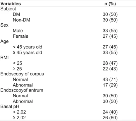
During the period of study, 60 dyspepsia patients were involved in the study. Of the 60 dyspepsia patients, there are 30 patients are diabetic patients, while the remaining 30 patients are non-diabetic patients. All of the 60 patients underwent endoscopic examination of the gastrointestinal tract and basal gastric pH. Furthermore, the 30 dyspepsia patients with DM also underwent microalbuminuria and HbA1c tests. Characteristics of 60 dyspepsia patients with DM and without DM who participated in the study are as follows:
Table 1.General characteristics of research samples
Variables n (%)
Before the data analysis, the normality test was done using Kolmogorov-Smirnov test and Shapiro-Wilk test in both study groups. Results of normality test results showed that the data are not normally distributed. Because the data was not normally distributed, then data transformation was done in order to normalize the data, but the result of data transformation is still not normally distributed. Therefore, the data analyzed using the comparative test of unpaired data with Mann-Whitney test.
Results of Mann-Whitney test found that basal gastric pH in dyspepsia patients with DM vs. dyspepsia patients without DM were: 2.30 ± 0.83 vs. 2.19 ± 0.52, p = 0.538 (p > 0.05), which means there is no VLJQL¿FDQWGLIIHUHQFHLQEDVDOJDVWULFS+LQG\VSHSVLD patients with DM compared to dyspepsia patients without DM.
%HFDXVHLWZDVIRXQGQRVLJQL¿FDQWGLIIHUHQFHIURP t-test results, Chi-square test was performed. Before the Chi-square test done, intersection point must be GHWHUPLQHG¿UVWXVLQJWKHDQDO\VLVRIUHFHLYHURSHUDWLQJ characteristic (ROC). From ROC analysis, it found that the intersection point of basal gastric was 2.02, with 70% VHQVLWLYLW\DQGVSHFL¿FLW\7KHUHIRUHIURP52& analysis, the groups divided as group of pH < 2.02 and JURXSRIS+$IWHUZDUGVWKH&KLVTXDUHWHVWZDV performed. Result of Chi-square test showed that there is VLJQL¿FDQWGLIIHUHQFHS EHWZHHQWKHG\VSHSVLD with DM group and dyspepsia without DM group. For more details, it can be seen in the table below.
are not normally distributed. Because the data was not normally distributed, then data transformation was done in order to normalize the data, but the result of data transformation is still not normally distributed. Therefore the correlation test was done using the Spearman test. Results of correlation test can be seen in the table below:
The results of this study found that basal gastric pH was 2.30 ± 0.83 for dyspepsia patients with DM and 2.19 ± 0.52 for dyspepsia patients without DM. The results obtained in this study found that gastric pH was higher in dyspepsia patients with DM than in dyspepsia patients without DM. Previous study that in accordance with this results is study from Hasler et al, who found that basal gastric pH in dyspepsia patients with DM was higher than basal gastric pH in dyspepsia patients without DM. Basal gastric pH in dyspepsia patients with DM was 3.64 ± 0.41 and basal gastric pH in dyspepsia patients without DM was 1.90 ± 0.18. In the Hasler study, it was obtained basal gastric pH higher (3.64 ± 0.41) than those in this study (2.30 ± 0.83). In Hasler study, it was turned out that all DM patients who participated in the study had experienced neuropathy complications in the form of gastric retention (diabetic gastroparesis).
Gastric acid secretion is regulated by two mechanisms: neuropathic factors and non-neuropathic factors. In several studies, prandial gastric acid secretion will decreased in 2/3 diabetic patients, which indicates a disruption of the vagus nerve response (neuropathy factor).4,15 Beside the neuropathy factors, the
non-neuropathy factors also contribute to impairment of gastric acid secretion, for example: gastric retention and parietal cells antibodies.15,16 Gastric retention is a
non-factor neuropathy associated with impaired secretion of gastric acid in diabetic patients. Although gastric retention contributed to impaired gastric secretion, the gastric retention itself was caused by autonomic neuropathy factor. A correlation study between gastric acidity with severity of gastric retention in diabetes, found increase in basal and post prandial pH that in line with severity of gastric retention.15
This study examined basal gastric acid secretion in diabetic and non-diabetic group, which found basal gastric pH was 2.30 ± 0,83 in diabetic group and 2.19 ± 0,52 in non-diabetic group. Study from Hasler et al found that basal gastric pH in diabetic group was 3.64 ± 0,41 and basal gastric pH was 1,90 ± 0,18. With cut-off RIEDVDOJDVWULFS+VHQVLWLYLW\DQGVSHFL¿FLW\ LW ZDV IRXQG VLJQL¿FDQW GLIIHUHQFH EHWZHHQ dyspepsia with diabetes group and dyspepsia without diabetes group. This result is same as result from Hasler HWDOZKLFKDOVRIRXQGWKHUHLVVLJQL¿FDQWGLIIHUHQFH
7DEOH'LIIHUHQFHRIJDVWULFS+LQG\SHSVLDZLWK'0DQGG\SHSVLDZLWKRXW'0XVLQJ&KLVTXDUHWHVW
Variable Basal gastric pH 1XPEHUSHUFHQWDJH p
Dypepsia with DM 14 (38,9%) 16 (66,7%) 30 (50%) 0,035 Dypepsia without DM 22 (61,1%) 8 (33,3%) 30 (50%)
7DEHO&RUUHODWLRQEHWZHHQLQGHSHQGHQWYDULDEOHVZLWKEDVDO
gastric pH
Variables r p Correlation
Ages (years old) 0,123 0,348 Spearman BMI (kg/m2) 0,785 0,036 Spearman
Duration of DM (years) 0,179 0,344 Spearman HbA1c (%) 0,590 0,757 Spearman Microalbuminaria (μg/mg) 0,470 0,009 Spearman
Statistical analysis of the correlation between the value of microalbuminuria and HbA1c with basal gastric pH in dypepsia patients with DM was done using Spearman test because the data distribution was not normal. There is a positive correlation between microalbuminuria with basal gastric pH with a FRUUHODWLRQFRHI¿FLHQWU DQGS:KLOHLQ HbA1c, there is no correlation with gastric pH, whereas p > 0.05, as shown in the table below.
7DEOH&RUUHODWLRQEHWZHHQPLFURDOEXPLQXULDDQG+E$FZLWK
basal gastric pH
Variable r p Correlation
Microalbuminaria (μg/mg) 0,470 0,009 Spearman HbA1c 0,590 0,757 Spearman
DISCUSSION
Secretion of gastric acid will decreased by itself in diabetic patients because of the diabetic factors itself (neuropathy factor) and also because of gastric retention, anti-parietal cell antibodies, gastric distension (non-neuropathy factor).15 Several studies
those conducted before found that acid secretion both basal and prandial will be decreased in diabetic patients compared to non-diabetic patients.4,15 In this study,
between dyspepsia with diabetes group and dyspepsia without diabetes group. The only difference is in Hasler et al study, gastroparesis diabetic complication was already happened.
Several previous studies as was done by Parkman et al and Nakamura et al found that gastric acid secretion decreased in diabetic patients who have experienced autonomic neuropathy complications. Whereas Meier et al found different results, which is there is no difference in gastric acid secretion in diabetic patients and normal subjects. On this study, the subject have not experienced diabetic autonomic neuropathy.9,14,15
In this study because of several limitations, gastric retention was not measured, but instead microalbuminuria measured. Gastric retention and microalbuminuria are late state diabetes’ complications. Mechanisms of gastric retention and microalbuminuria are similar, which caused by microangiopathy.15,17,18
In previous studies, it was found that the severity of the gastric acid secretion declining is in line with severity of gastric retention. In this study, the severity of microalbuminuria did not describe the severity of gastric retention, but only illustrate the severity of diabetes complications. This study measured microalbuminuria. Microalbuminuria is a sign of systemic endothelial dysfunction. In this study, microalbuminuria is used as a marker for diabetes patients who are already experiencing complications. )URPWKLVVWXG\LWZDVIRXQGWKDWWKHUHLVDVLJQL¿FDQW correlation between microalbuminuria and basal gastric pH. These data indicated that the impairment of acid secretion in patients with DM is correlated with the severity of diabetes complications that occur. This is consistent with Hasler et al study, which found that the gastric acid secretion in DM decreased in line with increasing severity of gastric retention.15
However, some early studies found normal gastric acid secretion in diabetic patients, as in studies conducted by Sasaki et al and Meier et al. This is possible due to diabetic patients who studied by Sasaki and Meier are the diabetic patients who have not developed any complications such as autonomic neuropathy and gastroparesis.10,11
This study also assessed the blood glucose control in the past 3 months through HbA1c, which later not found any correlation between levels of poor blood glucose control with basal gastric acid secretion impairment. It can be explained that the impairment of gastric acid secretion is associated with complications of the diabetes itself, whereas HbA1c does not describe the severity of complications of DM.
From the results of this study, we found several important things, which are: 1) The gastric pH in G\VSHSVLDSDWLHQWVZLWK'0VLJQL¿FDQWO\GLIIHUHQWZLWK dyspepsia patients without DM; 2) The management of dyspepsia patients is based on the balance theory of aggressive factors and defensive factors, therefore management of dyspepsia-DM is still to control aggressive factors and to increase defensive factors, this is due to even though there was an increasing gastric pH in diabetic group, it was not too high; 3) In the population of diabetic patients in Nagan Raya Aceh, it was found that basal gastric pH 2.02 is the limit for the occurrence of gastric secretion impairment in diabetic patients.
Some of the limitations in this study are gastric retention, which is a complication of DM that directly caused dyspepsia complaints, was not examined, and although in this study the gastric retention examination replaced by measuring microalbuminuria, because the similar pathophysiological mechanisms of gastric retention and microalbuminuria is through the same pathway. Furthermore, many factors were not evaluated in this study such as autonomic neuropathy, dyspepsia complaints, Helicobacter pylori, therefore the results of this study can not be generalized generally.
CONCLUSION
From the results of this study, the basal gastric pH on dyspepsia patients with DM was 2.30 ± 0.83, whereas in dyspepsia patients without DM was 2.19 ± 0.52. This research also found that when the basal gastric pH is 2.02, then it impairment on gastric acid secretion is already happened in dyspepsia patients with DM. Further research is needed which connects the gastric pH with gastroparesis, autonomic neuropathy, and dyspepsia complaints.
REFERENCES
1. Bytzer P, Talley NJ, Leemon M, Young LJ, Jones MP, Horowitz M. Prevalence of Gastrointestinal Symptoms Associated With Diabetes Mellitus. A Population-Based Survey of 15000 Adults. Arch Intern Med 2001;161:1989-96. 2. Mashimo H, May RJ, Goyal RK. Effect of Diabetes Mellitus
on the Digestive System. In: Joslin’s Diabetes Melitus 14th ed.
Lippincott Williams & Wilkins, USA 2006.p.332-41. 3. Bytzer P, Talley NJ, Hammer J, Young LJ, Jones MP, Horowitz
M. GI symptomps in Diabetes Mellitus are associated with both poor glycemic control and diabetic complications. Am J Gastroenterol 2002;97:604-11.
5. 6LPDGLEUDWD03DWR¿VLRORJLSHUDQKLSHUVHNUHVLDVDPGDQ pepsin pada sindroma dispepsia In: Rani Aziz A, Manan Chudahman, Djojoningrat, Simadibrata Marcellus, Makmun D, Abdullah M, Syam Fahrial A, eds. Buku Dispepsia: Sain dan Aplikasi Klinik. Jakarta: Interna Publ.2004.p.45-54. 6. Ma Jing, Rayner CK, Jones KL, Horowitz M. Diabetic
Gastroparesis Diagnosis and Management. Drugs 2009;69:971-86.
7. Park MI, Camilleri M. Clinical Reviews Gastroparesis: Clinical Update. Am J Gastroenterol 2006;101:1129-39. 8. Schubert ML, Peura DA. Review in Basic and Clinical
Gastroenterology, Control of Gastric Acid Secretion in Health and Disease. Gastroenterology 2008;134:1842-60.
9. Parkman HP, Schwartz SS. Esophagitis and gastroduodenal disorders associated with diabetic gastroparesis. Arch Intern Med 1987;147:1477-80.
10. Sasaki H, Nagulesparan M, Dubois A, Straus E, Samloff IM Lawrence WH, et al. Hypergastrinemia in obese
noninsulin-GHSHQGHQWGLDEHWHVDSRVVLEOHUHÀHFWLRQRIKLJKSUHYDOHQFHRI
vagal disfunction. J Clin Endocrinol Metab 1983;56:744-50. 11. Meler JJ, Nauck MA, Kask B, Holst JJ, Deacon CF, Schmidt
:(*DOOZLW]%,QÀXHQFHRIJDVWULFLQKLELWRU\SRO\SHSWLGHRQ
pentagastrin stimulated gastric acid secretion in patient with type 2 diabetes and healthy controls. World J gastroenterol 2006;12:1874-80.
12. Tashima K, Nishijima M, Fujita A, Kawauchi S, Takeuchi K. Gastric acid secretion in streptozotocin-diabetic rats-different responses to various secretagouges. J Physiol 2000;94:11-7. 13. Nabavizadeh RF, Vahedian J. The effect of insulin-dependent
diabetes mellitus on basal and distention-induced acid and pepsin secretion in rat. Diabetes Res Clin Pract 2004;66:1-6. 14. Nakamura T, Takebe K, Imamura K, Miyazawa T, Ishii M,
Kudoh K, et al. decreased gastric secretory functions in diabetic patients with autonomic neuropathy. Tohoku J Exp Med 1994;173:199-208.
15. Hasler WL, Coleski R, Chey WD, Koch KL, McCallum RW, Wo JM, et al. Differences in intragastric pH in diabetic vs idiopathic gastroparesis:relation to degree of gastric retention. Am J Physiol Gastrointest Liver Physiol 2008;294:1384-91. 16. Meier JJ, Nauck MA, Kask B, Holst JJ, Deacon CF, Schmidt
:(*DOOZLW]%,QÀXHQFHRIJDVWULFLQKLELWRU\SRO\SHSWLGHRQ
pentagastrin-stimulated gastric acid secretion in patients with type 2 diabetes and healthy controls. World J Gastroenterol 2006;12:1874-80.
17. Yaqoob M, Mc Clellend P, Patrick AW, Evidence of oxidend injury and tubular damage in early diabetic nephropathy. Cardiovasc Res 1997;34:55-68.