Dermatological
and Transdermal
Formulations
zyxwvutsrqponmlkjihgfedcbaZYXWVUTSRQPONMLKJIHGFEDCBA
edited
by
Kenneth A.
Walters
zyxwvutsrqponmlkjihgfedcbaZYXWVUTSRQPONMLKJIHGFEDCBA
An-eX Analytical Services Ltd.
Cardiff, Wales
zyxwvutsrqponmlkjihgfedcbaZYXWVUTSRQPONMLKJIHGFEDCBA
M A R C E L
MARCEL
DEKKER,
INC.
ISBN: 0-8247-9889-9
This book is printed on acid-free paper.
Headquarters
Marcel Dekker, Inc.
270 Madison Avenue, New York, NY 10016 tel: 212-696-9000; fax: 212-685-4540
Eastern Hemisphere Distribution
Marcel Dekker, AG
Hutgasse 4, Postfach 812, CH-4001 Basel, Switzerland tel: 41-61-261-8482; fax: 41-61-261-8896
World Wide Web http://www.dekker.com
The publisher offers discounts on this book when ordered in bulk quantities. For more information, write to Special Sales/Professional Marketing at the headquarters address above.
Copyright䉷2002 by Marcel Dekker, Inc. All Rights Reserved.
Neither this book nor any part may be reproduced or transmitted in any form or by any means, electronic or mechanical, including photocopying, microfilming, and recording, or by any infor-mation storage and retrieval system, without permission in writing from the publisher.
Current printing (last digit):
10 9 8 7 6 5 4 3 2 1
DRUGS AND THE PHARMACEUTICAL SCIENCES
Executive Editor
James Swarbrick
zyxwvutsrqponmlkjihgfedcbaZYXWVUTSRQPONMLKJIHGFEDCBA
PharmaceuTech, Inc. Pinehurst, North Carolina
Advisory Board
zyxwvutsrqponmlkjihgfedcbaZYXWVUTSRQPONMLKJIHGFEDCBA
Larry L. Augsburger University of Maryland Baltimore, Maryland Douwe D. Breimer Gorlaeus Laboratories Leiden, The Netherlands Trevor M. Jones The Association of the British Pharmaceutical Industry London, United Kingdom Hans E. Junginger LeidedAmsterdarn Center for Drug Research Leiden. The Netherlands Vincent H. L. Lee University of Southern California Los Angeles, California
David E. Nichols Purdue University West Lafayette, Indiana Stephen G. Schulman University of Florida Gainesville, Florida Jerome P. Skelly Alexandria, Virginia
Felix Theeuwes Alza Corporation Palo Alto, California
Geoffrey T. Tucker University of Sheffield Royal Hallamshire Hospital Sheffield, United Kingdom Peter G. Welling
DRUGS AND THE PHARMACEUTICAL SCIENCES
zyxwvutsrqponmlkjihgfedcbaZYXWVUTSRQPONMLKJIHGFEDCBA
A Series of Textbooks and Monographs
1. Pharmacokinetics, Milo Gibaldi and Donald Pern*er
zyxwvutsrqponmlkjihgfedcbaZYXWVUTSRQPONMLKJIHGFEDCBA
2. Good Manufacturing Practices for Pharmaceuticals: A Plan for Total Quality Control, Sidney H. Willig, Mumy M. Tuckerman, and William S. Hitchings lV
3. Microencapsulation, edited by J.
zyxwvutsrqponmlkjihgfedcbaZYXWVUTSRQPONMLKJIHGFEDCBA
R.
Nixon4. Drug Metabolism: Chemical and Biochemical Aspects, Bernard Testa and Peter Jenner
5. New Drugs: Discovery and Development, edited by Alan A. Rubin
6. Sustained and Controlled Release Drug Delivery Systems, edited by Joseph
R.
Robinson
7. Modem Pharmaceutics, edited by Gilbert S. Banker and Christopher T. Rhodes
8. Prescription Drugs in Short Supply: Case Histories, Michael A. Schwartz
9. Activated Charcoal: Antidotal and Other Medical Uses, David 0. Cooney
10. Concepts in Drug Metabolism (in two parts), edited by Peter Jenner and Bernard
Testa
11. Pharmaceutical Analysis: Modern Methods (in two parts), edited by James W. Munson
12. Techniques of Solubilization of Drugs, edited by Samuel H. Yalkowsky
13. Orphan Drugs, edited by Fred €. Karch
14. Novel Drug Delivery Systems: Fundamentals, Developmental Concepts, Biomedical Assessments, Yie W. Chien
15. Pharmacokinetics: Second Edition, Revised and Expanded, Milo Gibaldi and Donald Penier
16. Good Manufacturing Practices for Pharmaceuticals: A Plan for Total Quality Control, Second Edition, Revised and Expanded, Sidney H. Willig, Murray M. Tuckerman, and William S. Hitchings IV
17. Formulation of Veterinary Dosage Forms, edited by Jack Blodinger
18. Dermatological Formulations: Percutaneous Absorption, Brian W. Barry
19. The Clinical Research Process in the Pharmaceutical Industry, edited by Gary M. Matoren
20. Microencapsulation and Related Drug Processes, Patrick B. Deasy
21. Drugs and Nutrients: The Interactive Effects, edited by Daphne A. Roe and T. Colin Campbell
22. Biotechnology of Industrial Antibiotics, €rick J. Vandamme
23. Pharmaceutical Process Validation, edited by Bernard T. Loflus and Robert A. Nash
24. Anticancer and Interferon Agents: Synthesis and Properties, edited by Raphael M. Offenbrite and George B. Butler
25. Pharmaceutical Statistics: Practical and Clinical Applications, Sanford Bolton
26. Drug Dynamics for Analytical, Clinical, and Biological Chemists, Benjamin J. Gudzinowicz, Burrows T. Younkin, Jr., and Michael J. Gudzinowicz
27. Modern Analysis of Antibiotics, edited by Adjoran Aszalos
28. Solubility and Related Properties, Kenneth C. James
29. Controlled Drug Delivery: Fundamentals and Applications, Second Edition, Revised and Expanded, edited by Joseph R. Robinson and Vincent Lee
31. Transdermal Controlled Systemic Medications, edited by Yie W. Chien
zyxwvutsrqponmlkjihgfedcbaZYXWVUTSRQPONMLKJIHGFEDCBA
32. Drug Delivery Devices: Fundamentals and Applications, edited by Praveen Tyle 33. Pharmacokinetics: Regulatory Industrial Academic Perspectives, edited by
Peter G. Welling and Francis L. S. Tse
34. Clinical Drug Trials and Tribulations, edited by Allen E. Cato
35. Transdermal Drug Delivery: Developmental Issues and Research Initiatives,
edited by Jonathan Hadgrat? and Richard H. Guy
36. Aqueous Polymeric Coatings for Pharmaceutical Dosage Forms, edited by
James W. McGinity
37. Pharmaceutical Pelletization Technology, edited by lsaac Ghebre-Sellassie 38. Good Laboratory Practice Regulations, edited by Allen F. Hirsch
39. Nasal Systemic Drug Delivery, We W. Chien, Kenneth S. E. Su, and Shyi-Feu Chang
40. Modern Pharmaceutics: Second Edition, Revised and Expanded, edited by
Gilbert S. Banker and Christopher T. Rhodes
41
.
Specialized Drug Delivery Systems: Manufacturing and Production Technology,edited by Praveen Tyle
42. Topical Drug Delivery Formulations, edited by David W. Osborne and Anton H. Amann
43. Drug Stability: Principles and Practices, Jens T. Carstensen
44. Pharmaceutical Statistics: Practical and Clinical Applications, Second Edition, Revised and Expanded, Sanford Bolton
45. Biodegradable Polymers as Drug Delivery Systems, edited by Mark Chasin and
Robert Langer
46. Preclinical Drug Disposition: A Laboratory Handbook, Francis L. S. Tse and
James J. Jaffe
47. HPLC in the Pharmaceutical Industry, edited by Godwin W. Fong and Stanley K.
zyxwvutsrqponmlkjihgfedcbaZYXWVUTSRQPONMLKJIHGFEDCBA
Lam
48. Pharmaceutical Bioequivalence, edited by Peter G. Welling, Francis L. S. Tse, and Shrikant V. Dinghe
49. Pharmaceutical Dissolution Testing, Umesh V. Banakar
50. Novel Drug Delivery Systems: Second Edition, Revised and Expanded, Yie W. Chien
51. Managing the Clinical Drug Development Process, David M. Cocchefto and
Ronald V. Nardi
52. Good Manufacturing Practices for Pharmaceuticals: A Plan for Total Quality Control, Third Edition, edited by Sidney H. Willig and James R. Stoker
53. Prodrugs: Topical and Ocular Drug Delivery, edited by Kenneth B. Sloan
54. Pharmaceutical Inhalation Aerosol Technology, edited by Anthony J. Hickey
55. Radiopharmaceuticals: Chemistry and Pharmacology, edited by Adrian D. Nunn
56. New Drug Approval Process: Second Edition, Revised and Expanded, edited by
Richard A. Guarino
57. Pharmaceutical Process Validation: Second Edition, Revised and Expanded,
edited by Ira R. Berry and Robert A. Nash
58. Ophthalmic Drug Delivery Systems, edited by Ashim K. Mitra
59. Pharmaceutical Skin Penetration Enhancement, edited by Kenneth A. Walters
and Jonathan Hadgraft
60. Colonic Drug Absorption and Metabolism, edited by Peter R. Bieck
61 . Pharmaceutical Particulate Carriers: Therapeutic Applications, edited by Alain
Rolla n d
62. Drug Permeation Enhancement: Theory and Applications, edited by Dean S. Hsieh
63. Glycopeptide Antibiotics, edited by Ramakrishnan Nagarajan
66. Colloidal Drug Delivery Systems,
zyxwvutsrqponmlkjihgfedcbaZYXWVUTSRQPONMLKJIHGFEDCBA
edifed by Jorg Kreufer67. Pharmacokinetics: Regulatory Industrial Academic Perspectives, Second Edition, edifed by Peter G. Welling and Francis L. S. Tse
68. Drug Stability: Principles and Practices, Second Edition, Revised and Expanded,
Jens
zyxwvutsrqponmlkjihgfedcbaZYXWVUTSRQPONMLKJIHGFEDCBA
T. Carsfensen69. Good Laboratory Practice Regulations: Second Edition, Revised and Expanded,
edited by Sandy Weinberg
70. Physical Characterization of Pharmaceutical Solids, edifed by Harry G. Briffain 71. Pharmaceutical Powder Compaction Technology, edited by Goran Alderbom
and Chrisfer Nysfrom
72. Modern Pharrnaceutics: Third Edition, Revised and Expanded, edited by Gilbert
S. Banker and Christopher T. Rhodes
73. Microencapsulation: Methods and Industrial Applications, edited by Simon
Benifa
74. Oral Mucosal Drug Delivery, edited by Michael J. Rafhbone
75. Clinical Research in Pharmaceutical Development, edited by Barry Bleidt and
Michael Monfagne
76. The Drug Development Process: Increasing Efficiency and Cost-Effectiveness,
edited by Peter G. Welling, Louis Lasagna, and Umesh V. Banakar
77. Microparticulate Systems for the Delivery of Proteins and Vaccines, edited by
Smadar Cohen and Howard Bemsfein
78. Good Manufacturing Practices for Pharmaceuticals: A Plan for Total Quality Control, Fourth Edition, Revised and Expanded, Sidney H. Willig and James R.
Stoker
79. Aqueous Polymeric Coatings for Pharmaceutical Dosage Forms: Second Edition, Revised and Expanded, edited by James W. McGinify
80. Pharmaceutical Statistics: Practical and Clinical Applications, Third Edition,
Sanford Bolfon
81. Handbook of Pharmaceutical Granulation Technology, edited by Dilip M. Parikh 82. Biotechnology of Antibiotics: Second Edition, Revised and Expanded, edited by
William R. Sfrohl
83. Mechanisms of Transdermal Drug Delivery, edifed by Russell 0. Poffs and
Richard H. Guy
84. Pharmaceutical Enzymes, edifed by Albert Lauwers and Simon Scharpe
85. Development of Biopharmaceutical Parenteral Dosage Forms, edifed by John A.
Bonfempo
86. Pharmaceutical Project Management, edited by Tony Kennedy
87. Drug Products for Clinical Trials: An International Guide to Formulation 0
Production 0 Quality Control, edifed by Donald C. Monkhouse and Christopher T.
Rhodes
88. Development and Formulation of Veterinary Dosage Forms: Second Edition, Revised and Expanded, edifed by Gregory E. Hardee and J. Desmond Baggof
89. Receptor-Based Drug Design, edifed by Paul Let7
90. Automation and Validation of Information in Pharmaceutical Processing, edited
by Joseph F. deSpautz
91. Dermal Absorption and Toxicity Assessment, edited by Michael S. Roberts and
Kenneth A. Walfers
92. Pharmaceutical Experimental Design, Garefh A. Lewis, Didier Mafhieu, and
Roger Phan-Tan-Luu
93. Preparing for FDA Pre-Approval Inspections, edited by Martin D. Hynes lllv
94. Pharmaceutical Excipients: Characterization by IR, Raman, and NMR Spectroscopy, David E. Bugay and W. Paul Findlay
95. Polymorphism in Pharmaceutical Solids, edifed by Harry G. Briffain
96. Freeze-Drying/Lyophilization of Pharmaceutical and Biological Products, edifed
97. 98. 99. 100. 101. 102. 103. 104. 105. 106. 107. 108. 109. 110. 111. 112. 113. 114. 115. 116. 117. 118. 119. 120.
Percutaneous Absorption: Drugs-Cosmetics-Mechanisms-Methodology,
Third Edition, Revised and Expanded,
zyxwvutsrqponmlkjihgfedcbaZYXWVUTSRQPONMLKJIHGFEDCBA
edited by Robert L. Bronaugh and Howard 1. MaibachBioadhesive Drug Delivery Systems: Fundamentals, Novel Approaches, and Development, edited by Edith Mathiowitz, Donald E. Chickering 111, and Claus- Michael Lehr
Protein Formulation and Delivery, edited by Eugene J. McNally
New Drug Approval Process: Third Edition: The Global Challenge, edited by
Richard A. Guarino
Peptide and Protein Drug Analysis, edited by Ronald E. Reid
Transport Processes in Pharmaceutical Systems, edited by Gordon Amidon,
Ping 1. Lee, and Elizabeth M. Topp
Excipient Toxicity and Safety, edited by Myra L. Weiner and Lois A. Kotkoskie The Clinical Audit in Pharmaceutical Development, edited by Michael R.
Hamrell
Pharmaceutical Emulsions and Suspensions, edited by Francoise Nielloud
and Gilberfe Marfi-Mestres
Oral Drug Absorption: Prediction and Assessment, edited by Jennifer B.
Dressman and Hans Lennernas
Drug Stability: Principles and Practices, Third Edition, Revised and Expanded,
edited by Jens T. Carstensen and C. T. Rhodes
Containment in the Pharmaceutical Industry, edited by James Wood
Good Manufacturing Practices for Pharmaceuticals: Fifth Edition, Revised and Expanded, Sidney H. Willig
Advanced Pharmaceutical Solids, Jens T. Carstensen
Endotoxins: Pyrogens, LAL Testing, and Depyrogenation, Second Edition, Revised and Expanded, Kevin L. Williams
Pharmaceutical Process Engineering, Anthony J. Hickey and David Gander-
ton
Pharmacogenics, edited by Werner Kalow, Urs
zyxwvutsrqponmlkjihgfedcbaZYXWVUTSRQPONMLKJIHGFEDCBA
A. Meyer, and Rachel F. TyndaleHandbook of Drug Screening, edited by Rarnakrishna Seefhala and Prab-
havathi B. Fernandes
Drug Targeting Technology: Physical Chemical Biological Methods, edited
by Hans Schreier
Drug-Drug Interactions, edited by A. David Rodrigues
Handbook of Pharmaceutical Analysis, edited by Lena Ohannesian and
Anthony J. Streeter
Pharmaceutical Process Scale-Up, edited by Michael Levin
Dermatological and Transdermal Formulations, edited by Kenneth A. Walters Clinical Drug Trials and Tribulations: Second Edition, Revised and Expanded,
edited by Allen Cato, Lynda Suffon, and Allen Cato 111
zyxwvutsrqponmlkjihgfedcbaZYXWVUTSRQPONMLKJIHGFEDCBA
ADDITIONAL VOLUMES IN PREPARATION
Modern Pharmaceutics: Fourth Edition, Revised and Expanded, edited by
Gilberf S. Banker and Christopher T. Rhodes
Preface
The past two decades have witnessed brilliant discoveries regarding the structure and func-tions of the stratum corneum.
—Albert Kligman, 2000
An immense amount of research has been carried out over the past two decades on the micromorphology of the skin, in particular of the stratum corneum, and the important role that this organ plays in the maintenance of human life. It has also been nearly two decades since the publication of Brian Barry’s bookDermatological
Formulations — Percutaneous Absorption. This book remains one of the most widely
and frequently cited references in the field of skin transport and also has been used extensively as an introduction to the complexities surrounding the theory and de-velopment of topical pharmaceutical products.
The introduction and subsequent success of transdermal therapeutic systems have advanced our understanding of the structure of the skin and the mechanisms of transport through the barrier membrane. In addition, technological developments in molecular biology and pharmacology have led to an increased understanding of the biochemistry of skin diseases. The result is the introduction of new therapeutic strategies that use both existing and new chemical entities to treat skin diseases. This volume serves as a useful addition to the literature in the dermatopharmaceutics field. The rational treatment of skin diseases, based on the biochemical mechanisms underlying the pathology, is discussed inChapter 2. For example, vitamin D3
Preface
that some compounds of this type possess a high binding affinity to specific cellular receptors and are potent regulators of cell differentiation and inhibitors of cell pro-liferation in human keratinocytes. Cosmetic scientists have long known the epidermal advantages of another vitamin, retinol (vitamin A). Deficiency of this vitamin has been implicated in squamous metaplasia and keratinization of epithelial tissue, and several derivatives have been synthesized and evaluated for their effects in such diseases as acne, psoriasis, and hyperkeratosis. The results have been somewhat variable; however, the recent identification of several receptor proteins for retinoic acid should lead to the development of more potent analogs with fewer side effects. The ability to enhance skin penetration and permeation has been the subject of considerable research over the past two decades and is reviewed inChapter 6.The science of penetration enhancement has expanded considerably over the past few years, and it is now possible to increase drug delivery across the skin using both chemical and physical means. Various synthetic (e.g., SEPA威and Azone威) and nat-ural (e.g., terpenes) compounds have proved useful in this respect. Moreover, there is evidence that the skin penetration of large molecules such as insulin can be in-creased using physical methods of enhancement, such as iontophoresis.
The use of the skin as a drug delivery route for both topical and systemic therapy is covered inChapter 7. Transdermal drug delivery using patches or semi-solid formulations is now a reality with products available for travel sickness, hy-pertension, angina, postmenopausal symptoms, male hypogonadism, pain, inflam-mation, and smoking cessation. Problems of irritation are being overcome with the development of skin-compatible materials, such as some of the newer pressure-sen-sitive adhesives. The success of such systems has been achieved only by means of a greater understanding of the physical and biochemical nature of the permeation routes through the skin, especially in relation to the intercellular lipid lamellae of the stratum corneum (as discussed inChapters 1, 3,and4).In addition, the methods of studying percutaneous absorption, both in vivo and in vitro, have become more standardized thanks to the efforts of the American Association of Pharmaceutical Scientists (AAPS), the U.S. Food and Drug Administration (FDA), and other indus-trial and regulatory bodies.Chapter 5provides a complete description of the AAPS/ FDA guidelines for such experimentation, together with a full evaluation of the in vivo tape-stripping procedure. This chapter also gives a description of the use of cultured skin membranes for the study of irritation and other toxic responses to materials applied to the skin.
Preface
9. Chapter 8 covers bioequivalence of dermal and transdermal systems. Safety con-siderations are outlined inChapters 10and11.
This book will be useful to pharmacy students and practitioners and cosmetic and veterinary scientists. It may also prove useful to toxicologists working in the field of risk assessment and dermatologists requiring a deeper understanding of the mechanisms of drug transport through the skin. The chapters have been authored by international experts in their fields and provide a comprehensive review of current dermatopharmaceutics.
I acknowledge, and am extremely grateful for, the hard work and infinite pa-tience shown by the contributors to this volume. I would also like to acknowledge the unreserved help provided by my colleague Dr. Keith Brain. Finally, since the written word cannot fully express my love and gratitude to Peggy for her support and encouragement during many hours of word processing and reference hunting, this absentee husband will find other ways.
Contents
Preface Contributors
1. The Structure and Function of Skin
Kenneth A. Walters and Michael S. Roberts
2. Common Skin Disorders and Their Topical Treatment
C. Colin Long
3. Basic Mathematical Principles in Skin Permeation
Adam C. Watkinson and Keith R. Brain
4. Skin Transport
Michael S. Roberts, Sheree Elizabeth Cross, and Mark A. Pellett
5. Methods for Studying Percutaneous Absorption
Keith R. Brain, Kenneth A. Walters, and Adam C. Watkinson
6. Formulation Strategies for Modulating Skin Permeation
7. Dermatological Formulation and Transdermal Systems
Kenneth A. Walters and Keith R. Brain
8. Bioavailability and Bioequivalence of Dermatological Formulations
Christian Surber and Adrian F. Davis
9. Scale-up of Dermatological Dosage Forms: A Case for Multivariate Optimization and Product Homogeneity
Orest Olejnik and Bruce A. Firestone
10. Safety Considerations for Dermal and Transdermal Formulations
Peter J. Dykes and Anthony D. Pearse
11. Transdermal Delivery and Cutaneous Reactions
Contributors
Keith R. Brain, Ph.D. An-eX Analytical Services Ltd., Cardiff, Wales
Sheree Elizabeth Cross, Ph.D. Department of Medicine, University of Queens-land, Princess Alexandra Hospital, Brisbane, QueensQueens-land, Australia
Adrian F. Davis, Ph.D. GlaxoSmithKline Consumer Healthcare, Weybridge, Kent, England
Peter J. Dykes, B.Sc., Ph.D. Cutest (Skin Toxicity Testing Company), Cardiff, Wales
Bruce A. Firestone, Ph.D. Allergan, Inc., Irvine, California
Robert J. Gyurik MacroChem Corporation, Lexington, Massachusetts
Jonathan Hadgraft, D.Sc., F.R.S.C. Medway Sciences, University of Greenwich, Chatham Maritime, Kent, England
C. Colin Long, F.R.C.P. Department of Dermatology, Cardiff and Vale NHS Trust, Cardiff, Wales
Orest Olejnik, Ph.D., M.R.Pharm.S. Allergan, Inc., Irvine, California
Anthony D. Pearse, M.Sc., M.I.Biol., C.Biol., F.I.Sc.T. Department of Dermatol-ogy, University of Wales College of Medicine, and Cutest (Skin Toxicity Testing Company), Cardiff, Wales
Mark A. Pellett, Ph.D., M.R.Pharm.S. Whitehall International, Havant, England
Michael S. Roberts, Ph.D., D.Sc. Department of Medicine, University of Queens-land, Princess Alexandra Hospital, Brisbane, QueensQueens-land, Australia
Jagdish Singh Department of Pharmaceutical Sciences, College of Pharmacy, North Dakota State University, Fargo, North Dakota
Christian Surber, Ph.D., Priv.-Doz. Dr. Institute of Hospital-Pharmacy, University Clinics, Kantonsspital Basel, Basel, Switzerland
Kenneth A. Walters, F.I.Biol., Ph.D. An-eX Analytical Services Ltd., Cardiff, Wales
Adam C. Watkinson, Ph.D., M.B.A.* An-eX Analytical Services Ltd., Cardiff, Wales
1
The Structure and Function of Skin
KENNETH A. WALTERS
An-eX Analytical Services Ltd., Cardiff, Wales
MICHAEL S. ROBERTS
University of Queensland, Princess Alexandra Hospital, Brisbane, Queensland, Australia
I. INTRODUCTION
Walters Roberts
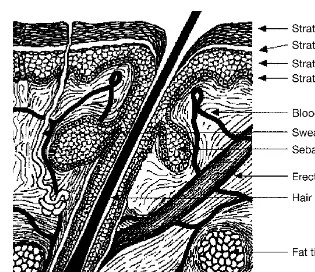
Figure 1 Components of the epidermis and dermis of human skin.
>
Figure 2 (A) Structure of the skin and processes of percutaneous absorption and trans-dermal delivery. Absorption can occur through sweat ducts (1), intercellular regions of the stratum corneum (2), and through the hair follicles (3). (B) Dermal absorption, sites of action and toxicity.
each of these functions, the skin must be tough, robust, and flexible, with effective communication between each of its intrinsic components.
Walters Roberts
was thought to (a) avoid the problems of stomach emptying, pH effects, and enzyme deactivation associated with gastrointestinal passage; (b) to avoid hepatic first-pass metabolism; and (c) to enable control of input, as exemplified by termination of delivery through removal of the device. In practice, as discussed later in this book, delivery of solutes through the skin is associated with various difficulties, such as (a) the variability in percutaneous absorption owing to site, disease, age, and species differences; (b) the skin’s ‘‘first-pass’’ metabolic effect; (c) the reservoir capacity of the skin; (d) irritation and other toxicity caused by topical products; (e) heterogeneity and inducibility of the skin in both turnover and metabolism; (f) inadequate definition of bioequivalence criteria; and (g) an incomplete understanding of the technologies that may be used to facilitate or retard percutaneous absorption. However, the con-trolled delivery of solutes through the skin continues to be of interest, with the further development of technologies, such as chemical penetration enhancement, sonopho-resis, transferosomes, and electroporation. The extent to which these are translated into practice will be defined by time.
II. GROSS STRUCTURE AND FUNCTION OF THE SKIN
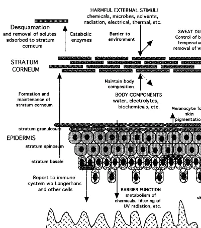
WhereasFigure 1 provides an overview of the gross structure of the skin,Figure 3 represents the skin components in terms of the various functions they perform. It needs to be emphasized that the protection, homeostatic, and sensing functions of the skin are both overlapping and integrated. For instance, barrier properties to a chemical entity involves resistance to its entry (barrier provided by stratum corneum), metabolism for that proportion of entity bypassing the stratum corneum (in viable epidermis), sensing of and attention to damage caused by entry (inflammatory me-diator release in epidermis, with involvement of dermis), and removal of entity from site by dermal blood supply and distribution into those body organs specifically responsible for elimination of the entity by metabolism (liver) and excretion (kidney). Heat regulation occurs through the use of the subcutaneous fat pad, physiological regulation of blood flow to effect, for instance, heat loss by vasodilation, and cooling by perspiration. We now consider the structure and functions provided by each skin component in some detail.
A. The Epidermis
The epidermis performs a number of functions, as shown in Figure 3, one of the most important being the generation of the stratum corneum, as described later. The stratum corneum is the heterogeneous outermost layer of the epidermis and is ap-proximately 10–20m thick. It is nonviable epidermis and consists, in a given cross-section, of 15–25 flattened, stacked, hexagonal, and cornified cells embedded in a mortar of intercellular lipid. Each cell is approximately 40m in diameter and 0.5
m thick. The thickness varies, however, and may be a magnitude of order larger in areas such as the palms of the hand and soles of the feet, areas of the body associated with frequent direct and substantial physical interaction with the physical environment. Not surprisingly, the absorption of solutes, such as methyl salicylate, is slower through these regions than through the skin of other parts of the body. The stratum corneum barrier properties may be partly related to its very high density (1.4 g/cm3
Function of Skin
Figure 3 Skin components and their function.
Walters Roberts
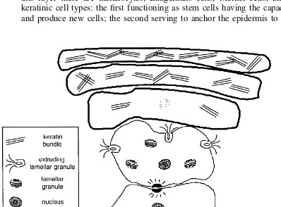
Figure 4 Epidermal differentiation: major events include extrusion of lamellar bodies, loss of nucleus, and increasing amount of keratin in the stratum corneum. The diagram is not to scale and only a few cells are shown for clarity.
with a total turnover of the stratum corneum occurring once every 2–3 weeks. Ac-cordingly, very lipophilic agents, such as sunscreens and substances binding to the horny layer (e.g., hexachlorophane), may be less well absorbed into the body than would be indicated by the initial partitioning of the agents into the horny layer from an applied vehicle. The stratum corneum also functions as a barrier to prevent the loss of internal body components, particularly water, to the external environment. It is estimated that the efficiency of this barrier is such that water loss from ‘‘insensible perspiration’’ is restricted to 0.5L/cm2
/h⫺1
, or 250 mL of water per day for a normal adult. Disorders of epithelization, such as psoriasis, lead to a faster skin turnover, sometimes being reduced to 2–4 days, with improper stratum corneum barrier func-tion formafunc-tion.
Function of Skin
membrane (2). The basement membrane is 50–70 nm thick and consists of two layers—the lamina densa and lamina lucida—which comprise mainly proteins, such as type IV collagen, laminin, nidogen, and fibronectin. Type IV collagen is respon-sible for the mechanical stability of the basement membrane, whereas laminin and fibronectin are involved with the attachment between the basement membrane and the basal keratinocytes.
The cells of the basal lamina are attached to the basement membrane by hemi-desmosomes, which are found on the ventral surface of basal keratinocytes (3). Hemidesmosomes appear to comprise three distinct protein groups: two of which are bullous pemphigoid antigens (BPAG1 and BPAG2), and the other epithelial cell-specific integrins (4–6). BPAG1 is associated with the organization of the cytoskel-etal structure and forms a link between the hemidesmosome structure and the keratin intermediate filaments. The integrins are transmembrane receptors that mediate at-tachment between the cell and the extracellular matrix. Human epidermal basal cells contain integrins ␣21, ␣31, and ␣64. Integrin␣64 and BPAG2 appear to be the
major hemidesmosomal protein contributors to the anchoring of the keratinocyte, spanning from the keratin intermediate filament, through the lamina lucida, to the lamina densa of the basement membrane (7). In the lamina densa, these membrane-spanning proteins interact with the protein laminin-5 which, in turn, is linked to collagen VII, the major constituent of the anchoring fibrils within the dermal matrix. It has also been suggested that both BPAG2 and integrin␣64mediate in the signal
transductions required for hemidesmosome formation (8) and cell differentiation and proliferation. Integrin␣31is associated with actin and may be linked with
laminin-5. Epidermal wounding results in an up-regulation of these proteins that appears to be involved with cell motility and spreading. The importance of maintaining a secure link between the basal lamina cells and the basement membrane is obvious, and the absence of this connection results in chronic blistering diseases such as pemphigus and epidermolysis bullosa.
In addition to hemidesmosome cell–matrix binding, another site for adhesion of the cells of the epidermal basal layer and the basal membrane is the adherens junction (9). The adherens junction expresses a protein profile different from des-mosomes and hemidesdes-mosomes (10,11) and contains talin, vinculin, and cadherins, and with the possible participation of type XIII collagen (12). Whereas the desmo-somes and hemidesmodesmo-somes are linked to cytoplasmic keratin, the adherens junctions are linked to cytoplasmic actin microfilaments.
Walters Roberts
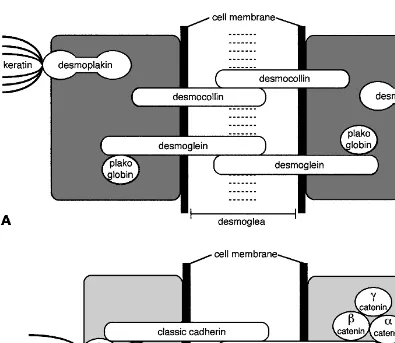
Figure 5 Epidermal cell cohesion and communication is provided by desmosomes and adherens junctions. Different functions are attributed to these junctions. See text for details.
The importance of the calcium ion in cell regulation and intercellular com-munication has been known for some time (17). It is not surprising, therefore, that the formation of desmosomes and hemidesmosomes appears to be induced by Ca2⫹ and mediated by protein kinase C (PKC) activation (18). The presence of Ca2⫹ activates the metabolism of inositol phospholipids, resulting in the generation of diacylglycerol and inositol-1,4,5-triphosphate. The diacyglycerol subsequently acti-vates protein kinase C, which plays an important role in keratinocyte differentiation (19), and the inositol-1,4,5-triphosphate generates further Ca2⫹ influx into the cyto-plasm. The epidermal distribution and function of Ca2⫹, and several other physio-logical elements, has been recently reviewed (20). As discussed later, Ca2⫹also plays a role in proteolysis and desquamation.
Function of Skin
main function appears to be to pick up contact allergens in the skin and present these agents to T lymphocytes in the skin-draining lymph nodes; thus, they play an im-portant role in contact sensitization. Cell surface moieties on the Langerhans cells are modified and the cells increase in size following topical application of hapten. The activated cells migrate from the epidermis to the dermis and from there to the regional lymph nodes where they sensitize T cells. The ability of Langerhans cells to migrate from bone marrow, localize in a specific region of the epidermis, and further migrate when activated, suggests that there is some mechanism for accu-mulation in the epidermis, adhesion to keratinocytes and the basement membrane, and for disruption of the adhesive bond. Migration into the epidermis may be me-diated by granulocyte–macrophage colony-stimulating factor (GM-CSF), tumor ne-crosis factor-␣ (TNF-␣), interleukin-6 (IL-6), transforming growth factor-
(TGF-), chemotactic cytokines, such as monocyte chemotactic protein (MCP), and cutaneous lymphocyte-associated antigen (CLAA) (24). The adhesive bonds within the epidermis appear to be formed by interaction of Langerhans cells with extracel-lular matrix proteins, such as fibronectin and laminin, through 1-integrins (25).
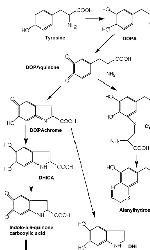
Detachment of Langerhans cells from keratinocytes and the basement membrane following skin sensitization may be mediated by epidermal cytokines, including GM-CSF and TNF-␣ (26), whereas cell maturation, which occurs during transit to the local lymph nodes, may be mediated by GM-CSF. Recently, it was shown that IL-1and TNF-␣acted directly on Langerhans cells to reduce adhesion to keratinocytes and the basement membrane by down-regulating the binding protein E-cadherin (27). Melanocytes are a further functional cell type of the epidermal basal layer and are also present in hair and eyes. The main function of these cells is to produce melanins, high molecular weight polymers of indole quinone, which affect pigmen-tation of the skin, hair, and eyes (28,29). Melanin is produced in the melanosomes: membrane-bound organelles that are transferred to keratinocytes, probably by a pro-cess involving phagocytosis (30), to provide a uniform distribution of pigmentation. Intracellular movement of melanosomes is possibly mediated by actin and microtu-bules (31). Visible pigmentation is dependent not only on the number, shape, and size of the melanosomes, but also on the chemical nature of the melanin. Hair color is governed by melanocytes which reside in the hair bulbs within the dermis (32). Melanosomes are transferred to the growing hair shaft. The major function of skin pigmentation is to provide protection against harmful environmental effects, such as ultraviolet (UV) radiation, especially in the proliferating basal layers where the mu-tagenic effects of this type of insult have particularly serious implications. Melano-cytes remain attached to the basal layer and are thought to exist in a nonproliferative state when in contact with undifferentiated keratinocytes. Recent studies have indi-cated, however, that melanocytes can proliferate if they are separated from the basal layer and surrounded by differentiated keratinocytes (33).
-melanocyte-Walters Roberts
Function of Skin
stimulating hormone (␣-MSH) and agouti signal protein are responsible for govern-ing the type of melanogenesis pathway followed and that, in conditions in which
␣-MSH dominates, eumelanins are produced. UV radiation appears to increase pro-duction of the precursor hormone proopiomelanocortin, which increases␣-MSH pro-duction, resulting in increased levels of eumelanins (35,36).
The final type of cell found in the basal layer of the stratum corneum is the Merkel cell. These cells can be distinguished from the keratinocytes by their clear cytoplasm and lack of tonofilaments. The cells are closely associated with nerve endings, present on the other side of the basement membrane, which suggests they function as sensory receptors of the nervous system. Although histochemical evi-dence demonstrating the presence of acetylcholinesterase suggests a sensory role for Merkel cells, there has been no direct evidence for the release of neurotransmitters. Indeed, acetylcholinesterases have been found in keratinocytes (37). Despite this lack of confirmation, most researchers in the field agree that Merkel cells play a role (a) in the mechanosensory system; (b) in trophic action on peripheral nerve fibers; (c) in stimulating and maintaining proliferation and keratinocytes; and (d) in release of bioactive substances to subepidermal structures (38,39).
B. The Dermis
The dermis, a critical component of the body, not only provides the nutrative, im-mune, and other support systems for the epidermis, through a thin papillary layer adjacent to the epidermis, but also plays a role in temperature, pressure, and pain regulation. The main structural component of the dermis is referred to as a coarse reticular layer. The dermis is about 0.1–0.5 cm thick and consists of collagenous fibers (70%), providing a scaffold of support and cushioning, and elastic connective tissue, providing elasticity, in a semigel matrix of mucopolysaccharides. In general, the dermis has a sparse cell population. The main cells present are the fibroblasts, which produce the connective tissue components of collagen, laminin, fibronectin, and vitronectin; mast cells, which are involved in the immune and inflammatory responses; and melanocytes involved in the production of the pigment melanin.
Contained within the dermis is an extensive vascular network providing for the skin nutrition, repair, and immune responses and, for the rest of the body, heat exchange, immune response, and thermal regulation. The blood flow rate to the skin is about 0.05 mL min⫺1
cc⫺3
Walters Roberts
The lymphatic system is an important component of the skin in regulating its interstitial pressure, mobilization of defense mechanisms, and in waste removal. It exists as a dense, flat meshwork in the papillary layers of the dermis and extends into the deeper regions of the dermis. Cross and Roberts (40) have shown that whereas blood flow determines the clearance of small solutes, such as water and lidocaine, lymphatic flow is an important determinant in the dermal removal of larger solutes, such as interferon. Also present in the dermis are a number of different types of nerve fibers supplying the skin, including those for pressure, pain, and temperature.
C. The Subcutis
The deepest layer of the skin is the subcutaneous tissue or hypodermis. The hypo-dermis acts as a heat insulator, a shock absorber, and an energy storage region. This layer is a network of fat cells arranged in lobules and linked to the dermis by interconnecting collagen and elastin fibers. As well as fat cells (possibly 50% of the body’s fat), the other main cells in the hypodermis are fibroblasts and macrophages. One of the major roles of the hypodermis is to carry the vascular and neural systems for the skin. It also anchors the skin to underlying muscle. Fibroblasts and adipocytes can be stimulated by the accumulation of interstitial and lymphatic fluid within the skin and subcutaneous tissue (41).
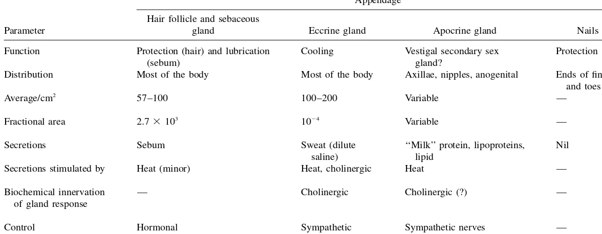
D. Skin Appendages
There are four skin appendages: the hair follicles with their associated sebaceous glands, eccrine sweat glands, apocrine sweat glands, and the nails. Each appendage has a different function as outlined inTable 1.The hair follicles are distributed across the entire skin surface with the exception of the soles of the feet, the palms of the hand and the lips. A smooth muscle, the erector pilorum, attaches the follicle to the dermal tissue and enables hair to stand up in response to fear. Each follicle is as-sociated with a sebaceous gland that varies in size from 200 to 2000m in diameter. The sebum secreted by this gland (Table 2), consisting of triglycerides, free fatty acids, and waxes, protects and lubricates the skin as well as maintaining a pH of about 5. The fractional area for these is slightly more than 1/1000 of the total skin surface (see Table 1). Also described in Table 1 are the eccrine or sweat glands and apocrine glands, accounting for about two-thirds and one-third of all glands, respec-tively. The eccrine glands are epidermal structures that are simple, coiled tubes aris-ing from a coiled ball, of approximately 100m in diameter, located in the lower dermis. It secretes a dilute salt solution with a pH of about 5, this secretion being stimulated by temperature-controlling determinants, such as exercise and high en-vironmental temperature, as well as emotional stress through the autonomic (sym-pathetic) nervous system (see Table 1). These glands have a total surface area of about 1/10,000 of the total body surface. The apocrine glands are limited to specific body regions and are also coiled tubes. These glands are about ten times the size of the eccrine ducts, extend as low as the subcutaneous tissues and are paired with hair follicles.
F
u
nction
o
f
Ski
n
Table 1 Appendages Associated with the Skin
Parameter
Appendage
Hair follicle and sebaceous
gland Eccrine gland Apocrine gland Nails
Function Protection (hair) and lubrication
(sebum)
Cooling Vestigal secondary sex
gland?
Protection
Distribution Most of the body Most of the body Axillae, nipples, anogenital Ends of fingers
and toes Average/cm2
57–100 100–200 Variable —
Fractional area 2.7⫻103
10⫺4
Variable —
Secretions Sebum Sweat (dilute
saline)
‘‘Milk’’ protein, lipoproteins, lipid
Nil
Secretions stimulated by Heat (minor) Heat, cholinergic Heat —
Biochemical innervation of gland response
— Cholinergic Cholinergic (?) —
Control Hormonal Sympathetic
nerves
Sympathetic nerves —
Walters Roberts
Table 2 Lipid Composition of Human Sebum
Lipid Lumen of glanda
Skin surfacea
Squalene Wax esters Cholesteryl esters Triglycerides Fatty acids Cholesterol 15 25 2 57 0 1 15 25 2 42 15b 1 a
Expressed as weight%.
b
Most abundant fatty acid in human sebum is C16:1. Source: Courtesy of P. Wertz.
mass, will afford some protection to the highly sensitive terminal phalanx. The cells of the nail plate originate in the nail matrix and grow distally at a rate of about 0.1 mm/day. In the keratinization process the cells undergo shape and other changes, similar to those experienced by the epidermal cells forming the stratum corneum. This is not surprising because the nail matrix basement membrane shows many biochemical similarities to the epidermal basement membrane (42). Thus, the ex-pression of integrins␣21and␣31within the nail matrix basement membrane zone
is indicative of a highly proliferative tissue. The structure of the keratinized layers is very tightly knit but, unlike the stratum corneum, no exfoliation of cells occurs. Given that it is a cornified epithelial structure, the chemical composition of the nail plate is not remarkable, and there are many similarities to that of the hair (Table 3) (43,44). Thus, the major components are highly folded keratin proteins (containing many disulfide linkages) with small amounts (0.1–1.0%) of lipid, the latter presum-ably located in the intercellular spaces. The principal plasticizer of the nail plate is water, which is normally present at a concentration of 7–12%.
The nail plate comprises two major layers (the dorsal and intermediate layer) with, possibly, a third layer adjacent to the nail bed (45,46). The dorsal nail plate is harder and thinner than the intermediate plate, suggesting that there are differences in the chemical composition of the two layers, which further suggests that applied drugs may possess differing partitioning tendencies between the layers. The latter is a particularly important consideration for the topical treatment of fungal infections of the nail (onychomycoses) (47), and mechanisms for enhancing solubility and diffusivity of drugs within these layers have been suggested (48).
III. DEVELOPMENT OF THE STRATUM CORNEUM
Function of Skin
Table 3 Amino Acid Composition of Nail, Hair, and Stratum Corneum
Amino acid Nail Hair Stratum corneum
Lysine Histidine Arginine 3.1a 1.0 6.4 2.5a 0.9 6.5 4.2a 1.5 3.9 Aspartic acid Threonine Serine 7.0 6.1 11.3 5.4 7.6 12.2 7.9 3.0 13.6 Glutamic acid Proline Glycine 13.6 5.9 7.9 12.2 8.4 5.8 12.6 3.0 24.5 Alanine Valine Methionine 5.5 4.2 0.7 4.3 5.5 0.5 4.4 3.0 1.1 Isoleucine Leucine Tyrosine 2.7 8.3 3.2 2.3 6.1 2.2 2.7 6.9 3.4 Phenylalanine Half-cysteine Sulfur 2.5 10.6 3.2%b 1.7 15.9 4.5%b 3.2 1.2 1.4%b
aExpressed as residues per 100 residues. bExpressed as % dry weight.
Source: Ref. 43.
description and many events occur that indicate that ‘‘dead’’ does not necessarily indicate a lack of response.
A. Epidermal Differentiation
The development of the stratum corneum from the keratinocytes of the basal layer involves several steps of cell differentiation, which has resulted in a structure-based classification of the layers above the basal layer (the stratum basale). Thus the cells progress from the stratum basale through the stratum spinosum, the stratum granu-losum, the stratum lucidium, to the stratum corneum (49). Cell turnover, from stratum basale to stratum corneum, is estimated to be on the order of 21 days.
prolifer-Walters Roberts
Function of Skin
ation (52). Generation and activation of the serine protease, plasmin, from plasmin-ogen is induced by uPA and the activated plasmin instigates localized extracellular proteolysis of cell surface adherent proteins and eventual disruption of the hemides-mosomes. The increasing understanding of the role of cytokines in the maintenance of epidermal homeostasis has stimulated research into the possibility of using such compounds as active principles in cosmetic products (53).
The stratum spinosum (prickle cell layer), which lies immediately above the basal layer, consists of several layers of cells that are connected to each other and to the stratum basale cells by desmosomes and contain prominent keratin tonofila-ments. The cells of the stratum spinosum have a larger cytoplasm than those of the stratum basale. Within the cytoplasm are numerous organelles and filaments. It is clear that the␣-keratins of the stratum spinosum are somewhat different from those found in the stratum basale (54), indicating that, although mitosis has ceased and a phase of terminal differentiation has been initiated, the cell still maintains a capacity to alter the transcriptional expression of its genes. In the outer cell layers of the stratum spinosum, intracellular membrane-coating granules (100–300 nm in diam-eter) appear within the cytosol, marking the transition between the stratum spinosum and stratum granulosum.
Although further keratin differentiation occurs in the stratum granulosum (55,56), new keratin synthesis stops. The most characteristic features of this layer are the presence of many intracellular keratohyalin granules and membrane-coating granules, the assembly of the latter appearing to take place in the endoplasmic re-ticulum and Golgi regions (57,58). Within these granules lamellar subunits arranged in parallel stacks are observed. These are believed to be the precursors of the inter-cellular lipid lamellae of the stratum corneum (57,59). Also present in the lamellar granules are hydrolytic enzymes, the most important of which is stratum corneum chymotryptic enzyme (SCCE). SCCE is a serine protease that, because of its ability to locate at desmosomal regions in the intercellular space, has been implicated in the desquamation process (60–62). In the outermost layers of the stratum granulosum the lamellar granules migrate to the apical plasma membrane where they fuse and eventually extrude their contents into the intercellular space (57). At this stage in the differentiation process, as a result of the release of selective lysing enzymes, the keratinocytes lose their nuclei and other cytoplasmic organelles, become flattened and compacted to form the stratum lucidum, which eventually forms the stratum corneum. The extrusion of the contents of lamellar granules is a fundamental re-quirement for the formation of the epidermal permeability barrier (63,64), and dis-turbances in this process have been implicated in various dermatological disorders (65).
The entire process of epidermal terminal differentiation is geared toward the generation of the specific chemical morphology of the stratum corneum. Thus, the end products of this process are the intracellular protein matrix and the intercellular lipid lamellae.
B. Cornified Cell Envelope
en-Walters Roberts
velope consists of both protein and lipid components. The protein envelope (⬃10 nm thick) is a covalent, cross-linking of several proteins as a result of actions by sulfhydryl oxidases and transglutaminases; whereas the lipid envelope (⬃5 nm thick) are lipid attached covalently to the protein envelope. Sulfhydryl oxidases and trans-glutaminases lead to the formation of disulfide and isopeptide bonds, respectively (66). It has been suggested that cross-linking by the creation ofN-(␥-glutamyl) lysine isodipeptide bonds formed by epidermal transglutaminases is a reaction possibly mediated by cholesterol sulfate (67). The envelope lies adjacent to the interior surface of the plasma membrane. In addition to the predominant protein loricrin, several other envelope precursor proteins have been identified including cystatin-␣ (68), cornifin-␣(69), elafin, and filaggrin (70). The predominance of the structural proteins in the cornified envelope are as follows: involucrin (65 kDa; 2–5%), loricin (26 kDa; 80%), small proline-rich proteins (a family of 11–14 closely related proteins, in-cluding cornifins and pancornulins, 6–26 kDa; 3–5%), and cystalin A or keratolinin (12 kDa; 2–5%). There are also a range of proteins with an expression of less than 1%, including elafin, profilaggrin, keratin intermediate filaments, desmoplakin I and II, S100 proteins, and annexin I (also called lipocortin I) (66).
Formation of the envelope is believed to occur in two stages. In the first stage soluble proteins, such as involucrin and cystatin-␣, form a scaffold to which other insoluble precursors, including loricrin, are added in the latter stage. Thus, the cor-nified envelope is formed by the sequential deposition of consecutively expressed proteins starting with the fixation of involucrin as a scaffold on the intracellular surface of the plasma membrane in a calcium- and phospholipid-dependent manner. It is cross-linked to desmoplakin and envoplakin and also covalently bound to -hydroxyceramides. Other proteins then reinforce the envelope by attaching, including loricin and small proline-rich proteins (66). The cross-linked protein complex of the corneocyte envelope is very insoluble and chemically resistant. Cornified cell en-velopes are also present in the hair follicle and nail matrix but, although morpho-logically similar, the pattern and types of precursor are slightly different from those of the epidermis (71).
Function of Skin
Figure 8 The corneocyte protein envelope plays an important role in the structural assem-bly of the intercellular lipid lamellae of the stratum corneum. The corneocyte possesses a chemically bound lipid envelope comprised ofN--hydroxyceramides that are ester-linked to the numerous glutamate side chains provided by the-sheet conformation of involucrin in the envelope protein matrix. (From Ref. 74.)
C. Stratum Corneum Proteins
Walters Roberts
Table 4 Lipid Content of the Stratum Corneum Intercellular Space
Lipid % (w/w) mol %
Cholesterol esters Cholesterol Cholesterol sulfate 10.0 26.9 1.9 7.5a 33.4 2.0
Total cholesterol derivatives 38.8 42.9
Ceramide 1 Ceramide 2 Ceramide 3 Ceramide 4 Ceramide 5 Ceramide 6 3.2 8.9 4.9 6.1 5.7 12.3 1.6 6.6 3.5 4.2 5.0 8.6
Total ceramides 41.1 29.5
Fatty acids Others 9.1 11.1 17.0a 10.6b
aBased on C
16alkyl chain. bBased on MW of 500.
appears in keratohyalin granules in the stratum granulosum. The profilaggrin mole-cule is processed in a calcium-dependent manner by dephosphorylation and prote-olysis into individual filaggrin molecules that serve to aggregate keratin filaments. The term filaggrin (filamentaggregating protein) was used to name the keratin matrix proteins. The interaction between filaggrin and keratin is believed to be ionic (86). Evidence of a role for profilaggrin as a calcium binder in epidermal differentiation has also been presented (87). It is important to recognize that filaggrin does not exist beyond the lower layers of the stratum corneum. It is completely proteolyzed into constituent amino acids about 2–3 days after its formation from profilaggrin (88).
D. The Intercellular Lipids
The composition of the stratum corneum intercellular lipids is unique in biological systems (Table 4). These lipids exist as a continuous lipid phase; occupying about 20% of the stratum corneum volume, and arranged in multiple lamellar structures. A remarkable feature is the lack of phospholipids and the preponderance of choles-terol (27%) and ceramides (41%), together with free fatty acids (9%), cholesteryl esters (10%), and cholesteryl sulfate (2%) (89). This composition varies with body site. There are distinct alterations in the distribution of lipid type during the course of epidermal differentiation (Fig. 9). Phospholipids, which dominate in the basal layer, are converted to glucosylceramides and, subsequently, to ceramides and free fatty acids, and are virtually absent in the outer layers of the stratum corneum. Levels of sphingolipids and free fatty acids increase in the latter stages of terminal differentiation.
Function of Skin
Figure 9 There are distinct alterations in the distribution of lipid type during the course of epidermal differentiation. Polar phospholipids, which dominate in the basal layer, are vir-tually absent in the outer layers of the stratum corneum. Levels of ceramides and neutral lipids increase in the latter stages of terminal differentiation.
Walters Roberts
Function of Skin
stabilization of the lamellae (92). Wertz (93) and Nemes and Steinhardt (66) have suggested that the long-chain ceramides constituting the lipid envelope and attached covalently to the protein envelope function in a ‘‘Velcro-like’’ fashion by interdigi-tating with the intercellular lipids, allowing the structural integrity of the lipid la-mellae to be maintained.
The functions of the individual ceramide type are not fully understood, and the knowledge that has been accumulated is based mainly on examination of barrier function, lipid content, and lipid distribution in diseased skin. For example, acylcer-amides, isolated from acne comedones and the skin of patients with acne, contained higher proportions of saturated and monounsaturated C16and C18fatty acids, and less
linoleate than those isolated from control subjects (94). Distribution of free fatty acids showed a similar pattern. Reduction in dietary linoleate in experimental animals results in epidermal hyperproliferation and impaired skin barrier function (95). In patients with acne, epidermal hyperproliferation produces a keratinous follicular plug that results in the formation of a comedone. These observations suggested a potential role of ceramide 1 as an essential constituent of the skin barrier and, possibly, as a mediator of epidermal proliferation (96). Also, the distribution and amount of cer-amide types in psoriatic scales is different from that in normal skin (97), but the significance of this anomaly is unknown. Similarly, ceramide content was reduced in the stratum corneum of patients with atopic dermatitis (98).
In many biological membranes cholesterol acts as a stabilizer and reduces the mobility of the alkyl chains. The exact function of cholesterol and cholesterol esters in the stratum corneum intercellular lamellae are unknown, although it is likely that cholesterol acts to reduce fluidity of the ceramide alkyl chains. Cholesterol and cer-amides are present at almost equimolar proportions throughout the stratum corneum. Norle´n et al. (99) obtained values of 37%mol for ceramides and 32%mol for cho-lesterol using human forearm skin (interestingly, the molar distribution of chocho-lesterol esters and free fatty acids was also similar at 15%mol and 16%mol, respectively). This finding supports the suggestion that cholesterol and ceramide may interact on a molecular one-to-one basis in the stratum corneum intercellular lamellae (100). There is strong evidence that cholesterol interacts with phospholipids to form one-to-one molar complexes involving hydrogen bonding of the 3--hydroxyl of choles-terol with the glyceryl oxygen at the 2 position of the phospholipid (101). It is possible that a similar type of binding occurs between ceramides and cholesterol within skin lipids.
The exact functions of cholesterol esters within the stratum corneum lamellae are also elusive. It is theoretically possible that cholesterol esters may span adjacent bilayers and serve as additional stabilizing moieties. Similarly, the role of fatty acids is unclear. The recent work of Norle´n and colleagues (102) has indicated that the free fatty acids of the stratum corneum are composed entirely of saturated long-chain acids, the majority of which are lignoceric acid (C24, 39%mol) and hexacosanoic
acid (C26, 23%mol). The authors extracted lipid from the deeper layers of the stratum
corneum and concluded that the sometimes reported presence of shorter-chain sat-urated and unsatsat-urated fatty acids in the outer layers of the stratum corneum is the result of contamination from sebaceous gland lipid and the environment.
inter-Walters Roberts
cellular lipid lamellae are not known. Forslind and colleagues (103,104) proposed a domain mosaic model in which the long-chain ceramides are in a crystalline state, whereas short-chain and unsaturated free fatty acids are in the liquid state. The model proposes that large crystalline domains are surrounded by thin liquid crystalline chan-nels and suggests that any water present in the region is associated with the liquid crystalline phase or the corneocytes. Considerable information on lipid structure within the stratum corneum has been generated by Bouwstra and colleagues (105– 108) using small-angle X-ray diffraction and transmission electron microscopic tech-niques. These and earlier studies have shown that the lipid lamellae of the stratum corneum are orientated parallel to the corneocyte surface and have repeat distances of approximately 6.0–6.4 and 13.2–13.4 nm. Bouwstra et al. (107) have proposed that the broad band represents regions where ceramide moieties are partly inter-digitating, and the narrow band represents regions of full interdigitation.
In a more recent study on lipid packing (109), the Leiden group have evaluated lipid organization of the stratum corneum using electron diffraction. Whereas wide-angle X-ray diffraction techniques were able to demonstrate lattice spacings that were consistent with orthorhombic (crystalline) packing of the lipids (reflections at 0.415 and 0.375 nm), they cannot confirm the presence or absence of hexagonal (gel) packing, where only the 0.415 nm reflection occurs. On the other hand, electron diffraction technology can distinguish between orthorhombic and hexagonal packing. In this elegant study the authors found that, although the majority of lipids in the intercellular space were present in the crystalline state, there were some lipids ex-isting in the gel state that has a slightly looser hexagonal packing arrangement in the outer layers of the stratum corneum. It was suggested that the existence of the gel phase represents the influence of contaminating sebaceous lipid in this region, but it is tempting to speculate that the alteration in lipid states in these outer layers is somehow related to the process of desquamation. Fenske et al. (110) showed a similar lateral packing in model membrane systems made of stratum corneum lipids.
E. Desquamation
The mechanisms underlying the desquamation of stratum corneum cells are not fully understood. Suzuki et al. (111) suggested that, through the action of two types of serine protease, the degradation of desmosomes leads to desquamation. Certainly there has to be proteolysis of any intercellular adhesive structures between the ter-minal keratinocytes. Egelrud’s group have suggested that desquamation may be reg-ulated by the extent of activation of protease precursors and changes in the pH of the stratum corneum intercellular space (112–114). Tape strips of the outer layers of human stratum corneum contained precursors and active forms of both stratum cor-neum chymotryptic enzyme and stratum corcor-neum tryptic enzyme (113). Although both enzymes possessed maximum activity at pH 8.0, considerable activity was re-tained at pH 5.5 (the pH of the skin surface).
Other proteins that may play a role in desquamation include cathepsin D, a protease active in the acid range (115), desquamin (116), and stratum corneum ge-latinase (117).
Function of Skin
which is located in the extracellular part of the desmosomes and adjacent parts of the cornified cell envelope. It has been suggested that this protein is continuously degraded, providing an explanation for the gradient of increased corneocyte cohe-siveness from the skin surface toward deeper layers (118). It has been postulated that cell cohesion is lost through proteolytic degradation, which may be inhibited by calcium ions.
Scaly skin diseases may sometimes be a consequence of a disrupted desqua-mation process. Desquadesqua-mation is associated with a conversion of cholesterol sulfate to cholesterol (119). Interestingly, X-linked ichthyosis, a scaly disease characterized by a disrupted desquamation process, is identified with a lack of the enzyme cho-lesterol sulfatase (120). More recent work (121) has shown that hyperkeratosis at-tributable to desmosomes is associated with an increased content of cholesterol sul-fate in patients with X-linked ichthyosis. It is apparent that cholesterol sulsul-fate retards desquamation by acting as a serine protease inhibitor.
IV. EPIDERMAL REPAIR MECHANISMS
A. The Effects of Hydration
Hydration of the stratum corneum can lead to profound changes in its barrier prop-erties (122). The mechanisms involved in the hydration response are not full defined, although it is likely that it is the result of a combination of water-induced swelling of the corneocytes and some form of water-induced expansion of the intercellular lipid lamellae. In the normal state, the stratum corneum holds between 15 and 20% (dry weight) water, most of which appears to be associated with intracellular keratin (123). Stratum corneum water can be increased up to about 400% (dry weight) following excessive soaking. Swelling of corneocytes is possibly due to increased uptake of water, which then interacts with keratin to expand the spatial orientation of the protein. The observation that the corneocytes of the nail plate and hair do not swell to the same extent as those of the stratum corneum following excessive hy-dration indicates that the degree of interaction between water and keratin is a function of the positioning and stability of disulfide bonds in the peptide (44). Thus, where the␣-helix keratin filaments are loosely packed and more flexible, as in the stratum corneum keratinocytes, there is a greater ability to alter conformation to accommo-date water.
Walters Roberts
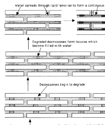
It has been suggested that the vesicle-like structures observed by van Hal et al. (126) may depict the lacunae that result from desmosomal degradation (127). Lacunae are discontinuous microdomains located in the extracellular space in the middle to outer layers of the stratum corneum (128). During hydration the lacunae provide an obvious site for water pooling and, during prolonged exposure to water, lateral expansion of the lacunae occurs through polar head regions of the intercellular lipids(Fig. 11) (127). Although the expansion of the individual lacunae may lead to a continuous lacunar system, this process does not appear to disrupt the lipid la-mellae. Menon and Elias (127) have proposed that the continuous lacunal system may represent a putative ‘‘poor’’ pathway through the stratum corneum.
It is well recognized that natural moisturizing factor (NMF) can make up to 10% of the corneocyte dry weight and, as humectants, these materials can sorb water extensively. There appears to be an absence of NMF in severe, dry flaking skin in both psoriasis and ichthyosis vulgaris. Rawlings et al. (88) have pointed out that the amino acids to which filaggrin is proteolyzed are themselves precursors for the nat-ural moisturizing factor. Glutamine is converted to the potent humectant, pyrrolidone carboxylic acid, a major component of NMF, whereas histidine is converted to uro-canic acid. Interestingly, filaggrin is converted to NMF only when the water activity is between 0.70 and 0.95, filaggrin being stable at higher water activities and pro-teolysis being impeded by low water activity. Hence, under occlusive conditions the stratum corneum NMF level decreases to close to zero, and all corneocytes contain filaggrin. The result of this homeostatic mechanism is that the skin has prevented itself from being ‘‘overhydrated.’’
In conclusion, the current observations suggest that stratum corneum hydration does not lead to an overall decrease in intercellular lipid order and only small amounts of water are present in the intercellular polar head group regions (89). Therefore, it is tempting to revisit a possible mechanism by which hydration pro-motes percutaneous absorption, which has been raised in an earlier review (122). In that model, swelling of the keratin is akin to the ‘‘bricks’’ becoming swollen in the ‘‘bricks-and-mortar’’ model of the stratum corneum, with a loosening of the inter-cellular lipid ‘‘mortar.’’ The overall effect should be an increase in the mobility of the chains and in permeability, without an effect on the lipid ordering.
B. Chemical Damage
Function of Skin
Walters Roberts
the induced perturbation. It is remarkable that the initial perturbation, which occurs in the outermost layers of the stratum corneum, can rapidly stimulate biochemical events in the stratum granulosum and lower levels of the epidermis.
Although the exact mechanisms stimulating these events are unknown, there is some indication that a change in the rate of transepidermal water loss (TEWL) in-duced by barrier alterations, may play a role (131). This increase in TEWL may lead to focal changes in the concentration of certain ions in the outer epidermis. In the normal state, the epidermis possesses a Ca2⫹ ion gradient such that there is more Ca2⫹in the outer layers than the inner (138). Following barrier disruption the Ca2⫹ gradient is lost. The presence of higher levels of intracellular Ca2⫹ in the outer epidermis is believed to block lamellar body secretion (139,140), and reduced levels will stimulate secretion. In addition, K⫹may play a role in this homeostatic mech-anism and may also influence barrier repair independently of Ca2⫹ (141). Thus, although there are still many uncertainties concerning the biochemistry of barrier repair, there is much evidence that suggests the role of ion concentration and the induction of lipid-producing enzymes; such as 3-hydroxy-3-methylglutaryl coenzyme A and serine palmitoyl transferase (142).
Perturbation of barrier function sometimes, but not always, also induces an inflammatory response that results in irritation. It is important to appreciate that
irritationis used to describe skin reactions that can range from a mild and transient
erythema or itch, to serious vesiculation (see Chaps. 12 and 13). Whereas the insults of solvent delipidation and tape-stripping of the stratum corneum result in barrier repair and epidermal hyperplasia, they do not necessarily lead to an irritant reaction. On the other hand, application of SDS almost always results in an irritant response (143,144). Although solvent delipidation and tape-stripping of the stratum corneum both physically remove the intercellular lipid lamellae, which results in considerable increases in TEWL, SDS intercalates with the lamellae and increases fluidity in this region (145), resulting in an increase in TEWL. Furthermore, although other surface-active agents, such as sodium laurate and polysorbates, can increase TEWL to levels similar to SDS, the resultant irritation is much less and, in some cases, not signifi-cantly different from untreated skin (146). It follows that irritation subsequent to exposure to SDS must be a result of factors other than an increase in water transport and the stimulation of lipogenesis.
That surface-active agents can cause skin irritation is well established and has been so for many years (147). Also, whereas ionic surfactants can cause severe irritation, nonionic surfactants are considered virtually nonirritant in normal use (148,149). Thus, much of the research on surfactant-induced skin irritation has in-volved studies on SDS. The collective data suggest that SDS can interact with both lipid and protein structures in the stratum corneum. Interaction with lipids will in-crease lipid fluidity and thereby enhance skin permeability. This alone, however, apart from increasing its own permeation, will not account for the irritation caused by SDS. Although SDS can penetrate into the corneocyte and interact with the protein structure such that␣-keratin is uncoiled (150), it is difficult to relate this aspect to an irritant response. A more likely explanation for the irritation induced by SDS is its capacity to stimulate keratinocyte production of inflammatory mediators such as IL-1 and PGE2 (151). Whether this induction is secondary to some interaction
Function of Skin
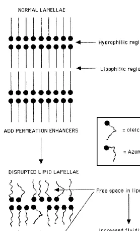
There is a range of mechanisms by which solvents may affect skin permeability, as proposed by Menon et al. (152). Suhonen et al. (89) recently reviewed chemical enhancement in terms of stratum corneum alterations. They