* Corresponding author.
CALLUS INDUCTION AND PHYTOCHEMICAL CHARACTERIZATION OF
Cannabis sativa
CELL SUSPENSION CULTURES
Tri J. Raharjo
a,*, Ogbuadike Eucharia
b, Wen-Te Chang
cand Robert Verpoorte
b aDepartment of Chemistry, Faculty of Mathematics and Natural Sciences, Gadjah Mada University, Yogyakarta Indonesia
b
Division of Pharmacognosy, Section Metabolemics, Institute of Biology, Leiden University, Gorlaeus Laboratories, PO Box 9502, 2300 RA, Leiden,The Netherlands
c
Department. of Applied Plant Sciences, TNO Voeding, Zernikedreef 8, Leiden, The Netherlands
Received 30 December 2005; Accepted 16 January 2006
ABSTRACT
Callus of Cannabis sativa has been successfully induced from C. sativa explants and seedings. It seems that flowers are the best explant for callus induction and induction under light also give better results than induction in dark. Four cell culture lines were established from flower induced callus. Phytochemical profiles of C. sativa suspension cell cultures were investigated using HPLC and 1H-NMR. Cannabinoids and phenolic compounds related to cannabinoids such as flavonoids could not be found in the cell suspension cultures and there is no major chemical difference between the cell lines though they can visually be distinguished by their colors. Only in one cell line some aromatic compounds in the water/methanol extract could be observed in the 1H-NMR. Further investigations showed that none of these compounds are flavonoids. It seems that lack of cannabinoids in the cell cultures is related to lack of polyketide synthase activity.
Keywords:Callus, Cannabis, phytochemical.
INTRODUCTION
Plant cell and tissue cultures, in several cases, have shown to be excellent model systems for studying secondary metabolite biosynthesis. Numbers of pathways have been unraveled with the aid of such systems, e.g. the terpenoid indole alkaloid biosynthesis in Rauvolfia serpentina [1,2], and Catharanthus roseus [3].
Cannabis sativa cell cultures induction have been studied for years and most studies were purposed to produce cannabinoids from cell cultures [4-6] but so far the results ware not satisfactory. Only the identification of 8-tetrahydrocannabinol from callus cultures growing in B5 medium with various regulators have been reported [7]. This product was believed to be an artifact of9-tetrahydrocannabinolic acid due to the fact that the product was only detected in an extract obtained after concentration at elevated temperature. However, neither
9
-tetrahydrocannabinol nor 9-tetrahydrocannabinolic acid has been found so far in the cell cultures. Addition of known cannabinoid precursors or related compounds such as olivetol, geraniol, nerol, and ethyl olivetolate did not result in the formation of cannabinoids instead it seems that oxidation of the precursors was the main route of transformation [4,8]. One report mentioned the transformation of cannabidiol to cannabielsoin in cannabis cell suspension cultures, but it was not specifical forC. sativacultures [4,5].
Besides cannabinoids, 19 flavonoids as well as 16 other phenolic compounds have been found in C. sativa plant [9]. In this study we present an effort to identify secondary metabolites produced in C. sativa cell cultures, particularly focusing on cannabinoids and flavonoids. Also we looked for other compounds from pathways that might compete with the cannabinoid biosynthesis. This information might be used to develop strategies to get the cell cultures to produce cannabinoids.
EXPERIMENTAL SECTION
Establishment of cell suspension cultures
Leaves, flowers and 4 days old seedlings of C. sativa “Four Way” (The Sensi Seed Bank, Amsterdam The Netherlands) were subjected to sterilization. Sterilization was started with washing with detergent, followed by washing with 70% ethanol for 3 min, and a rinsing period with sterilized distilled water for 10 min. Thereafter the explants were transferred to a solution of 2% sodium hypochloride in which the explants were kept for 20 min, after which they were again thoroughly rinsed three times with sterilized distilled water.
thiamine diHCl 10 mg/L pyridoxine HCl 1 mg/L and nicotinic acid 1 mg/L, 2,4-D 1 mg/L, sucrose 30 g/L and agar 10 g/L; pH 5.5 [10]. Some of the explants in the petri dishes were cultured under light others in the dark.
Callus obtained in the petri dishes were used to set up cell suspension cultures. Approximately 1.5 g of callus from each explant was chopped to smaller particles and suspended into 250 mL flask with 50 mL of liquid MS medium (as MS agar, without agar). The suspension cultures were grown at 25 oC under continuous light (intensity 1,200-1,800 lux) on a gyratory shaker at 110 rpm. After 2 weeks, the contents of each flask was divided into two, one part was maintained in the MS medium while the other was maintained in B5 medium (B5 components, 2,4-D 2.0 mg/L, IAA 0.5 mg/L, NAA 0.5 mg/L, K 0.2 mg/L and sucrose 30 g/L). These cell cultures were then maintained by sub-culturing weekly.
Phytochemical characterizaton Extraction
One gram of cell culture biomass was extracted in one step with 4 mL of chloroform and 4 mL of methanol/water (1/1) by vortexing for 30 sec and sonication for 1 min. The mixture was then centrifuged at 3,000 rpm, 25 oC for 20 min. The two solvent layers were collected separately. Extraction was repeated and the resulting extracts were combined with the first extraction. The combined extracts were evaporated until dry. The chloroform extract was subjected to HPLC and
1
H-NMR analysis, the water-methanol extract was subjected to 1H-NMR analysis. Dry extracts were resuspended with the solvent according to the method of analysis.
HPLC
The HPLC system consisted of a Waters 712 pump, a Water 600E system controler, a Waters 717plus autosampler and Waters 991 photo diode array detector (Waters Corp. Milford, MA, USA). The column was a Hypersil C18 240 x 4.6 mm separation column
(Phenomenex, Torrance, CA, USA). The solvent system consisted of solvent A: H2O/CH3CN (98/2) containing
0.01% H3PO4 and solvent B: H2O/CH3CN (2/98)
containing 0.01% H3PO4. The gradient profile was as
follows: an isocratic step of 100% A in B for 1 min, then a linear gradient from 0-100% of B in A for 34 min, followed by an isocratic step at 100 % of B in A for 10 min. After this gradient, the eluent was returned to 50% of B in A for 5 min and was finally kept for 10 min before injection of the next sample. The flow rate was 0.8 mL/min. The chromatogram was monitored for maximum absorbance.
1
H-NMR
NMR spectra were recorded in CDCl3and KH2PO4
buffer at 400 MHz using a Bruker AV-400 NMR
spectrometer (Bruker Daltonics Inc., Manning Park, Billerica, MA, USA) equipped with an Indy Silicon graphics computer. For each sample 128 scans were recorded using the following parameters: 0.17 Hz/points, pulse width (PW) = 0.4 µsec, and relaxation delay (RD) = 1.0 sec [11].
Column Chromatography
Thirty gram of cell biomass were extracted by methanol. After drying the extract was resuspended with minimum volume of water and poured into packed HP-20 resin column (25 cm with 25 g HP-20). Ethanol in water (25%, 50% 75%, and 100%) was used to elute the sample. The eluent was collected at intervals of 50 mL, and then evaporated. Each fraction was analyzed with TLC (thin layer chromatography) using silica plates and water/acetic acid/formic acid/ethyl acetate (37/11/11/11) as mobile phase.
RESULT AND DISCUSSION
Callus induction
Flowers and leaves from plants and four days old seedlings were used to induce callus. It was observed that the flowers gave more callus while the leaves were less callus productive. The shoots of the seedlings produced more callus than the stems and no callus was formed on the roots. Light and dark conditions were employed for callus induction. For all materials it seems that callus grows much better under light (Table 1).
It seems that cannabis callus is easily induced in a standard medium. As the callus material consisted of heterogenous cells it took several several subcultures to obtain cultures that seemed homogenous (stable) based on the visual inspection of the color. We obtained several cell suspension cultures. Cell lines grown at a light intensity of 1,200-1,800 lux showed a green (G cell) and greenish brown (GB cell) or brown color (B cell). Others grown at lower light intensity (400 lux) showed milky (M cell), brown (B cell) or light brown color (LB cell).
Table 1Comparison of callus growing
Materials Dark Light according to a subjective scale based on the amount of the callus, fast growth, color and friability
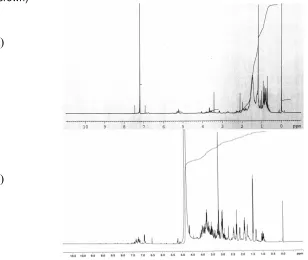
-Fig 1 HPLC profile and UV spectra of chloroform extract of cannabis cell suspension culture line GB (greenish brown)
Fig 21H-NMR spectra of chloroform (a) and water/methanol phase (b) of cannabis cell suspension cultures GB (greenish brown)
Phytochemical profile of cannabis suspension cell cultures
Metabolite profiling of cannabis cell suspension cultures aimed at the identification of secondary metabolic pathways that are operative and may be
linked with cannabinoid biosynthesis. We targeted both at polar and non-polar compounds. In case of cell cultures, sonication extraction using the same portion of water/methanol (1/1) and chloroform succeeds to extract most of the compounds present in the cells [11].
(a)
The extraction results in two phases, a water/methanol phase which contains polar compounds and a chloroform phase which contains non-polar compounds.
In our experiment HPLC analysis was used to analyze the chloroform fraction for cannabinoids. An HPLC profile is shown in Fig. 1. We checked the extracts by comparing the retention times and UV spectra of the 6 main cannabinoids (CBD, CBG, CBDA, CBN, 9-THC and THCA) with the peaks in the chromatogram. None of the cells did show any peak that has the same retention time and UV spectra as any of the cannabinoid references. We thus conclude that no cannabinoids are detectable in the cell cultures.
Using 1H-NMR spectrometry we targeted not only on cannabinoids but also on other phenolic compounds.
1
H-NMR spectra (both water/methanol and chloroform) of the extracts are shown in Fig. 2. In the chloroform extract, there are no specific signals of aromatic compounds present (6-8 ppm). Neither any specific aromatic signals were observed in the water/methanol extract of the cell lines except for the GB cell, which shows (Fig. 2 (b)) several weak signals at around 6-8 ppm.
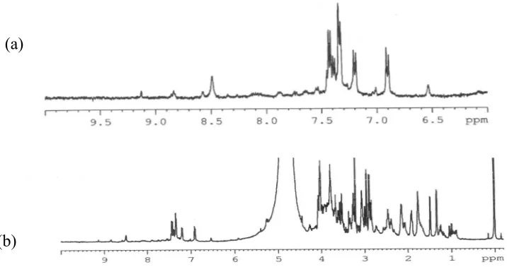
It seems interesting to see what aromatic compounds are present in the water/methanol extract. Sixteen non-cannabinoid phenols have been found inC. sativa such as eugenol, isoeugenol and anethol [9]. However, these groups of compounds are expected in the chloroform extract. On other hand, flavonoids are always present in C. sativain the glycoside form, which dissolve well in water/methanol. To see if the signals shown in Fig. 2(b) are from flavonoids, water/methanol extract was subjected of further fractionation using an HP-20 resin column. Fractions were then analyzed further using1H-NMR. Fractions eluted by 50% and 75% ethanol showed aromatic signals (Fig 3).
We compared these (Fig. 3) with characteristic spectra of flavonoids. In C. sativa plant 19 flavonoid glycosides derived from apigenin, luteolin and quercetin are present [9]. Apigenin and luteolin belong to the flavones while quercetin is a flavonol. Flavonoids usually give some signals at 6.5-8 ppm (aromatic protons of B ring), 5.0-6.8 ppm (aromatic protons ring A). Flavones give a singlet at 6.5 ppm of H3. The protons of the sugar of the glycoside will appear as a couple of multiplets at 3.5-4.5 ppm [12]. Using these criterias we can see clearly multiplets around 3.5-4.5 that could be due to a sugar. We can see two singlet at 6.5 and 6.9 ppm but they are not like aromatic ring A protons which should be multiplets. We also compare the spectra with the spectra of a series of flavonoids [13]. None of the flavonoid spectra have similarity with the spectra as shown in Fig. 3. Based on that we think that the aromatic compounds here are not flavonoids but could be phenylpropanoid primary metabolites. The spectra of aromatic amino acid, phenylalanine and tyrosine, are different from the spectra as shown in Fig. 3.
CONCLUSION
We confirmed that callus of cannabis can be induced from many parts of the plant as well as seedlings. This fact is interesting for further studies on the regeneration of cannabis plants from callus, needed for metabolic engineering studies. From the metabolic profiling, we learned that cannabinoids are not be produced in the cannabis cell cultures. No other secondary metabolites related to cannabinoid biosynthesis such as flavonoids were present in the cannabis cell cultures either. Phenolic compounds, maybe phenylpropanoids, were found in one culture.
Fig 31H-NMR spectra fraction 50% ethanol of water/methanol extract fractionated using HP-20 (a) and expanded picture at 6-8 ppm (b)
Lack of cannabinoids in the cell cultures might be due to lack of both the polyketide pathways. However, further confirmation is needed on the level of enzymes to prove this hypothesis.
REFFERENCES
1. Kutchan, T.M., Hampp, N., Lottspeich, F., Beyreuther, K., and Zenk, M.H., 1988, FEBS Lett, 237, 40-44.
2. De Luca, V., Marineau, C., and Brisson, N., 1989, Proc Natl Acad Sci USA, 86, 2582-2586
3. Pasquali, G., Goddijn, O.J.M., de Waal, A., Verpoorte, R., Schilperoort, R.A., Hoge, J.H.C., and Memelink, J., 1992, Plant Molecular Biol. 18, 1121-1131.
4. Loh, W.H.T, Hartsel, S.C. and Robertson, L.W., 1983,Z Pflanzenphysiol Bd,111, S.395-400
5. Hartsel, S.C., Loh, W.H.T., and Robertson, L.W., 1983,Planta Med.,48, 17-19
6. Veliky, I.A., and Genest, K., 1972, J Nat Prod, 35, 450-456
7. Heitrich, A., and Binder, M., 1982, Experentia, 38, 898-899
8. Itokawa, H., Takeya, K., and Mihashi, S., 1977, Chem Pharm Bull, 25, 1941-1946
9. Turner, C.E., Elsohly, M.A. and Boeren, E.G., 1980,J Nat Prod, 43,169-234
10. Murashige, T., and Skoog, F., 1962,Physiol Plant, 15, 473-497
11. Casas, E., 2003, Metabolic Profiling of Catharanthus roseus Leaves Infected by Pythoplasma using 1H-NMR and Principle Component Analysis, Departament de Productes Naturalis Facultat de Farmacia, Universitat de Barcelona. Barcelona.
12. Mabry, T.J., Markham, K.R., and Thomas, M.B., 1970, The Systematic Identification of Flavonoids, Springer-Verlag, Berlin