ST Elevation Myocardial Infarction in Young Women Caused by a Coronary Embolism Generated from Mitral Valve Disease: a Case Report
Budi Yuli Setianto1, Nahar Taufi q1, Muhamad Taufi k Ismail1
1 Department of Cardiology and Vascular Medicine, Faculty of Medicine, Gadjah Mada University–Sardjito Hospital.
Jl. Kesehatan no.1 Sekip, Yogyakarta 55284, Indonesia. Correspondence mail: [email protected].
Abstract
Coronary emboli causing ST elevation myocardial infarction (STEMI) is a rare condition. Intracardiac thrombus generated by valvular disease is the most common source of intracoronary embolism and mitral stenosis is frequently affected by intraatrial thrombus. Therapeutic strategy of coronary embolism is challenging because there were still no recommendation regarding coronary embolism published to day. We reported A 25 years old woman was admitted to the emergency department because of STEMI with complication of acute pulmonary edema and cardiogenic shock, and recurrent cardiac arrest. Angiographically showed total occlusion of left main artery with thrombotic lesions. Unfortunately after unfractionated heparin (UFH) was given and performed catheter cannulation, thrombus run to distal part of left coconary artery. Procedure is stopped and then patient transferred to cardiac care unit, but death shortly after arrival. Transthroracic echocardiography revealed mild to mild-modertae mitral stenosis with severe regurgitation due to mitral valve prolaps, aortic regurgitation, and left atrial and ventricular dilatation with no thrombus or spontaneous echo contrast, and failed to fi nd thrombus with in atrium. The presence af atrial dilatation and aortic regurgitation increased risk of thrombosis, meanwhile mitral regurgitation were reported as protective factor of atrial thrombosis. The pathophysiology of arterial thrombus or white thrombus involving platelet activation leads to double antiplatelets and GbIIb/IIIa inhibitior to be more cruciale in coronary embolism. Double antiplatelet dan UFH administration did not improve survival in these patients. So that further research was needed to make a consensus of therapy. Careful assessment of intracardiac thrombus and risk of thromboembolism were important to prevent systemic embolization.
Keywords: STEMI – coronary embolism – mitral valve disease.
Introduction
Atherosclerosis plaque rupture followed by thrombus formation is the main cause of acute myocardial infarction (AMI). Nevertheless, about 7% of patients who underwent coronary a n g i o g r a p h y h a v e n o n a r r o w i n g o f t h e atherosclerosis disease1.
Coronary embolism is a cause of non-atherosclerosis AMI. Prizel et al. (1978) reported incidence of coronary embolism about 13% in autopsy study of 419 AMI patients, which main cause is valvular heart disease (40%), atrial fibrillation (AF) (24%), and cardiomyopathy (20%)2.
Before 1960, coronary embolism more caused by infective endocaditis. Nevertheless, according to antibiotic development, recently it more caused by valvular heart disease such as mitral and aortic valve disease, AF, or in patients with prosthetic valve implantation2.
Embolism that come from heart usually originate from thrombus within left ventricle, heart tumor such as myxoma, dilatation of left atrium (LA), or paradoxical embolism come from patent with foramen ovale. Calcifi cation of mitral anulus as well as mitra valve prolaps were also associated with systemic embolism event, although remains controversial3. Mitral stenosis (MS) is most common valvular heart disease that causes thrombus formation within LA4, although very rare resulted in coronary embolism5.
In the following I want to presentate a young female patients 25 years old came to my hospital with STEMI and previous mild-modertae mitral stenosis and severe mitral rugurgitation.
Case Illustration
tenderness chest pain has suffered more than 20 minute, accompanies with cold sweating and short of breath, no pain radiation, palpitation and fever.
No history of injury, previous surgery, hypertension and diabetes mellitus. Otherwise, there was a hisory of smoking about lebih 1-2 pack a day. No history of hormonal contraception, leg edema or vein thrombosis and renal disease. No family history of coronary artery disease (CAD).
T h i s p a t i e n t c o m p l a i n i n g d y s p n o e on effort since 3 months ago. Trans thoracal echocardiography showed LA and LV dilatation, LV ejection fraction 57%, normokinetic, diastolic dysfunction pseudonormal type, mild-modertae mitral stenosis and severe mitral regurgitation due to anterior mitral valve prolaps. She routinely was administered digoxin 1x 0,125 mg, candesartan 2x40 mg, and furosemid 1x20 mg, and had planned to be performed mitral walve replacement.
On physical examination demonstrated her general condition bad, look short of breath and
stupor. Blood pressure not measurable, respiration
rate 36 x/ minute, pulse 100 x/ minute regular, no fever. There is no anemia and jaundice. Jugular vein pressure was 5+4 cm H2O. There was cardiomegaly with touchable apex in V intercostal space 1 finger left lateral midclavicularis line. Cardiac auscultation showed normal S1 and S2 heart sounds, the degree of pan-systolic murmur encountered 3/6 with punctum maximum in the apex and radiating to the axilla. There was no additional murmur. Lung examination found normal vesicular sounds, crackles at the bottom of both lungs. Abdominal examination within normal limits, and there was no peripheral edema.
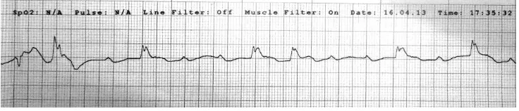
Electrocardiography (Figure 1) showed sinus rhythm with a heart rate of 68 x / min. Intermediate axis, with ST segment elevation in leads V4-V6, I, and aVL. Based on the results of history, physical examination, and ECG, patients clinically diagnosed as high lateral and anterolateral STEMI onset 2 hours, Killip IV, TIMI risk 9/14 score,
and Grace scores 233, in chronic heart failure with dilated left atrium and ventricle due to mild-modertae mitral stenosis and severe mitral valve regurgitation because of mitral valve prolapse.
Patients present with cardiogenic shock and acute pulmonary edema, and then the patient was given 40 mg of furosemide injection and inotropic support with dopamine to noradrenaline, and loading with dual antiplatelet. But the shock is not improved, the ECG monitor showed total AV block (Figure 2). Two hours later the patient fell in a state of pulseless electrical activity (PEA) and then performed cardiopulmonary resuscitation and intubation. After cardiac resuscitation performed for approximately 1-2 hours, patients got ROSC (return of spontaneous circulation) with ECG still showed ST segment elevation and blood pressure 90/60 mmHg. Patients were then planned to do coronary angiography immediately.
Laboratory results showed a hemoglobin level 11.9 g / dL; leukocyte count 23 rb / mmc; platelets 295 rb / mmc; hematocrit 36.7; blood urea nitrogen 9 mg / dl; creatinine 1.38 mg / dl; AST 63; ALT 51; albumin 4.2 g / dL, sodium 139; potassium 3.6; chloride 99. Cardiac enzymes performed 6 hours after the onset of chest pain showed CKMB 119, Troponin I 1.57.
[image:2.595.308.526.82.273.2]ECG on admision
[image:2.595.117.485.665.744.2]FIGURE 2. Total AV block seen on ECG monitor.
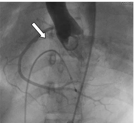
FIGURE 3. Angiography in the LAO-cranial view 30/0 showed normal RCA, the origin comes from the non-coronary sinus valvasa.
FIGURE 4. Angiography in the LAO-cranial view 30/0 (A) and LAO-caudal view 45/35 (B), thrombotic lesions seen in the distal left main artery (white arrow).
[image:3.595.115.461.553.712.2]After a temporary pacemaker (TPM) inserted, then performed coronary angiography. Cannulation of the right coronary artery (RCA) can not be done, so it was decided to do aortic angiography. Aortic angiography showed normal RCA, with origin from valvasa non-coronary sinus (Figure 3). Figure 4 showed total occlusion of the distal left main artery (LM), with thrombotic lesions.
Then the patient is given heparin and performed left coronary artery cannulation. However, when injected contrast, thrombus off toward the distal LAD and LCX. Based on the results of coronary angiography, there was also not found atherosclerotic lesions of LM (Figure 5). During coronary angiography, patients fell into cardiac arrest condition two times. Patient then transferred to the cardiac intensive care (ICCU). However when the patient arrives in ICCU, she
returned cardiac arrest and performed CPR, but no response and the patient died.
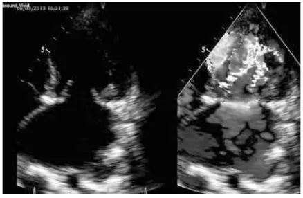
According to the evaluation results of transthoracic echocardiography (TTE) 3 months ago, found severe mitral valve regurgitation and mild-moderate mitral stenosis with a mean gradient of 9.5 mmHg and a peak gradient of 19 mmHg, mitral valve area of 2.3 cm2. Left atrial diameter of 57 mm (Figure 6). Presence of aortic regurgitation on aortic angiography (does not seem clear on TTE) can lead to underestimate of MVA value in these patients.
Discussion
Patient in this case was a young woman aged 25 years with no obvious risk factors for atherosclerosis except history of smoking. Osula et al.6 generally halve the IMA at a young age into the coronary arteries “normal” and “abnormal” on coronary angiography. Coronary arteries are “normal” include thrombosis, embolism, spasm, or a combination. While the “abnormal” may be premature atherosclerosis, spontaneous coronary dissection, aneurysm, ectasia, or anomalous origin of the coronary arteries. The presence of thrombus in the LM and the absence of atherosclerotic lesions, the etiology of this case is thrombosis or embolism.
Coronary artery thrombus in situ of the two cases reported, caused by a defi ciency of antithrombin, protein C, or protein S 7, 8. However this is a very rare occurrence, because, although these factors were strong for the occurrence of venous thrombosis, but no effect on arterial or very small 9. No history of deep vein thrombosis, or thromboembolic events in these patients and her families also support that the etiology of coronary thrombus in these patients is not due to factors such hypercoagulability. Therefore, coronary embolism originating from the heart valve disease such as mitral stenosis with regurgitation most likely the cause of AMI in these patients.
[image:4.595.68.288.327.438.2]Coronary embolism of the case reported is a rare disease, because the ostium of the coronary artery is located behind the aortic valve cusps and the presence of heavy systolic ejection of the left ventricle. Etiology of coronary embolism commonly associated with valvular heart disease, the use of prosthetic valve, infective endocarditis, dilated cardiomyopathy, and arrhythmias5. Paradoxical FIGURE 7. TTE apical four chambers position,
[image:4.595.69.288.550.693.2]which seemed to indicate a mosaic fl ow turbulence while diastolic transmitral fl ow that normally occur in mitral stenosis with a mean gradient of 9.5 mmHg MV. FIGURE 6. TTE parasternal long axis position
embolism can also occur as a result of venous thrombosis in atrial septal defect 10.
Coronary embolism of intracardiac thrombus may originate from the atria or the ventricles due to stasis of blood fl ow. Intraventricular thrombus generally caused ventricular aneurysm, severe LV systolic dysfunction, or kinetic disorders4. All-cause ventricular stasis is not found in this case. While the factors predisposing to thrombus was intraatrial atrial enlargement, mitral valve disease and atrial fi brillation which causes low blood fl ow in the left atrium. The highest incidence of left atrial thrombus is a rheumatic mitral stenosis and atrial fi brillation4.
Mitral stenosis associated with increased left atrial coagulation activity, thus increasing the incidence of intracardiac thrombus, although patients with sinus rhythm but found left atrial spontaneous echo contrast (LASEC). Atrial fi brillation increases the risk of thromboembolism 2-7 times 11. The incidence of systemic embolism in mitral stenosis was 9.1% in patients with sinus rhythm for 3 years. Based ECG of this case, heart rhythm is still sinus rhythm despite the possibility of the occurrence of atrial fi brillation are not detected, as reported by Samol et al.12 who reported prevalence in risk groups by 5.3%.
The age factors associated with increased risk of embolism (relative risk [RR] 1.12 [95% CI 1.04 to 1.21]). Similarly, the presence of thrombus (RR 37.1 [CI 2.82 to 487.8]), the severity of mitral stenosis (RR 16.9 [CI 1.53 to 187.0]), and the presence of aortic regurgitation (RR 22 , 4 [CI 2.72 to 184.8]) associated with an increased risk of embolism14. While the presence of mitral regurgitation is associated with a protective factor (OR 0.39) occurrence of thrombus because regurgitant fl ow lead no stasis in the left atrium 4,15. In fact, the degree of mitral regurgitation
especially severe is also a protective factor against the incidence of ventricular thrombus in dilated cardiomyopathy patients because it increases blood flow in the ventricular apex16. However, study Tse et al.17 mention that mitral regurgitation associated with systemic platelet activity, who may be associated with thromboembolic events. Protective effects in patients with rheumatic mitral regurgitation also be reduced if left atrial diameter is more than 60 mm18.
In these patients the dominance of the valve abnormality is mitral regurgitation than stenosis, and the results of TTE, not clearly found LASEC
or thrombus. However, the TTE examination has a low sensitivity (53-63%), so the TEE examination (sensitivity 99%) should be performed in these patients 4.
Age was associated with an increased risk of thrombosis exponentially. Mechanisms thought to be caused by the cumulative effect of factors of blood vessel walls, decreased physical activity, and increased activity of several coagulation factors such as factor V, VII, VIII, IX, and fi brinogen 9. However, the patient is is still young 25 years old,
so the risk for thromboembolism is small.
Currently, there are no clear recommendations about the management of coronary embolism. Raj and Singh19 reported, a woman 23 years after double valve replacement with infarction, and performed with streptokinase thrombolytic followed by administration of an anticoagulant, ACE inhibitors, and antiplatelet. The result of the evaluation of coronary angiography showed normal.
Thrombolysis in coronary embolism caused by infective endocarditis should be avoided because of the risk of cerebral embolism 20 or brain and systemic hemorrhage 21. Dual antiplatelet therapy and intravenous heparin is used in cases of coronary embolism with mitral stenosis 5 and in cases with atrial fi brillation 20.
Antiplatelet in case with prosthetic valve serves improve platelet survival and reduce the risk of thromboembolism when combined with anticoagulants13. Antiplatelet also associated with the prevention of platelet adhesion and activation in arterial thrombosis or intracardiac thrombus 22,23. However the role of antiplatelet therapy in
coronary embolism due to intracardiac thrombus needs further research.
antiplatlet discontinued after coronary embolism improved 5.
Conclusion
W o m e n 2 5 y e a r s o l d w i t h S T E M I presenting Killip IV, performed emergency coronary angiography procedures with results total occlusion of the distal LM caused by a thrombus, which suspected due to embolism of intraatrial thrombus due to mild-moderate mitral stenosis and severe regurgitation. Therapy of patients with coronary embolism due to intracardiac thrombus is recommended to administer double antiplatelet, heparin and GPIIb/IIIa inhibitors. Although there are no standard guidelines at this time. Transesophageal echocardiography examination is needed to ascertain whether any intraatrial thrombus in patients with mitral valve disease.
Acknowlegment
The authors would like to thanks doctor who performed Percutaneous Coronary Intervention and Catheterization Laboraratorium Staff for assistance to conduct this case report.
References
Rigatelli G. Normal angiogram in patients 1.
with acute coronary syndrome: searching for unusual substrates of myocardial ischemia. Int J Cardiol 2005;99:25–7.
Prizel KR, Hutchins GM. Bulkley BH., Coronary 2.
artery embolism & myocardial infarction – A clinicopathologic study of 55 patients. Ann Int Med 1978;88:155–61
Homma S, and Di Tullio MR. Cardiac Sources 3.
of Embolus, How To Find It. ACC Current Journal Review 2001;5-6
Otto CM. Textbook of Clinical Echocardiography 4.
4th edition. Chapter 15 - Cardiac Masses and Potential Cardiac “Source of Embolus”. Elsevier Saunders, 2009.
Liang M, Kelly D, Puri A, Devlin G. Mitral 5.
Stenosis as a Risk Factor for Embolic Myocardial Infarction—Anticoagulation for Some Patients, Individual Treatment for All. Heart, Lung and Circulation 2011;20:728–730
Osula S, Bell GM, Hornung RS. Acute 6.
myocardial infarction in young adults: causes and management. Postgrad Med J 2002;78:27–30.
Hacker SM, Williamson BD, Lisco S, Kure 7.
J, Shea M, Pitt B. Protein C defi ciency and acute myocardial infarction in the third decade. University of Michigan Medical Centre, Ann Arbor, Michigan 1991.
Conard J, Samama MM. Inhibitors of 8.
coagulation, atherosclerosis, and arterial thrombosis. Semin Thromb Hemost. 1986 Apr;12(2):87-90.
Previtali E, Bucciarelli P, Passamonti SM, 9.
Martinelli I. Risk factors for venous and arterial thrombosis. Blood Transfus 2011;9:120-38 Cuculi F, Togni M, Meier B. Myocardial infarction 10.
due to paradoxical embolism in a patient with large atrial septal defect. The Journal of Invasive Cardiology 2009;21:E184–186.
Peverill RE, Harper RW, Gelman J, Gan 11.
TE, Harris G, Smolich JJ. Determinants of increased regional left atrial coagulation activity in patients with mitral stenosis. Circulation 1996;94(August (3)):331–9.
Samol A, Masin M, Gellner R, Otte B, 12.
Pavenstädt HJ et al. Prevalence of unknown atrial fi brillation in patients with risk factors. Oxford Journals. 2013-04-26.
Charles RG, Epstein EJ. Diagnosis of coronary 13.
embolism: a review. Journal of the Royal Society of Medicine Volume 76 October 1983 863.
Chiang CW, Lo SK, Ko YS, Cheng NJ, 14.
Lin PJ, Chang CH. Predictors of systemic embolism in patients with mitral stenosis: a prospective study. Annals of Internal Medicine 1998;128(June (11)):885–9.
Wanishsawad C, Weathers LB, Puavilai W. 15.
Mitral Regurgitation and Left Atrial Thrombus in Rheumatic Mitral Valve Disease: A Clinicopathologic Study. CHEST 1995; 108:677-81
Kalaria VG, Passannante MR, Shah T, Modi K, 16.
Weisse AB, et al. Effect of mitral regurgitation on left ventricular thrombus formation in dilated cardiomyopathy. Am Heart J 1998;135:215-20. Tse HF, Lau CP, Cheng G. Relation between 17.
mitral regurgitation and platelet activation. J Am Coll Cardiol. 1997 Dec;30(7):1813-8. Kranidis A, Koulouris S, Filippatos G, Kappos 18.
Raj V, Singh, RK. Acute Myocardial Infarction 19.
in a Young Patient of Rheumatic Heart Disease. MJAFI 2011; 67: 77-79
Walle SV, Dujardin K. A case of coronary 20.
embolism in a patient with paroxysmal atrial fi brillation receiving tamoxifen. International Journal of Cardiology 123 (2007) 66–68. Roxas CJ, Weekes AJ. Acute myocardial 21.
infarction caused by coronary embolism from Infective endocarditis. The Journal of Emergency Medicine, Vol. 40, No. 5, pp. 509–514, 2011.
Nieswandt B, Pleines I, Bender M. Platelet 22.
adhesion and activation mechanisms in arterial thrombosis and ischaemic stroke. J Thromb Haemost. 2011 Jul;9 Suppl 1:92-104.
Uchiyama S, Yamazaki M, Iwata M, Maruyama 23.
S. Diagnosis of intracardiac thrombi by various imaging techniques and activation of platelets and coagulation-fi brinolysis in patients with cardioembolic stroke. Rinsho Shinkeigaku. 1996 Mar;36(3):429-35.
Steinwender S, Hofmann R, Hartenthaler B, 24.
Leisch F. Resolution of a coronary embolus by intravenous application of bivalirudin. International Journal of Cardiology 132 (2009) e115–e116
Yuce M, Yavuz F, Cakici M, Sari, Davutoglu V. 25.