ISSN: 2455-264X, Volume 4, Issue 1 (Jan. - Feb 2018), PP 27-32
www.iosrjournals.org
Isolation and Molecular Identification of Endophytic bacteria
from Red Betel Root (
Piper crocatum
Ruiz & Pav) as a Producer
of Anti-Bacterial Compounds
Lalu Wira Zain Amrullah
1, Dwi Soelistya Dyah Jekti
2, Lalu Zulkifli
2*1(Master Program of Science Education, University of Mataram, Indonesia) 2(Study Program of Biology Education, University of Mataram, Indonesia)
*(Corresponding author : [email protected])
Abstract :
This research conducted to find out the capabilities of endophytic bacteria isolate from root of red betel plants (Piper crocatum Ruiz & Pav) in inhibiting the growth of bacteria Streptococcus aureus, escherichia coli, Klebsiella pneumoniae, Streptococcus mutans, and to identify the endophytic potensial bacteria. this research was devide into 3 stages, endophytic bacteria isolation, antibacterial test, biochemical test and molecular identification upon the potensial endophytic bacteria. It was found 2 isolates from root of the red betel plants; only One (1) isolat can be inhibition S. aureus and E. coli. Based on 16S rRNA molecular identification, isolates of endophytic bacterial of red betel root which have inhibitory effect are closely related to Bacillus pumilus strain AUEC29.Keywords
-
Root, Red Betel, Isolation, inhibition, molecular test of 16S rRNAI.
I
NTRODUCTIONIndonesia is known as a source of raw materials tropical medicine that can be used to solve various kinds of diseases. Be side that Indonesia has been known of the world's largest medicinal herbs alongside other countries in Asia, such as China and India [1]. According to some research results, chemicals component in plants such as aliphatic, alkaloids, flavonoids, Peptide, Phenol, Quinon, and terpenoids, serves as an anti-microbial [2].
The search of bioactive compounds source continue to be done in line with the increasing number of new diseases including many kind of infectious diseases, cancer and other dangerous diseases. Bioactive compounds (secondary metabolites) can be obtained from several sources such as from plants and microbes. [3]. Endophytic bacteria normally found in living plant tissue, and give benefit for plant by, for instance, increase the availability of nutrients and produce growth hormone [4]. The endophytic bacteria was also able to increase plant resistance against a wide variety of microbial pathogens by inducing plant resistance, known as induced systemic resistance (ISR) in order to survive against the disease occurs.
It was proved that endophytic bacteria isolated from green betel plant have potential as an anti-bacterial, especially on the growth of Staphylococcus aureus [5]. In relation with that finding, it is important to investigate the ability of endophytic bacteria as antimicrobe from another spescies of betel plant, such as red betel plant. Red betel comes from the same genus as green betel, it is estimated that red betel has the same effect on the growth of endophytic microbes.
Red betel plant is a type of herb plants that contain flavonoids, polyphenols, alkaloids, tannins, astsiri oil, saponins, hydrosicaficol, chavicol, chavibetol, allylprokatekol, carvacrol, eugenol, P-cymene, cineole, coryofelen, extragol, terpene, steroid and phenyl propodate [6].
II.
M
ATERIALM
ETHODS2.1 Isolation of Endophytic Bacteria
2.2 Endophytic Bacteria Test
Three (3) ose endophytic bacteria were grown in 10 ml of sterilized nutrient broth and incubated for 48 hours at a temperature of 32oC which further isolates of endophytic bacteria was shaken by the shaker 150 cycles/1 min. Furthermore, the culture medium on centrifuge at 5000 g for 30 minutes and the supernatant was collected by filtration. The supernatant was used for antimicrobial activity and the material can be stored before use at a temperature of 4oC.
To determine the activity of anti-bacterial used diffusion method. Pathogenic bacteria to be tested was grown on NB medium with a concentration of 105cells/ml. Pathogenic bacteria of 100 mL plated on MHA medium evenly. Make pits with a diameter of 4 mm in order and pitting charged as much as ± 50 mL of the supernatant and then incubated 24 hours. As a control used antibiotics ciprofloxacin, inhibitory zone form of a clear zone formed around the wells was observed and compared with the positive control, then the diameter of inhibition zone was measured. Endophytic bacteria isolates were able to form a clear zone around the wells can be considered as a potential isolates.
2.3 Molecular Identification of 16S rRNA
The identification of endophytic bacterial isolates capable of forming the inhibitory zone diameter against microbial growth of clinical isolates was analysis with skuens 16S rRNA with a primer 63f (5’-CAG GCCTAA CACATG CAA GTC-3’) and1387r (5’-GGG CGG WGT GTA CAAGGC-3’). The results of the skuens were edited using Clustal W in the MEGA 7 program and the results were compared with the existing Gene Bank skuens on the NCBI website [7].
III.
RESULTS
AND
DISCUSSION
code ASM 2 for second isolate. Both of isolates will be observed morphological form, anti-bacteria ability, biochemical identification and molecular identification with 16S rRNA.3.2 Observation of Morphology
Endophytic bacteria isolates that have inhibitory show the diversity in colony morphology. Gram coloring results, show endophytic bacteria isolates from red betel root is a Gram-positive, basil-shaped and able to form spores on the second (2) and third (3) days after planted.
Table 2 Colony and cell morphology red betel plant endophytic bacteria
Isolate Code
Morphology Colonies
Gram
Cell Morphology
Pigmentation Form Edge Elevation Form Spore
formation
ASM 2 Creamy, Wet Round Flat Flat Positive Bacil
(Single) +
Description: + = forming spores
Gram staining is used to determine the morphology of bacteria and distinguish between of both Gram positive bacteria and Gram negative bacteria. Spore-produce bacteria are more resistant to extreme environmental pressures because of their stopping cell metabolism or dormancy if present in a poor environment [8]. Bacillus which forms spores cause resistant to extreme environmental pressures such as heat, UV light and y-radiation, mechanical disturbances, enzymatic enzymes and toxic chemicals. In addition, spores play a role in bacterial resistance to environmental stresses, spores can survive for very long periods in more stable environmental conditions [9].
3.3 Anti-Bacterial Activity Results
Isolated of Endophytic bacteria from red betel root, produced a supernatant to test the inhibitory effect on 4 test bacteria that are pathogenic, Two (2) Gram-positive bacteria (S. aureus and S. mutans) and Two (2) Gram-negative bacteria (E. coli and K. pneumoniae) to determine the nature of the antagonist.
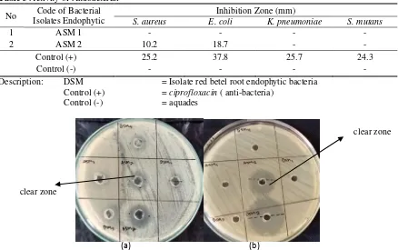
mm (Table 3). Isolates of Endophytic bacteria that have inhibitory characterized by the formation of clear zone (Fig. 1 (a) and (b)).
Table 3 Activity of Antibacterial
No Isolates Endophytic Code of Bacterial Inhibition Zone (mm)
S. aureus E. coli K. pneumoniae S. mutans
1 ASM 1 - - - -
2 ASM 2 10.2 18.7 - -
Control (+) 25.2 37.8 25.7 24.3
Control (-) - - - -
Description: DSM = Isolate red betel root endophytic bacteria Control (+) = ciprofloxacin ( anti-bacteria)
Control (-) = aquades
Figure 1 (a)Endophytic bacteria inhibition zone of red betel root (ASM) against S. aureus, (b) the inhibition zone of red betel root endophytic bacteria (ASM) to E. coli.
Anti-bacteria activity of endophytic bacteria isolates against 4 test bacteria are S. aureus, E. coli, S. mutans and K. pneumoniae. Of the 2 isolates , there are 1 isolate that have anti-bacteria against S. aureus
and E. coli. Isolat of endophytic bacteria have inhibitory characterized by the formation of clear zone. The formation of clear zone around the supernatant of endophytic bacteria indicate the possible presence antibacterial compounds capable of inhibiting the growth of pathogenic bacteria [5].
Shape of inhibition showed activity of secondary metabolites produced by the endophytic bacteria derived from red betel root. In general, anti-bacterial affect the formation of the cell wall or cell membrane permeability worked as bacteriosida, while antibacteria affecting of protein synthesis work as bacteriostatic [10]. Zone of Inhibitory is an activity of secondary metabolites of endophytic bacteria in inhibiting the growth of pathogenic bacteria by interfering with the metabolism, inhibiting of cell wall synthesis, disrupt the permeability of the cell membrane, inhibiting of protein synthesis and impair the synthesis of nucleic acid [11].
According to criteria of sensitivity from antibiotics classified into three criteria, in accordance with the National Committee for Clinical Laboratory Standards is criteria resistant (R) when the size of inhibitory 0-10 mm, intermediate (I) when the size of inhibitory of 11-19 mm and sensitive (S) when the size of inhibitory zone of ≥ 20 mm [12].
Zone of inhibitory is 18.7 mm on isolate of endophytic bacteria with code ASM 2 againts E. coli the indicates a sensitive (S), and the zone of inhibitory is 10.2 mm in isolate of endophytic bacteria with code ASM 2 against S. aureus the indicates criteria resistant (R). From the result of this research, Endophytic bacteria can be produce bioactive compounds that character is similar or identical to a compound produced by host plants. [13].
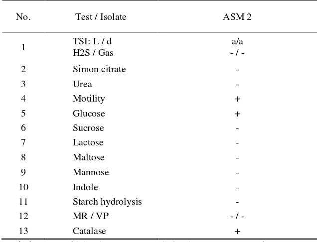
3.4 Biochemical Test Results
Biochemical tests on the study include testing, the TSI test, hydrolysis urea test, simon citrate test, motility test, carbohydrates test (glucose, sucrose, lactose, maltose, mannose), catalase test, indole test, starch hydrolysis and Methyl Red-Voges Proskauer (MR-PV) test. The bacteria tested with Biochemical are bacteria that have zone of inhibitory againts bacteria pathogen. The result of this research, Isolates of endophytic bacteria with code of ASM 2 on motility test, glucose and catalase showed positive results.
(a) (b)
clear zone
Table 4 Results of biochemical tests
No. Test / Isolate ASM 2
1 TSI: L / d
H2S / Gas
a/a - / -
2 Simon citrate -
3 Urea -
4 Motility +
5 Glucose +
6 Sucrose -
7 Lactose -
8 Maltose -
9 Mannose -
10 Indole -
11 Starch hydrolysis -
12 MR / VP - / -
13 Catalase +
Description: a = acid, b = base, MR = methyl red, VP= Voges Proskauer
Result of Motality test showed isolate of bacterial endophytic from red betel root showed positive result on ASM 2. Motility test is used to determine the ability of bacteria to moved. The results of positive test , if there is spread of bacterial colonies growth (visible turbidity) around inoculation [14]. Motility in bacteria can be caused by various mechanisms, but the most commonly involves flagella [15]. On Glucose testing showed that endophytic bacterial isolates with code ASM 2 showed positive results.
Result of catalase test on isolate of endophytic bacterial with code ASM 2 showed positive results in the catalase test, meaning that is bacterial isolates were capable of producing catalase enzymes. The catalase test is used to identify microbes capable of producing catalase enzymes, by breaking up hydrogen peroxide formed from the aerobic respiration process into dihydrogen oxide (H2O) and oxygen (O2) [16].
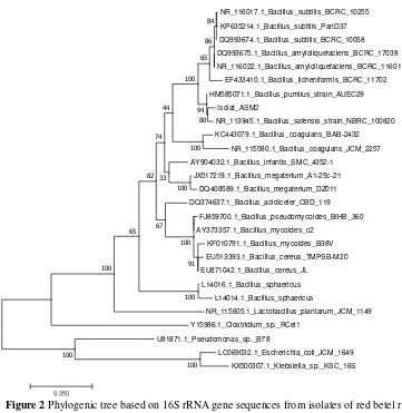
3.5 Molecular identification of 16S rRNA
Endophytic bacteria species identified molecularly by PCR using universal primers 63F and 1387r. The results of amplification on an agarose gel electrophoresis at a voltage of 100 V and a current of 400 A was observed under UV transluminator. Electrophoresis results showed DNA encoding 16S rRNA genes in parallel on the size of ± 1324 bp as compared with the DNA marker (100 bp DNA ladder).
After the identification followed by sequencing of 16S rRNA gene, results of sequencing were processed using Bioedit program. Then the 16S rRNA gene sequence data derived from isolate of endophytic bacteria compared with the existing data base on Gene Bank, the comparative results of sequencing then visualized in tree form phylogenic based genetic distance or evolutionary distance.
Table 5 Based Genetic distance of endophytic bacteria isolated from red betel root (ASM 2) acording the bacteria reference (standard) which is accessible from GenBank.
No Endophytic bacteria
isolates code bacteria reference genetic distance
1 ASM 2 Bacillus Safensis strain NBRC 100820 0.010
NR_116017.1_Bacillus_subtilis_BCRC_10255
Figure 2 Phylogenic tree based on 16S rRNA gene sequences from isolates of red betel root (ASM 2)
Phylogenetic is a method used in systematics to understand the diversity of living things through the reconstruction of kinship relations (phylogenetic relationship). The phylogenetic tree is a graph used to describe the kinship consisting of a number of point and branches with only one branch connecting the two closest points. Each point represents the taxonomic units and each branch represents the relationships between units that describe the hereditary relationship with the ancestors [17].
Endophytic bacteria isolated from red betel root (ASM 2) are one group with several strains of the species Bacillus Safensis strain NBRC 100820 and Bacillus pumilus strain AUEC29 (Fig. 2). Based on genetic distances (Table 5), isolated with code ASM 2 have The closest kinship with Bacillus pumilus strain
AUEC 29 the closest genetic distance of 0.007.
IV.
C
ONCLUSIONIsolate of endophytic bacteria from red betel root, obtained by 2 isolated, inhibitory test showed one (1) isolated which have inhibitory to S. aureus and E. coli. Biochemical result showed ASM 2 positive on glucose, motility and catalase. Molecular identification of 16S rRNA showed isolated with code ASM 2 that had a kinship relationship with Bacillus pumilus strain AUEC29 at the 0.007 gene distance.
Acknowledgements
This article paper can be accomplished with the help of lecturers, staff of biomedical laboratory from RSUD NTB .
R
EFERENCES[1]. Widjaja, E.A, Rahayuningsih, Y. Rahajoe, J.S. Ubaidillah, R. Maryanto, I. Walujo, E.B, Semiadi, G. 2014. Kekinian Keanekaragaman Hayati Indonesia. LIPI Press, Kementerian Lingkungan Hidup dan Bappenas.
[2]. Hongsheng, Y. Zhang, L. Li, L. Zheng, C. Guo, L. Li, W. Sun, P. & Qin, L. 2010. Recent developments and future prospects of entimicrobial metabolites produced by endophytes. Microbiological Research 165 437-449.
[4]. Hallman J, Halmann AQ, Miller WG, Sikora RA & Lindow SE. 2000. Endophytic colonization of plant by the biocontrol agent Rhizobium etli G12 in relation to Meloidogyne incognita infection. Phtopathol. 91:415-422
[5]. Purwanto, U.M.S. Pasaribu, F.M. & Bintang, M. 2014. Isolasi Bakteri Endofit dari Tanaman Sirih Hijau (Piper betle L.) sebagai penghasil senyawa anti bakteri. Current Biochemistry Volume 1 (1):51-57 e-ISSN.
[6]. Kristiningrum, E. & Meymurti. 2012. Dahsyatnya Khasiat Herbal Untuk Hidup Sehat. Jakarta Timur : Dunia Sehat.
[7]. Aris, M. Sukenda. Harris, E. Sukadi, M. F. Yuhana, M. 2013, Identifikasi molecular patogen dan desain PCR. Vol. 1 No. 3: 43-50.
[8]. Jenson I, Moir CJ. 2003. Bacillus cereus and other Bacillus spesies. CH 14 In: Hocking AD (ed) Foodborne microorganisms of public health significance.
[9]. Setlow peter. 2005. The bacterial spore: nature’s survival package. Culture. ISSN 0965-0989 Vol 26 No 2. [10]. Mutschler, E 1991. Dinamika Obat, Ed. 4, translated by Widjianto, M.B. dan Setiadi, A.R. Penerbit ITB: Bandung [11]. Brooks, Geo, Janet S. Butel, L. Nicholas Ornston. 2005. Medical Microbiology. Jakarta: EGC
[12]. Noviana, H. 2004. Pola kepekaan antibiotic Escherichia coli yang diisolasi dari berbagai specimen klinis. Mikrobiologi Fakultas Kedokteran Universitas Katolik Atma Jaya Jakarta. Vol. 23. No. 4
[13]. Tan, R.X and Zou, W.X. 2001. Endophytes: a Such Source of Funcional Metabolites. Natural Product Rep. 18:448-459. [14]. Brown, A 2001. Microbiological Applications Lab Manual. 8th Ed. The McGraw-Hill Companiens, New York.
[15]. Shield P, Cathcart L. 2013. Motility test medium protocol. American Society for
Microbiology.hhtp://www.microbelibrary.org/library/laboratory-test/2871-motility-test-medium-protocol. [16]. Djide, N. Sartini. 2006. Dasar-dasar Microbiology. Universitas Hasanudin, Makassar.