Original Article
The effect of an
Annona muricata
leaf extract on
nutritional status and cytotoxicity in colorectal cancer:
a randomized controlled trial
Lili Indrawati
MKes1, Purwantyastuti
Ascobat
SpFK2, Budiman Bela
Sp.MK(K)3,
Murdani Abdullah
SpPD-KGEH4, Ingrid S Surono
PhD51Department of Pharmacotherapy, Faculty of Medicine, Universitas Kristen Indonesia, Indonesia 2Department of Pharmacology, Faculty of Medicine, Universitas Indonesia, Indonesia
3Department of Microbiology, Faculty of Medicine, Universitas Indonesia, Indonesia 4Department of Internal Medicine, Faculty of Medicine, Universitas Indonesia, Indonesia 5Food Technology Department, Faculty of Engineering, Bina Nusantara University, Indonesia
Background and Objectives:Annona muricata leaf infusion has traditionally been consumedto maintain health, but is now considered for use in treating cancer patients. The objective of this study was to elucidate the effects of
A. muricata leaf extract in humans and human cell lines. Methods and Study Design: Thirty outpatients with colorectal cancer who had undergone primary tumor resection were enrolled in a randomized double-blind place-bo-controlled pre–post-trial. They were divided into two groups: those who ingested A. muricata leaf extract (n=14) and those who ingested a placebo (n=14) daily for 8 weeks. Twenty-eight subjects completed the trial; they were equally distributed between the two groups. Serum from patients of both groups was compared for cy-totoxicity against colorectal cancer cell lines. The nutritional status of patients was monitored throughout the study. Results: Ex vivo and clinical studies showed higher cytotoxicity in the supplemented group compared with the placebo group. Further research is required to investigate the long-term effect of A.muricata leaf extract, par-ticularly on parameters directly related to cytotoxic activity toward colorectal cancer cells and nutrition status.
Key Words: Annona muricata, leaf extract, nutrition status, colorectal cancer, cytotoxicity
INTRODUCTION
The prevalence of colorectal cancer is rising in Asia1, where it is now the third most common malignant disease in both men and women. Data from the International Agency for Research shows that the incidence of colorec-tal cancer in many affluent Asian countries is similar to that in the West. In recent decades, Eastern Asian coun-tries such as China, Japan, South Korea, and Singapore have experienced a two-to-four fold increase in the inci-dence of this disease,2 and data from 13 cancer registries in Indonesia show that colorectal cancer is one of the five most prevalent cancers in males and females.3
Annona muricataL. (Soursop) belongs to the An-nonaceae family. It has been investigated as a potential source of biologically active annonaceousacetogenins, some of which have demonstrated powerful antitumor activity.4 The cytotoxicity of acetogenins was found to be stronger in tumorous cells than in normal cells.5 One study showed that application of an ethyl acetate extract of A. muricata leaf (EEAML) significantly reduced the formation of colonic aberrant crypt foci (ACF) compared with that in a cancer control group. When isolated from EEAML, annomuricin E inhibited the growth of HT-29 cells with an IC50 value of 1.62±0.24 μg/mL after 48 h. The cytotoxic effect of annomuricin E was further sub-
stantiated by G1 cell cycle arrest and early apoptosis in-duction in HT-29 cells. Annomuricin E triggered mito-chondria-initiated events, including the dissipation of the mitochondrial membrane potential and leakage of cyto-chrome c from the mitochondria. Prior to these events, annomuricin E activated caspase 3/7 and caspase 9, and upstream it induced time-dependent upregulation of Bax and downregulation of Bcl-2 at the mRNA and protein levels.6
The nutrition status of patients with gastrointestinal cancer is related to both a systemic inflammatory re-sponse and an advanced stage of disease. Results of an animal study have suggested that ethanolic extract of A. muricata leaves can be considered a potential source of bioactive compounds with antinociceptive and anti-inflammatory activity, such as alkaloids, essential oils, and acetogenins.7 The extract was observed to be effect-
Corresponding Author: Lili Indrawati, Faculty of Medicine, Christian University of Indonesia. Jalan Mayjen Sutoyo no 2. Cawang-Jakarta, Indonesia.
Tel: +62-21-29362033, 081513303424; Fax: +62-21-29362038 Email: [email protected]
Manuscript received 19 January 2016. Initial review completed 29 January 2016. Revision accepted 28 March 2016.
ive in overcoming acute and chronic inflammation,8 and its antioxidative properties were reported.9
The reported evidence motivated this human study, which explored the effect of A. muricata on ex vivo pa-rameters and evaluated its effect on colorectal cancer through an assessment of nutrition status.
METHODS
Subjects and trial criteria
The subjects were colorectal cancer (CRC) outpatients at the CiptoMangunkusumo teaching hospital, Faculty of Medicine, University of Indonesia, Jakarta, Indonesia, after having undergone surgery for complete primary tu-mor resection. Thirty patients were recruited and random-ly assigned to either the A. muricata leaf extract group or to a placebo group for 8 weeks. The study protocol was approved by Ethics Committee, Faculty of Medicine, University of Indonesia (406/H2.F1/ETIK/2013), and registered on ClinicalTrials.gov under the identifier NCT02439580. Written informed consent was obtained prior to the study, with voluntary participation.
Inclusion criteria
Male and female CRC patients older than 30 years who had undergone primary tumor resection and were willing to take one capsule per day of A. muricata extract or a placebo as an additional therapeutic treatment throughout the study period were included in the study. In addition, the patients were required to have satisfactory hematolog-ical and biochemhematolog-ical function and a Karnofsky perfor-mance status of ≥60%.
Exclusion criteria
Patients with the following conditions were excluded from the study: uncontrolled hypertension (untreated sys-tolic blood pressure >160 mm Hg, or diassys-tolic blood pres-sure >95 mm Hg); serious heart problems; kidney, liver, endocrine, or neurologic or psychiatric disease; a disabil-ity rendering them unable to communicate verbally; or a history of cancers other than colorectal (such as non-melanoma skin cancer, basal cell carcinoma, and squa-mous cell carcinoma) in the past 5 years. Pregnant or lac-tating women, and those not using adequate contraception, were also excluded. In addition, patients taking other in-vestigational drugs, patients with HNPCC (Lynch syn-drome), and patients taking probiotic supplementation during the study period were also excluded to avoid po-tentially conflicting conditions and treatments.
Annona muricata L. extract
The A. muricata extract used in this study is ethanol-soluble fraction of A. muricata leaves water extract (ESFAM). ESFAM contains 0.36% acetogenin (w/w) or 3.6 mg/g, and a 10 g water extract is equivalent to a 2 g ethanolic fraction.
In this study, the CRC patients consumed either 300 mg of ESFAM, or maltose as a placebo, in the form of a cap-sule after breakfast.
Protocol
A randomized double-blind placebo-controlled pre–post trial (RCT) was conducted. The patients were assigned
consecutively into either ESFAM or placebo, through block randomization (four patients per block); supple-mentation was administered for 8 weeks.
Anthropometry and dietary intake measurements were conducted every 2 weeks throughout the study period (Figure 1). The patients recorded their food intake as measured using a food scale, and energy and nutrient in-takes were calculated using average values calculated over 2 days.
Peripheral blood samples were drawn from the patients through venipuncture at the baseline and at the end of the study period; the hemoglobin level was analyzed using an automated biochemical analyzer (Sysmex).
Ex vivo study
Venous blood samples used in ex vivo study were centri-fuged at 3,000 rpm for 10 min to obtain serum and then labeled and maintained at -80°C until analysis. Ex vivo study was performed by treating colorectal cell lines with the serum of patients from both groups. The cytotoxic activity of serum was assessed using an MTT assay; cells were cultured in 96-well microtiter plates, where each well contained 2 ×10⁴ cells, and treated for 48 h. Cytotox-icity was assessed using the MTT test (Trevigen’s TACS® MTT Cell Proliferation Assay), in triplicate.10-11
The human colorectal cell line types used in this study were COLO 205 and DLD-1 and were purchased from ATCC® (Catalog No. 222 and Catalog No. CCL-221, respectively); they were maintained according to supplier guidelines (American Type Culture Collection, ATCC, Manassas, VA). In addition, the human embryon-ic kidney (HEK) cell line was used as the normal control cell line; it were obtained from the Institute of Human Virology and Cancer Biology, University of Indonesia.
DLD-1 is derived from the Dukes’ type C human colo-rectal adenocarcinoma tissue of a male subject, and CO-LO 205 is derived from the Dukes’ type D human colo-rectal adenocarcinoma tissue of a 70 year old Caucasian male. Both lines were isolated from metastatic sites. Cells were incubated with 95% air and 5% CO2 at 37°C; all cells were maintained below passage 20 and used in ex-periments during the linear phase of growth. The human colorectal cancer cell lines DLD-1 and COLO 205 were maintained in RPMI1640 medium and HEK was main-tained in DMEM, both mediums were supplemented with 10% FCS, 2 mM L-glutamine, 100 units/mL penicillin, and 100 mg/mL streptomycin and maintained in a humid-ified environment containing 5% CO2 at 37°C.
Statistical analysis
during supplementation. A value of p<0.05 was consid-ered significant.
RESULTS
Nutrient intakes
Of 253 patients from the two centers, 30 met the inclusion criteria and consented to take part in the trial; they were randomly allocated into two groups (n=15), those taking an ESFAM supplement and those taking a placebo (control group). Although 30 patients participated, only 28 aged 30–80 years (x=50.2 years) completed the study, and this number was divided equally between the placebo (n=14) and the ESFAM (n=14) group. The baseline characteristics of the patients are shown in Table 1. Table 2 shows the nutrition status and hemoglobin
concentra-tion of the patients at the baseline, which was comparable between the groups.
Ex vivo experiment
The ex vivo experiment on cytotoxic activity was con-ducted using serum from the CRC patients. Table 3 shows significant growth inhibition of DLD-1 colorectal cancer cells line (p=0.013) after ESFAM administration compared with before ESFAM administration (baseline).
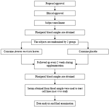
Serum from the ESFAM group also showed cytotoxici-ty against COLO 205 (p=0.08) compared with the base-line serum, but there was no significant difference com-pared with the placebo group. In consideration of the aforementioned results, a value of p≤0.08 can be used instead of p<0.05 to show a significant difference be-Proposal approval
Ethical approval
Subject enrollment
The subjects are randomized by 2 groups Pheriperal blood samples are obtained
Consume Annona muricata leaves Consume placebo
Pheriperal blood samples are obtained Followed up every 2 weeks during
supplementation
Serum obtained from blood sample were used to treat cell lines in ex vivo study
Data analysis and final examination
Figure 1. Flow chart of study design
Table 1. Medical history of patients in each group
Groups Between group
difference p value† ESFAM (n=150) Placebo (n=15)
Gender (%) Men 66.7 66.7 1.00
Women 33.3 33.3
Age (yr) 51.0±15.0 52.2±10.2 0.79
Chemotherapy (%) Received 80.0 73.3 1.00
Not 20.0 26.7
Tumor stage (%) I-II 26.7 33.3 1.00
III-IV 73.3 66.7
tween groups.
The serum obtained from the ESFAM group signifi-cantly increased HEK cells (p=0.006) compared with the serum from the placebo group, which showed significant cytotoxic activity against HEK cells compared with the baseline (p=0.04). HEK cells are normal cells, hence, serum was not expected to have cytotoxic activity.
Nutrient intake and anthropometry
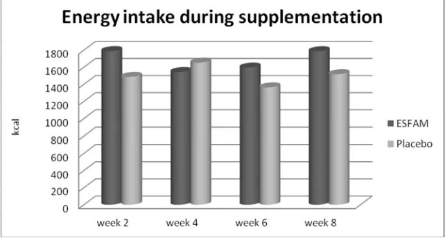
In general, the total energy intake (Figure 2) of the pa-tients was lower than the recommended daily allowance for Indonesia (Permenkes No.75, 2013) in all age groups. There was no significant difference in nutrient intake be-tween groups throughout the 8 weeks indicating that sup-plementation had no effect on the energy intake of either the ESFAM group or the placebo group. However, there was significant increase within group as time point. In the ESFAM group, energy intake at week 8 (1787.55±620.2 kcal) significantly increased compared with week 4 (1544.31±560.43 kcal) and week 6 (1596.6±531.11 kcal),
at p=0.04 and p=0.02, respectively. In placebo group, there was no significant differences in energy intake throughout the 8 weeks of measurement, and the energy intake was stable at all points of observation.
There was also no significant difference in the anthro- pometric status between groups. Comparing the endline values of both groups with the baseline values revealed that the nutrition status of both groups was maintained. In addition, the BMI of the patients during supplementation was stable among the measurement points, with the BMI difference being lower than one unit between the groups throughout the 8 weeks (Figure 3). Furthermore, hemo-globin concentrations before and after supplementation were within a normal range of values, showing that ESFAM did not reduce the hemoglobin concentration (Table 4).
Combining human and ex vivo studies
Nutrition status and hemoglobin concentrations are con-sidered as quality of life indicators. Data obtained from
Table 2. Nutrition status and haemoglobin levels of patients at the baseline
Nutrition status ESFAM (n=15) Groups† Placebo (n=15) Between group difference p value‡ Total energy intake (kcal/day) 1788.9±494.1 1487.6±564.5 0.14
BMI 23.3±0.0 21.0±3.2 0.17
Hemoglobin 12.7±1.6 12.6±1.5 0.88
†Data are expressed as the mean±SD. ‡Independent sample t test was performed.
Table 3. Cytotoxicity of serum against cell lines in each group
ESFAM (n=10) Living cells (%)
Within group difference p
value
Placebo (n=10) Living cells (%)
Within group difference
p value
Between group difference
p value‡ HEK Before 70.46 (58.33-78.27) 0.08 71.1 (58.4-98.2) 0.04 0.006
After 78.63 (64.32-91.29) 66.0 (58.5-71.7)
DLD-1 Before 56.28 (33.54-105.06) 0.01 32.7 (15.6-93.6) 0.86 0.08 After 52.16 (36.70-74.92) 49.7 (33.3 -71.7)
COLO 205 Before 83.5±29.81 0.08 81.6 (66.6-130.1) 0.09 0.47 After 72.28±24.03 72.7 (57.5-88.4)
†Data are expressed as the mean ± SD for normally distributed data and the median (25th, 75th percentiles) for non-normally distributed
data.
‡Independent sample t test was performed for normally distributed data, Mann-Whitney U test for non-normally distributed data.
the ESFAM group revealed that energy intake, BMI, and hemoglobin concentrations remained stable or increased (Table 5). Moreover, the results from the placebo group were comparable (Table 5), meaning that the quality of life of the patients in both the ESFAM and placebo groups was maintained throughout the 8 weeks as shown by stable or increased energy intake and BMI. Ninety seven percent of the serum obtained from ESFAM group exhibited an inhibitory property against one or all of the CRC cell lines. By contrast, only 58.3% of the serum in placebo group showed inhibitory properties against one or all of the CRC cell lines (Table 5). While 72% of the se-rum obtained from ESFAM group increased normal HEK cells and only 14.3% of the serum in placebo group showed an ability to increase normal HEK cells (Table 5).
DISCUSSION
This study is the first to expose CRC cell lines to the se-rum of patients extracted after supplementation with ESFAM. Serum from patients supplemented with ESFAM was used because it currently is not possible to conduct bioavailability studies as a result of the uncer-tainty regarding the bioactive compounds in the extract. In vitro study on ESFAM may reflect the direct effect of the extract on tumor cells as it passes through tumors in the colon; ex vivo study using patient’s serum is consid-ered to represent the effect of ESFAM after it is absorbed and reaches the plasma. In non-gastrointestinal cancer,
the amount of extract that reaches the blood (bioavailabil-ity) is the fraction considered able to exert an effect on cancer cells.
The previous in vitro cytotoxicity against colorectal cancer cell lines12 are in agreement with the present ex
vivo study, indicating that the bioavailability of the ex-tract was adequate, enabling it to reach the blood and in-hibit cancer cells. The results of this ex vivo study show that the serum had a higher inhibition capacity toward DLD-1, which may be due to the difference in cell line origins: DLD-1 is derived from Dukes’ type C, whereas COLO 205 is derived from Dukes’ type D colorectal ade-nocarcinoma, which is a more advanced stage colorectal cancer than Dukes’ type C. Before using cell lines de-rived from ATCC, efforts to develop cell line dede-rived from CRC patients (primary tumor cell line) was highly contaminated since these are colorectal tissue.
The results of the present study also showed that serum from the ESFAM group exhibited selectivity in exclusive-ly inhibiting the growth of colorectal cancer cells and not inhibiting the normal cells. The cytotoxicity of acetogen-ins was reported to be stronger in tumorous cells than in normal cells:5 The primary site of action of acetogenins is complex I of the electron transport chain in mitochon-dria.13
Another study evaluated the chemopreventive proper-ties of EEAML on azoxymethane-induced colonic ACF in rats. The cytotoxic compound of EEAML
(annomuri-Figure 3. Changes in BMI throughout trial
Table 4. Haemoglobin concentrations in each group
Baseline Endline Within-group difference p-value Between-group difference p-value‡
ESFAM (n=15) 12.7 ±1.7 12.8±2.1 0.582 0.867
Control (n=15) 12.4±1.4 12.6±1.7 0.539
†Data are expressed as the mean±SD. ‡independent sample t test was performed.
Table 5. Combined human and ex vivo studies Group Energy intake
stable/increased stable/increased BMI stable/increased Hemoglobin (normal cell) increased HEK DLD-1 or COLO 205 (CRC cell) decreased
ESFAM 66.7% 91.7% 100% 72% 97%
cin E) was isolated, and its apoptosis-inducing effect was investigated against the HT-29 colon cancer cell line.6 The cytotoxic activity in this in vitro study was consistent with the present ex vivo study, implying that active ingre-dients were able to reach the blood and inhibit the colo-rectal cancer cells in the present study.
The patients in this study were asked to record their food intake, mainly to assess their appetites. The inhibi-tion of tumorous cells was expected to improve the gen-eral condition of the patients, including their appetite and nutrition status. However, no significant changes in the food intake or anthropometry data at any of the four measurement points were observed between the groups, indicating that the nutrition status of patients in both groups was stable throughout the 8 weeks of supplemen-tation. However, there was a significant energy intake increase ESFAM group but this improvement was not observed in the placebo group. Longer supplementation period may improve the nutrition status of patients, and a larger and longer cohort study is required to test this hy-pothesis.
Previous studies have suggested that nutrition is a ma-jor concern in oncology, and a decline in nutrition status may ensue from both the course of the disease and its treatment. Cancer-associated malnutrition and a progres-sive loss of body weight are common features in most cancer patients, although prevalence rates vary from9% to 85% depending on the tumor type and disease stage.14
Some patients in this study took the extract in parallel with chemotherapy. However, their compliance remained acceptable despite the adverse effects of chemotherapy, and there was neither bodyweight loss nor a reduction in nutrition intake during supplementation of A. muricata extract, confirming there was no detrimental effect.
The most common adverse effects of chemotherapy on nutrition status are anorexia, altered perception of taste and smell, food aversion, nausea and vomiting, mucositis, xerostomia, constipation, diarrhea, and early satiety. Chemotherapy may also cause abdominal cramps and bloating, paralytic ileus, and even malabsorption. Some antineoplastic agents such as fluorouracil, adriamycin, methotrexate, and cisplatin may induce severe gastroin-testinal complications.14 In this study, some of the pa-tients undergoing chemotherapy felt better while consum-ing the A. muricata leaves extract supplement, and one subject felt that the mass in her abdomen was less uncom-fortable and she experienced reduced stimulation to strain.
Many findings on phytochemicals are inconclusive be-cause the studies are conducted either in vitro or by using animals in vivo. This is partly due to our limited under-standing on phytochemical bioavailability, on which health benefits depend. In addition, the transport mecha-nisms for delivering phytochemicals to target sites, the phytochemical metabolism of the human body, and bi-omarkers exerting health benefits are poorly understood. Using subjects’s serum who have ingested the A. muri-cata leaves extract to examine the effect of its bioactive compounds on cells is a more suitable method in elucidat-ing the effect of the bioactive compounds on the human body than directly treating cells in vitro by the extract. This approach is more relevant, because bioavailability data on phytochemicals in vivo are limited.
The present RCT provides evidence that serum from patients taking the extract showed cytotoxicity against human colorectal cancer cell lines. The reduced viability of cancer cells may improve the nutrition status and quali-ty of life of cancer patients, revealed that an adequate amount of extract reaches the blood to kill cancer cells. Therefore, an optimal dose should be determined for ad-ministration to provide the adequate amount required to reach the blood. However, the present RCT cannot be expected to produce results that are directly relevant to all patients in all settings.
More importantly, the effect of A. muricata leaf extract should be studied in relation to the outcomes of various cancers over a study period longer than the human cell lifespan. For example, to examine the effect on red blood cell counts, a study should be conducted over a period of at least 120 days; the longer the study period, the more cells will benefit from the treatment, which may then im-prove the quality of life. Moreover, future studies should be performed using a more comprehensive study design.
More extensive studies are also required on the absorp-tion and bioavailability of the extract and its fracabsorp-tions. In addition, most dietary compounds contain a mixture of acetogenins and other bioactive compounds may present, and these should be identified in A. muricata extract. Some phytochemicals may have synergistic effects, alt-hough they are not individually effective as chemopre-ventative agents. Thus other bioactive compounds, in addition to acetogenins, should also be explored to pro-vide a thorough understanding on the chemopreventive effect of the extract and its fractions. Additional extensive studies are thus required.
Finally, the amount of extract used in this study, in terms of the equivalent concentration of the active ingre-dient, was extrapolated from the amount traditionally consumed. This was performed to find an effective and affordable treatment for cancer patients. Thus, the amount of A. muricata used traditionally, which is in line with the results of in vitro, ex vivo, and pilot human studies, can be used as an adjunct to standard treatment for colorectal cancer patients at a dose of 7–15 cups daily prepared as an infusion which is equivalent to 1030 mg annonacin. However, a study with a large cohort of colorectal cancer patients and a thorough evaluation of the efficacy and safety of each fraction of the water extract is required to confirm this finding and recalculate the dose.
ACKNOWLEDGEMENTS
This study was supported by a grant in aid from the Centre for Ageing Studies, Universitas Indonesia (contract number 036/H2. R12.5/PPM.01.04/2012).
AUTHOR DISCLOSURES
The authors have no financial or commercial conflicts of inter-est in this work.
REFERENCES
1. Yee YK, Tan VPY, Chan P, Hung IFN, Pang R, Wong BCY. Epidemiology of colorectal cancer in Asia. J Gastroenterol Hepatol. 2009;24:1810-6. doi: 10.1111/j.1440-1746.2009.06 138.x.
4251/wjgo.v4.i4.68.
3. Soeripto, Indrawati, Indrayanti. Gastro-intestinal cancer in Indonesia. Asian Pac J Cancer Prev. 2003;4:289-96. 4. Yu D-Q. Recent works on anti-tumor constituent from
Annonaceae plants in China. Pure and Applied Chemistry. 1999;71:1119-22. doi: 10.1351/pac199971061119.
5. García-Aguirre KK, Zepeda-Vallejo LG, Ramón-Gallegos E, Alvárez-González I, Madrigal-Bujaidar E. Genotoxic and cytotoxic effects produced by acetogenins obtained from Annona cherimolia Mill. Biol Pharm Bull. 2008;31:2346-9. doi: 10.1248/bpb.31.2346.
6. Moghadamtousi SZ, Rouhollahi E, Karimian H, Fadaeinasab M, Firoozinia M, Abdulla MA, Kadir HA. The chemopotential effect of Annona muricata leaves against azoxymethane-induced colonic aberrant crypt foci in rats and the apoptotic effect of Acetogenin Annomuricin E in HT-29 cells: a bioassay-guided approach. PLoS One. 2015; 10:e0122288. doi: 10.1371/journal.pone.0122288.
7. Orwa C, Mutua A, Kindt R, Jamnadass R, Simons A. Agroforestree Database: a tree reference and selection guide version 4.0; 2009. [cited 2011/02/15]; Available form: http://www worldagroforestry org/af/treedb/.
8. Foong CP, Hamid RA. Evaluation of anti-inflammatory activities of ethanolic extract of Annona muricata leaves. Revista Brasileira de Farmacognosia. 2012;22:1301-7. doi: 10.1590/S0102-695X2012005000096.
9. Baskar R, Rajeswari V, Kumar TS. In vitro antioxidant studies in leaves of Annona species. Indian J Exp Biol. 2007; 45:480.
10.Alley MC, Scudiero DA, Monks A, Hursey ML, Czerwinski MJ, Fine DL, Abbott BJ, Mayo JG, Shoemaker RH, Boyd MR. Feasibility of drug screening with panels of human tumor cell lines using a microculture tetrazolium assay. Cancer Res. 1988;48:589-601.
11.Vistica DT, Skehan P, Scudiero D, Monks A, Pittman A, Boyd MR. Tetrazolium-based assays for cellular viability: a critical examination of selected parameters affecting formazan production. Cancer Res. 1991;51:2515-20. 12.Kim G-S, Zeng L, Alali F, Rogers LL, Wu F-E,
Sastrodihardjo S, Mclaughlin JL. Muricoreacin and murihexocin C, mono-tetrahydrofuran acetogenins, from the leaves of annona muricata in honour of professor G. H. Neil Towers 75th birthday. Phytochemistry. 1998;49:565-71. doi: 10.1016/S0031-9422(98)00172-1.
13.Gupta A, Pandey S, Shah D, Yadav J, Seth N. Annonaceous acetogenins: the unrevealed area for cytotoxic and pesticidal activities. Systematic Reviews in Pharmacy. 2011;2:104. doi: 10.4103/0975-8453.86299