190 December 2012
Accredited by DGHE No: 66b/DIKTI/Kep/2011 DOI: 10.5398/medpet.2012.35.3.190
*Corresponding author:
Phone: +62 651 7551536; E-mail: [email protected]
The Ability of Immunoglobulin Yolk Recognized the Antigen in the Tissue of
Ascaridia galli
Darmawia,*, ��� �al�is, ��� �al�isa, M�� Hambala, R�� Tiuriab, ��� P�� Priosoeryantob, & E�� Handharyanib aFaculty of Veterinary Medicine, Syiah Kuala University
Jln. Tgk. H. Hasan Krueng Kale No. 8 Darussalam, Banda Aceh 23111, Indonesia bFaculty of Veterinary Medicine, Bogor Agricultural UniversityBogor Agricultural University
Jln. Agatis, Kampus IPB Darmaga Bogor 16680, Indonesia (�ece��e�� 2��10�2012�� �e��e�e�� 1��01�201��� �cce��e�� 2��01�201���ece��e�� 2��10�2012�� �e��e�e�� 1��01�201��� �cce��e�� 2��01�201�� 2��10�2012�� �e��e�e�� 1��01�201��� �cce��e�� 2��01�201��
A�STRACT
Antigen-antibody reaction is an important tool for the analysis of localization of target molecules, including antigenic protein within worm tissues�� The purpose of the present research was to demonstrate the ability of immunoglobulin yolk (IgY) anti-excretory/secretory recognized the antigen in the tissue of Ascaridia galli by mean of immunohistochemistry method�� The excretory/ secretory protein was procured from A. galli and concentrated by mean of �i�aspin ��,��� M�C���and concentrated by mean of �i�aspin ��,��� M�C��� IgY was produced by egg yolks of immunized chickens with excretory/secretory, and purified using fast protein li�uid chromatography (FPLC) method�� A. galli adult worms were cut in trans�ersal and longitudinal section of the center and anterior region�� Slides were incubated with both primary IgY for o�ernight at 4 oC and secondary antibody rabbit anti-chicken IgY HRP-conjugate for one hour at room temperature�� The slides were stained with �-amino, �-ethylcarbazole (AEC) chromogen,�-amino, �-ethylcarbazole (AEC) chromogen, (AEC) chromogen, counterstained with Lillie Mayer Haematoxylin, and mounted in glyserin a�ueous mount�� Antigen-antibody reaction was in�estigated under a microscope�� The result showed that antigen was appearedin�estigated under a microscope�� The result showed that antigen was appeared The result showed that antigen was appeared in the tissues such as cuticle, epicuticle, buccal ca�ity, and eggs inside the uterine of A. galli�� This research concluded that IgY stimulated by the excretory/secretory was able to recognized the antigen scattered in the tissues of A. galli so the IgY could be applied for immunodiagnostic��
Key words: Ascaridia galli, excretory/secretory, IgY, immunohistochemistry
A�STRAK
Reaksi antigen-antibodi adalah salah satu cara yang penting untuk menganalisis lokasi target molekul, termasuk protein antigen di dalam jaringan cacing�� Tujuan riset ini adalah untuk menunjukkan kemampuan immunoglobulin yolk (IgY) anti-ekskretori/sekretori mengenal antigen di dalam jaringan cacing Ascaridia galli melalui metode imunohistokimia�� Protein eksretori/sekretori diperoleh dari A. galli dan dipekatkan melalui �i�aspin ������� M�C��� IgY dihasilkan oleh kuning�i�aspin ������� M�C��� IgY dihasilkan oleh kuningIgY dihasilkan oleh kuning telur dari ayam yang di�aksinasi dengan ekskretori/sekretori, dan dimurnikan dengan menggunakan metode fast protein liquid chromatography (FPLC)�� Cacing A. galli dewasa dipotong secara melintang dan memanjang pada bagian tengah dan atas�� Slide dieramkan dengan kedua antibodi, yaitu antibodi primer IgY selama satu malam pada temperatur 4 oC dan antibodi sekunder IgY HRP-conjugate selama satu jam pada temperatur kamar�� Slide diwarnai dengan kromogen 3-amino, 9-ethylcarbazole (AEC), dilatar-belakangi oleh Lillie Mayer Haematoxylin, dan direkatkan di dalam gliserin�� Reaksi antigen-antibodi diamati di bawah mikroskop�� Hasil menunjukkan bahwa antigen ditampilkan di dalam jaringan seperti kutikula, epikutikula, rongga mulut, dan telur di dalam uterus cacing A. galli�� Riset ini menyimpulkan bahwa IgY yang dirangsang oleh ekskretori/sekretori mampu mengenal antigen yang tersebar di dalam jaringan A. galli sehingga IgY dapat diaplikasikan untuk imunodiagnostik��
December 2012 191 INTR�D�CTI�N
Ascaridiosis, caused by a nematode parasite
so-called �scar����a gall�, is one of the major poultry intestinal
roundworm infections. The disease is an important poultry health problem in many parts of the world, may give rise to serious economic problems in laying hens husbandry (Anwar & Zia-ur-Rahman, 2002). According
to some reports, �. gall� has a worldwide distribution
wich predominates in temperate zones (Schou e� al.,
2003). �. gall� is also found on most continents, but primarily in the tropical regions of Asia (Lalchhandama e� al., 2009), and Africa (Siamba e� al., 2007). Laying hens are usually infected by ingestion of feed and water that contain infective larvae. �. gall� infection is prevalent in Aceh (Fahrimal & Raflesia, 2002) and some parts of Java (Zalizar e� al., 2007; Balqis e� al., 2009; Suharti e� al., 2010).
The parasitological diagnosis of poultry ascaridi
-osis is often unreliable because the parasite’s eggs are not found in the stool during the early phase of infec
-tion. Even when the worms have matured the diagnosis may still be difficult since eggs are only intermittently released. The classic diagnosis of ascaridiosis usually depends on the demonstration of �. gall� eggs in the
feces (Fahrimal & Raflesia, 2002; Suharti e� al., 2010).
However, this method as described by many
investiga-tors, is not sufficiently sensitive. Diagnosis of worms infection by mean of immunodiagnostic methods during prepatency utilizing excretory/secretory antigen in some serologic tests have already been reported. The immu
-nodiagnostic tests could be utilized for the detection of disease even at the prepatent stages before eggs begin to appear in the feces. Prasad e� al. (2008) suggested that early diagnosis of ascaridiosis is necessary for prompt treatment before irreparable damage to the intestine occurs. Thus, serological tests are the most dependable diagnostic methods (Rokni & Kia, 2005; Hassan & Aziz, 2010).
Serological techniques such as immunodiffusion test using excretory/secretory and somatic antigen of the worm for the detection of antibodies against this parasite are sensitive and have been exploited for its
serodiagnosis. Karimi e� al. (2008) reported that excre
-tory/secretory and somatic antigen of Orn��hob�lharz�a �urkes�an�cum had strong cross reaction with each other
in agar gel diffusion test. Smith e� al. (2009) analyzed that excretory/secretory product identi��ed in the larvalidenti��ed in the larval secretome ofof Tela��orsag�a c�rcumc�nc�a, an importantan important
parasitic nematode of domestic small ruminants, were potentially involved in immunity so targets of local im
-munoglobulin A (IgA) responses in mucus from sheep rendered immune to infection.
Immunoglobulin yolk (IgY) antibody produced by chickens is considered to be a potent antibody for im
-munodiagnostic. These polyclonal and monospeci��c IgY antibodies are of higher-titer and speci��cally recognize
recombinant Hel�cobac�er �ylor� urease puri��ed from Escher�ch�a col� (Kazimierczuk e� al., 2005). Futhermore, application of IgY was extended for immunotherapy (Dias da Silva & Tambourgi, 2010). Diraviyam e� al.
(2011) described that the speci��c activity of IgY anti
-bodies against speci��c bacterial pathogens have been suggested as the mode of action of speci��c IgY to inhibit E. col� and Salmonella en�er�ca growth. Previous investi
-gation showed that passive immunization of chickens with anti-coccidia IgY antibodies provided protective immunity against coccidiosis caused by multiple spe
-cies of E�mer�a in broiler infection (Lee e� al., 2009). The authors described generation of IgY antibodies directed against various antigens to tested antigen-antibody reaction. However the standardization of test under different conditions and with different types of antigens is essential before its practical application and utility under ��eld condition. In this study, tissue sources of the antigen were investigated by immunohistochemistry in the body of adult worm sections using the IgY-anti ex -cretory/secretory �. gall� puri��ed from chicken egg yolk
of immunized laying hens.
MATERIALS AND METH�DS
IgY Anti-Excretory/Secretory Ascaridia galli Production
Excretory/secretory crude protein was procured from �. gall� cultured in flasks containing RPMI 16�0cultured in flasks containing RPMI 16�0 media. The protein was poured and concentrated byrotein was poured and concentrated bypoured and concentrated by mean of vivaspin 30,000 MWCO, and quantities deter-
deter-mined by Bradford method (λ= 280 nm) as described
by Darmawi e� al. (2009). Twowenty four-week old IsaTwowenty four-week old Isa
brown chickens were vaccinated with the crude protein of excretory/secretory, applied intra muscularly with an applied intra muscularly with an initial dose of 80 µg. The immunizations were repeated three times with dose of each 60 µg with an interval of one week. The ��rst immunizations were excretory/secre
-tory protein mixed with Fruend Adjuvant Complete (Sigma, USA) and subsequently mixed with Freund Adjuvant Incomplete (Sigma, USA). The chicken’s eggs were collected after ELISA test shown antibody titre increased signi��cantly (Darmawi e� al., 2008).
The antibody enrichment procedure was performed
as described by Darmawi e� al. (2010), a serial precipita
-tions was used. Chicken IgY was precipitated from egg yolks by adding 1 volume of �0% polyethylene glycol (PEG) 8000 (Sigma, USA) in PBS to 3 volumes of egg yolk, followed by centrifugation at 13,000×g for 20 min. IgY was extracted from egg yolks by means of ammo
-nium sulphate. The crude extraction suspension was mixed with 50% (V/V) saturated ammonium sulfate solution and stirred at � °C for 2 h. After centrifugation,
the precipitate was collected and dissolved in 0.01 M
thiophilic, 2-mercaptopyridine bond to sepharose high
performance, Hi TrapTM IgY Puri��cation (Amersham
192 December 2012
Ascaridia galli Parasite
�. gall� adult worms were procured from the intestine of freshly slaughtered chickens. They were brought to the laboratory from local restaurant. Worms were washed sometimes with phosphate buffered saline (PBS).
Tissue Preparation for Immunohistochemistry Protocol
The parasite tissues aimed for immunohistochemis
-try were ��xed in Bouin’s ��xer for 2� h at room tempera
-ture and embedded in paraffin block until use. �. gall� adult worms were preparated by mean of dehydration, clearing, in��ltration, embedding, cutting, and staining. The tissues were blocked with paraffin, and saved in re
-frigerator in order to cut off the paraffin for easy cutting. Paraffin blocks of the worm were prepared in a sequence of 10% neutral formalin ��xation, dehydration in graded alcohol series and paraffinization. The paraffin embed
-ded tissues of �. gall� adult worm were cut longitudi
-nally and transversally using a microtome. Thin sections (3-5 µm) of the worm tissues were floated and warmed3-5 µm) of the worm tissues were floated and warmed) of the worm tissues were floated and warmed
at 6060 oC and coated on object glasses. The sections were The sections were deparaffinised with three changes of xylene (III, II, and I) for 3 min of each. The sections were rehydrated through graded alcohols, namely: 95%, 90%, 80%, and 70% for95%, 90%, 80%, and 70% for for 3 min of each. The sections were rinsed (clearing) with(clearing) withwith
diionized water for 15 min. The endogenous peroxidasefor 15 min. The endogenous peroxidase activity was inactivated by incubating these sections in 3% HH2O2 for 20 min and skim milk for 30 min at room
temperature, and rinsed with three changes of diio-
diio-nized water and PBS for 5 min of each as described byPBS for 5 min of each as described by
Claeys e� al. (200�) and Pokharel e� al. (2006) with certain
modi��cations.
After rinsing, the slides were reacted with primary antibody procured from puri��ed IgY by mean of fast performans liquid chromatogra�� (FPLC) method. The slides were put on PBS-based in humidity chamber at � oC overnight, rinsed with three changes of PBS for 5 minrinsed with three changes of PBS for 5 min of each. The slides were dropped with secondary anti- The slides were dropped with secondary
anti-body (IgY conjugate HRP rabbit anti-chicken, Promega, USA). Peroxides complex were reacted for 30 min atfor 30 min at room temperature, and rinsed with three changes of PBS for 5 min of each. The reaction was colored by AEC The reaction was colored by AEC
chromogen (Sigma, USA) for 3 min at room temperature,at room temperature,,
and rinsed with three changes of PBS for 5 min of each.rinsed with three changes of PBS for 5 min of each. Finally, the tissue sections were counterstained with Lillie Mayer hematoxylin for 1 min, and rinsed with diionized water. The slides were attached with glyserin
and covered cover glasses. Positively stained with
AEC chromogen was investigated under a microscope (Olympus) with �0 objective magni��cations and video micrometer (Video measuring gauge IV – 560, FOR A Company limited) as described by ClaeysClaeys e� al. (200�) and Pokharel e� al. (2006) with certain modi��cations..
RES�LTS AND DISC�SSI�N
The results of the present study showed that egg yolk antibody IgY had strong reaction with antigen
some parts of the round worm’s tissue of �. gall� as seen
in Figure 1. Excretory/secretory product released by �. gall� contained antigenic substance(s) could stimulate speci��c antibody. This reflect that the antigenic materials are common between somatic and excretory/secretory products of parasite and there is no difference between the antigenicity of somatic and excretory/secretory antigens of �. gall�. This hypothesis supported by many previously reports exist about the role of excretory/se
-cretory released by nematode in generating immune
response mechanisms. Prasad e� al. (2008) suggested that
the puri��ed fraction of excretory/secretory antigen may be utilized for early diagnosis against Haemonchus con� �or�us in sheep. Smith e� al. (2009) analyzed of excretory/
secretory products released by Tela��orsag�a c�rcumc�nc�a,
where antigens that were targets of local IgA responses in mucus from sheep rendered immune to infection. The similar phenomenon observed by Hassan & Aziz (2010), excretory/secretory antigen of Toxocara ���ulorum infective larvae was the most diagnostic antigen and was successful in the detection of high infection percentage of toxocariasis among buffalo calves. Excretory/secre
-tory antigen has also been reported to be a better antigen for a serodiagnosis of clonorchiasis than crude antigen (Choi e� al., 2003).
Excretory/secretory as described by many investiga-tors, is more sufficiently sensitive and speci��c compared with somatic antigen. It has been shown that using the excretory/secretory antigens of worms by enzyme linked immunosorbant assay (ELISA) method could be
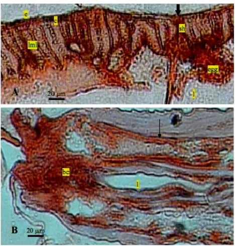
Figure 1. Positive reaction in AEC staining of �.gall� adult worm. A= tranversal section of the distal region (20x). B= lon
-gitudinal section of the anterior region (10x). (c= cu
December 2012 193 a good diagnostic tool for the illness. Rokni & Kia (2005)
have been used the ELISA test that overall excretory/ secretory antigen showed a more convincing diagnosis in comparison with somatic antigen of S�rongylo���es s�ercoral�s in human intestinal nematode infection,
al-though every interpretation of the results should be in accompany with clinical manifestations and a history of the disease. Karimi e� al. (2008) prepared and compared
the somatic and excretory/secretory antigens of O. �urke� s�an�cum in gel diffusion test. According to the results,
somatic antigens showed strong reaction with antisera raised against excretory/secretory antigens and excre-tory/secretory antigens also showed strong reaction with antisera raised against somatic antigens. Antigenicity of O. �urkes�an�cum excretory/secretory and somatic
antigens is the same in gel diffusion test. ZhengZheng e� al. (2011) found that the components of the excretory/secre-the components of the excretory/secre-tory products from a food-borne zoonotic parasite that resides in bile ducts and causes clonorchiasis, Clonorch�s s�nens�s localizes to the intestinal cecum and vitellarium
in adult worms. Hewitson e� al. (2011) described that
gastrointestinal nematode Hel�gmosomo���es �olygyrus re-veals dominance of venom allergen-like (VAL) proteins, which should allow functional testing of the various po-tentially immunomodulatory proteins. Rica e� al. (2005)
analyzed that amphidial glands, excretory/secretoryamphidial glands, excretory/secretory gland cells, pharyngeal glands secreted proteins in adult females and males of Syngamus �rachea, a cosmopolitan nematode parasite of the trachea of wild and domesti-cated galliformes and some passeriformes,
Excretory/secretory released by �. gall� was able to trigger humoral immune responses by mean of IgY an-tibody formation. It is a well-known concept that IgY is the major antibody produced by laying hens. IgY is con-tinually synthesized, secreted into the blood and trans-ferred to the egg yolk, where it is accumulated. Various authors revealed that IgY speci��c antibody formation in egg yolk could be stimulated by Hel�cobac�er �ylor�
(Kazimierczuk e� al., 2005), bacterial enteric (Chalghoumi e� al., 2009; Diraviyam e� al., 2011), and against E�mer�a acer�ul�na (Lee e� al., 2009).
IgY antibodies from egg yolk chickens should be commonly used for immunodiagnostics regarding the advantages of the IgY. Dias da Silva & Tambourgi (2010) supported that application IgY can be developed in im-munodiagnostic tests that provide more accurate results. IgY antibodies are therefore the easiest way to eliminate the errors that arise due reactions from the use of mammalian IgG. Previously, Hau & Hendriksen (2005) reported that the antibodies from the egg yolk have great potential for active implementation of the three Rs (replacing, reducing, and re��ning the use of laboratory animals to the extent possible) in polyclonal antibody production schemes. In the part of animal welfare, it is also ethically more attractive to produce chicken an-tibodies, as they are puri��ed from the yolk. Unlike the production of mammalian IgG antibodies, IgY produc-tion does not require bleeding of animals. Dias da Silva & Tambourgi (2010) explained that the amount of IgY that can be obtained from a hen is also much larger than from e.g. a similar sized mammals. So, chicken egg yolk IgY antibodies offer a practicable alternative to
mam-malian serum antibodies because of their feasibility for large-scale commercial production and the relatively noninvasive methods used for their preparation.
When the IgY anti-excretory/secretory, reacting to epitopes, was used as a primary antibody. The appear-ance of dark brown color of AEC was considered as the basis for evaluation of positive staining in the parasite tissues. As seen in Figure 1, very strong immunostaining was observed in the epicuticle and cuticle followed by syncytial hypodermis. A part of scattered parenchymal cells in the worm were faintly stained. The positive im-munohistochemical ��ndings of IgY bindings suggested strongly that these antigenic proteins were produced at the cuticle cells and transported to the syncytial via connecting tubules or pore canals. The positive reaction in AEC staining to the antigenic proteins were especially strong et the distal margin of cuticle. This histologic ��nding suggested also that proteins might released out-side of the worm.
IgY antibody puri��ed from yolk of laying hens immunized with excretory/secretory antigen showed positive reaction with somatic antigen that appeared in the body of �. gall�. Cuticle and epicuticle cells were
positively stained with AEC chromogen. Good staining was observed in longitudinal muscle layers and syncy-tial hypodermis. The cuticle in all nematode parasites is an extracellular hydroskeleton that is relatively inert, structurally robust, and selectively permeable. Antigenic protein of �. gall� were localized at egg inside the uterus
of the worm when observed by immunohistochemistry using the polyclonal IgY antibodies reacting to these pro-teins. Polyclonal IgY anti-excretory/secretory antibodies in immunized chicken yolks were reacting to almost all structures of �. gall� such as muscle, parenchymal cells,
lining cells of excretory canal. Buccal cavity also reacted with these polyclonal antibodies. It is possible that ex-cretory/secretory is synthesized from the oesophageal glands secreted into buccal cavity. Different stained abil-ity of worm structures con��rmed the excretory/secretory might represent from the some tissues of �. gall�.
Immunohistochemistry provides the most direct method for identifying both the cellular and subcellular distribution of protein, including the protein antigen of worms. The sensitivity of the immunohistochemistry test against protein antigen that appeared in the somatic of worms have been conducted by some researches. Immunohistochemical techniques employed by Claeys
e� al. (200�) showed that the formation of the cuticle,
microvilli in the embryonic intestine and adult nema-todes, Caenorhab�����s elegans. Pokharel e� al. (2006) found
that proteases were present in the adult female Se�ar�a cer�� tissues mainly localized in the body wall, outer
body surface of the parasite, epicuticle, cuticle, syncy-tial hypodermis and longitudinal muscle layers. This enzyme were also distributed in the nerve cord region, intestine, growing embryos, uterus and mature eggs of the parasite.
194 December 2012
(2009) described that the speci��city of polyclonal IgY antibody produced in chicken egg yolk was able to capture live organisms and has potential in the develop-ment of an immunocapture assay in a variety of clinical samples for Mycobac�er�um a��um subsp. �ara�uberculos�s, a chronic inflammatory bowel disease of both domestic and wild ruminants. An immunomagnetic bead ELISA using IgY against soluble egg extract as a capture anti-body was applied to detect circulating antigens parasitic disease caused by trematode flatworms in serum of mice infected with Sch�s�osoma ja�on�cum (Lei e� al., 2009).
Furthermore, Lei e� al. (2011) also described that the
same method appeared to be sensitive and speci��c by using serum samples for diagnosis of schistosomiasis of persons with schistosomiasis. Recently, Lu e� al. (2012)
successfully developed a novel method of immunoassay based on IgY is an effective approach to discriminate between previous exposure and current infection of S. ja�on�cum by mean of identi��cation and pro��ling
circu-lating antigens in sera of S. ja�on�cum infected patients.
C�NCL�SI�N
IgY stimulated by the excretory/secretory was able to recognized the antigen scattered in the tissues of �. gall� so the IgY could be applied for immunodiagnostic.
ACKN��LEDGMENT
We wish to thank the Directorate General of Higher Education, Ministry of Education and Culture Republic Indonesia for funding of the work grant from the Hibah Bersaing (No. 019/SP2H/PP/DP2M/III/2007). We also thank Mr. Sulaeman, Mr. Kosasih, and Mr. Kasnadi, Laboratory of Faculty of Veterinary Medicine of BogorBogor Agriculture Institute, for their expert technical help in, for their expert technical help in the materials preparation.
REFERENCES
Anwar, H�� & Zia-ur-Rahman�� 2002. Effect of �scar����a gall� infes
-tation on electrolytes and vitamins in chickens. J. Biol. Sci. 2: 650–651. http://dx.doi.org/10.3923/jbs.2002.650.651
�al�is, ���, Darmawi, M�� Hambal, & R�� Tiuria���� 2009.
Develop-ment of infective eggs of �scar����a gall� in �n ���ro cultiva -tion. J. Ked. Hewan 3: 183-189.3: 183-189.
Chalghoumi, R��, Y�� �eckers, D�� Portetelle, & A�� Théwis�� 2009.
Hen egg yolk antibodies (IgY), production and use for passive immunization against bacterial enteric infections in chicken: a review. Biotechnol. Agron. Soc. Environ. 13: 295-308.
Choi, M�� H��, I�� C�� Park, S�� Li, & S�� T�� Hong�� 2003. Excretory-se
-cretory antigen is better than crude antigen for the serodi
-agnosis of clonorchiasis by ELISA. The Korean J. Parasitol. �1: 35-39. http://dx.doi.org/10.33�7/kjp.2003.�1.1.35
Claeys, M��, D�� Vanhecke, M�� Cou�reur, T�� Tytgat, A�� Coomans, & G�� �orgonie�� 200�. High-pressure freezing and freeze
substitution of gravid Caenorhab�����s elegans (Nematoda: Rhabditida) for transmission electron microscopy. Nema
-tology 6: 319-327. http://dx.doi.org/10.1163/15685�10�2360
564
Darmawi, ��� �al�is, R�� Tiuria, M�� Hambal, & Samadi�� 2008.
Study of antibody titre in yolk from immunized chickens with excretory/secretory of stage L3�scar����a gall�. J. Agric.
8: 21-26.
Darmawi, ��� �al�is, R�� Tiuria, R�� D�� Soejoedono, & F�� H�� Pas-aribu�� 2009. Protein concentration and molecule weigh
determination of excretory/secretory of stage Lof excretory/secretory of stage L3�scar����a
gall�. J. Ked. HewanJ. Ked. Hewan. 3: 197-202.
Darmawi, ��� �al�is, R�� Tiuria, M�� Hambal, & Samadi�� 2010.
Puri��cation of immunoglobulin yolk from immunized chi
-ckens against excretory/secretory of stage L3�scar����a gall�. J. Agric. 10: 9-15.
Dias da Sil�a, ��� & D�� V�� Tambourgi�� 2010. IgY: A promising
antibody for use in immunodiagnostic and in immuno
-therapy. Vet. Immunol. Immunopathol. XXX: 1-8.
Dira�iyam, T��, T�� Jee�itha, P�� Sara�anan, A�� Michael, & S�� Meenatchisundaram�� 2011. Preparation of Chicken (IgY)
Antibodies Consortium for the Prevention of Enteric Infec
-tions in Poultry. J. Microbiol. Biotech. Res. 1:95-103.
Fahrimal, Y. & R. Raflesia. 2002. Infestation stage of gastroin -testinal Nematode in domestic chickens kept
semi-inten-sively and traditionally. J. Med. Vet. 2: 11� – 118.
Hassan, S�� E�� & M�� M�� A�� Aziz�� 2010. Detection of antibody to
excretory/secretory antigen of Toxocara ���ulorum infective larvae in buffalo calves by ELISA. Glob. Vet. 4: 97-102. Hau, J�� & C�� F�� M�� Hendriksen�� 2005. Re��nement of polyclonal
antibody production by combining oral immunization of chickens with harvest of antibodies from the egg yolk. ILAR J. �6(3) (online issues).
Hewitson, J�� P��, Y�� Harcus, J�� Murray, M�� �an Agtmaal, K�� J�� Filbey, J. R. Grainger, S. Bridgett, M. L. Blaxter, P. D. Ashton, D�� A�� Ashford, R�� S�� Curwen, R�� A�� �ilson, A�� A�� Dowle, & R�� M�� Maizels�� 2011. Proteomic analysis of secre
-tory products from the model gastrointestinal nematode Hel�gmosomo���es �olygyrus reveals dominance of Venom Allergen-Like (VAL) proteins. J. Proteom. 74: 1573-1594.
http://dx.doi.org/10.1016/j.jprot.2011.06.002
Karimi, G�� R��, M�� Abdigoudarzi, M�� Valizadeh, & H�� Miran-zadeh�� 2008. Comparison of Excretory-Secretory and So
-matic Antigens of Orn��hob�lharz�a �urkes�an�cum in Agar
Gel Diffusion Test. Iranian J. Parasitol. 3: 19-22
Kazimierczuk, K��, L�� Co�a, ��� Ndeboko, ��� Szczyrk, A�� Targosz, T�� �rzozowski, & A�� Sirko�� 2005. Genetic immunization of
ducks for production of antibodies speci��c to Hel�cobac�er �ylor� UreB in egg yolks. Acta Biochim. Polon. 52: 261-266. Lalchhandama, K��, ��� Roy, & ��� K�� Dutta. 2009. Anthelmintic
activity of �cac�a oxy�hylla stem bark against �scar����a gall�.
Pharm. Biol. �7: 578-583. http://dx.doi.org/10.1080/1388020
0902902463
Lee, S�� H��, H�� S�� Lillehoj, D�� ��� Park, S�� I�� Jang, A�� Morales, D�� Garcia, E�� Lucio, R�� Larios, G�� Victoria, D�� Marrufo, & E�� P�� Lillehoj�� 2009. Induction of passive immunity in broiler chickens against E�mer�a acer�ul�na by hyperimmune egg yolk immunoglobulin Y, B�o�echnol. Agron. Soc. Environ.
13: 295-308.
Lei, J�� H��, ��� Q�� Liu, C�� S�� Sun, C�� L�� Tang, M�� J�� Li, Y�� L�� Chen, & Y�� L�� Li�� 2009. Detection of circulating antigen in serum
of mice infected with Sch�s�osoma ja�on�cum by immuno
-magnetic bead ELISA based on IgY. Acta Trop. 111:39-�3. http://dx.doi.org/10.1016/j.actatropica.2009.02.012
Lei, J�� H��, ��� T�� Su, H�� Xu, J�� L�� Shen, X�� H�� Guan, Z�� Q�� Feng, Y�� L�� Li, M�� X�� Xu, & ��� Q�� Liu�� 2011�� Evaluation of an IgY-based immunomagnetic enzyme-linked immunosorbent assay system for detection of circulating Sch�s�osoma ja�on�� cum antigen in serum samples from patients in China. Am.
J. Trop. Med. Hyg. 85: 1054-1059.http://dx.doi.org/10.�269/
ajtmh.2011.11-0051
Lu Y��, ��� Xu, C�� Ju, X�� Mo, S�� Chen, Z�� Feng, X�� �ang, & ��� Hu�� 2012. Identi��cation and pro��ling of circulating an
-tigens by screening with the sera from Sh�s�osom�as�s ja� �on�ca patients. Parasites & Vectors 5:115. http://dx.doi.
December 2012 195 Pokharel, D�� R��, R�� Rai, P�� Kumar, C��M�� Chatur�edi, & S��
Rathaur�� 2006. Tissue localization of collagenase and leu
-cine aminopeptidase in the bovine ��larial parasite Se�ar�a cer��. Filar. J. 5: 1-8.
http://dx.doi.org/10.1186/1�75-2883-5-7
Prasad, A��,A�� Nasir, & N�� Singh�� 2008. Detection of anti-Hae� monchus con�or�us antibodies in sheep by dot-ELISA with immunoaffinity puri��ed fraction of ES antigen during pre
-patency. Indian J. of Exp. Biol. �6: 9�-99.
Rica, E��,R. N. Perry, J. Barrett, & M. R. L. Johnston. 2005. Bio
-chemical analyses on single amphidial glands,
excretory-eecretory gland cells, pharyngeal glands and their
secre-tions from the avian Nematode Syngamus �rachea. Inter. J. Parasitol. 25: 1151-I 158.
Rokni, M����� & E�� ��� Kia�� 2005. Evaluation of enzyme- linked immunosorbaent assay, using somatic and excretory-se
-cretory antigens of S�rongylo���es s�ercoral�s for the serodiag
-nosis of Strongyloidosis. Iranian J. Publ. Health 3�: 8-12. Schou, T��,A. Permin, A. Roupstorff, P. Sørensen, & Kjǽr. 2003.
Comparative genetic resistance to �scar����a gall� infections of different commercial layer-lines. Brit. Poult. Sci. 44: 182
– 185. http://dx.doi.org/10.1080/0007166031000088335 Shin S��J��, S�� S�� Lee, E�� J�� ��� Manning & M�� T�� Collins�� 2009.
Pro-duction of and applications for a polyclonal IgY diagnostic reagent speci��c for Mycobac�er�um a��um subsp. �ara�uber� culos�s. The Journal of Microbiology �7: 1-10. http://dx.doi.
org/10.1007/s12275-009-0052-7
Siamba, D��N��, L�� ��� �kitoi, M�� K�� �atai, A�� M�� �achira, F�� ��� Lukibisi, & E�� A�� Mukisira�� 2007. Efficacy of Te�hros�a
�ogell� and Vernon�a amyg��al�na as anthelmintics against �s� car����a gall� in indigenous chicken. Livestock Res. for Rural
Develop. 19: 1-6.
Smith, S��K��, A�� J�� Nisbet, L�� I�� Meikle, N�� F�� Inglis, J�� Sales, R�� J��
Beynon, & J. B. Matthews. 2009. Proteomic analysis of ex
-cretory/secretory products released by Tela��orsag�a c�rcum� c�nc�a larvae early post-infection. J. Parasite Immunol. 31:
10–19. http://dx.doi.org/10.1111/j.1365-302�.2008.01067.x Suharti, S��,K�� G�� �iryawan, R�� Tiuria, Y�� Ridwan, S�� Fitriana,
& N�� Sumarni�� 2010. Effectiveness of Ja�ro�ha curcass L�nn leaves as an anthelmintic for �scar����a gall� and its effect on native chicken performance. Med. Pet. 33:108-11�. http:// dx.doi.org/10.5398/medpet.2010.33.2.108
Tangvarasittichai, S.,O. Tangvarasittichai, & N. Jermnim. 2009.
Comparison of fast protein liquid chromatography (FPLC) with HPLC, electrophoresis & microcolumn chromatogra
-phy techniques for the diagnosis of β-thalassaemia. Indian J Med Res. 129: 2�2-2�8.
Zalizar, L��,F�� Satrija, R�� Tiuria, & D�� A�� Astuti�� 2007. Respon ayam yang mempunyai pengalaman infeksi �scar����a gall� terhadap infeksi ulang dan implikasinya terhadap produk
-tivitas dan kualitas telur. J. Prod. Ternak 9: 92-98.
Zheng, M��,K�� Hu, ��� Liu, X�� Hu, F�� Hu, L�� Huang, P�� �ang, Y�� Hu, Y�� Huang, ��� Li, C�� Liang, X�� Yin, Q�� He, & X�� Yu�� 2011. Proteomic analysis of excretory secretory products from Clonorch�s s�nens�s adult worms: molecular charac
-terization and serological reactivity of a excretory–secre