VOL. 5, NO. 2, pp. 98-104, May, 2015
Gametophyte Morphology and Development of Six Species of Pteris (Pteridaceae)
from Java Island Indonesia
Dwi Sunarti Puspitasari1
, Tatik Chikmawati2*
, Titien Ngatinem Praptosuwiryo3
1
Plant Biology Graduate Program, Department of Biology, Faculty of Mathematics and Natural Sciences, Bogor Agricultural University, Darmaga Campus, Bogor, Indonesia
2Department of Biology, Faculty of Mathematics and Natural Sciences Bogor Agricultural University,
Darmaga Campus, Bogor, Indonesia
3Center for Plant Conservation- Bogor Botanical Gardens, Indonesian Institute of Sciences, Bogor, West Java, Indonesia
ABSTRACT
The morphology of sporophyte, the type of reproduction, and cytology of Pteris had been reported, while the gametophyte morphology of Pteris in Java island has not been studied yet. The objective of this study was to describe the gametophyte morphology and development of P. biaurita, P. ensiformis, P. exelsa, P. longipinnula, P. tripartita, and P. vittata in Java island. Spores were obtained from fertile leaves of Pteris plants originated from several locations in Java island. The number of spores per sporangium was counted from fresh fertile leaves with mature sporangia. As much as 0.002 g spores was sown in a transparent box with sterile medium contain of ver -miculite, sphagnum moss, and perlite with ratio 2:2:1. The gametophyte development of each species was observed under a microscope every 7 days. The spores of P. ensiformis were germinated faster, ten days after sowing, while the spores of P. longipinnula were germinated slower, 18 days after sowing. The pattern of spore germination is Vittaria-type. The development of gametophyte is Ceratopteris-type in common, but in a few cases is the Adi-antum-type. The gametophyte development of observed Pteris species is varied in six characters including the number of filament cell, germinated time, the formation time of notch and gametangia, margin shape, and devel -opment type.
Keywords: development, gametophyte, Java, morphology, Pteris
Pteris L. is a large fern genus belong to the family Pteridaceae, that are distributed in tropical and sub-tropical regions [1]. It is estimated about 250 Pteris species in the world [1,2] and 19 species of them dis-tributed in Java island, Indonesia [3]. The species of Pteris grow in warmer temperature areas, but they also found in cold temperatures. They grow either terrestri-ally or lithophyticterrestri-ally on rocks in several habitats, shaded canopy, and open areas, forests, coastal, and xeric niches [1,4]. Some species also survive in soil contaminated arsenic or other metals [5].
The Pteris species are easily distinguished from their sori characters. Their sori are linear and located in marginal of leaves, but are usually not reaching the
apices of segments. A false indusium protected each sorus [1,6].
Several studies on gametophyte morphology of Pteris have been done. The gametophyte development of several Pteris species including, P. vittata, P. finotii, P. fauriei, P. exelsa, P. wallichiana, P. ensiformis, P. cretica, P. multifida, P. deflexa, P. denticulata, P. tris-ticula, P. faurirei, P. incompleta, P. berteroana, P. chilensis, and P. tripartita were reported having some unique characters [7-13]. The similarity of these species was on spore germination, the development gameto-phyte, and gametangia type. Among these species are differed in some gametophytic characters such as the number cell of the filament, germinated time, the for-mation time of notch, the forfor-mation time of gamet-angia and type of gametophyte development.
Pteris is taxonomically very interesting taxon since it is a species complex [14]. The morphology of the sporophyte, the type of reproduction and cytology of some Pteris species in Java island such as P. biaurita,
INTRODUCTION
*Corresponding author: Tatik Chikmawati
P.ensiformis, P. multifida, P. vittata, and P. tripartita, were reported by several authors [15-19], while the ga-metophyte morphology of Pteris has not been studied before. The information on gametophyte phase is im-portant to study the evolution, phylogeny and repro-ductive biology of ferns [20,21]. Therefore, it is neces-sary to examine the development and morphology of fern gametophyte, especially on the genus Pteris.
The purpose of this study is to describe the gameto-phyte morphology and development of six species of Javanese Pteris, P. biaurita, P. ensiformis, P.exelsa, P. longipinnula, P. tripartita, and P. vittata.
Spores were obtained from fertile leaves of Pteris plants originated from several locations in Java island (Table 1). Fertile leaves were put in paper envelopes under the dry condition to release spores from spora-ngia. After 6-7 days, the contents of the envelopes were sifted to eliminate sporangial fragments and other de-bris [22].
The number of spores per sporangium was counted from fresh fertile leaves with mature sporangia, and five to 10 sporangia were observed for it [23]. Spo-rangium with 64 normal spores were treated as sexual reproduction type while the sporangium with 32 spores treated as apogamous one [14].
As much as 0.002 g spores was sown in a transpar-ent box with sterile medium contain of vermiculite, moss and perlite with ratio 4:4:2. Medium were covered with paper filter [24]. Before planting, the medium was soaked in hot water for a day. All cultures were maintained at the temperature ranged from 27-30°C. The gametophyte development of each species
was observed under a microscope Olympus Stemi 1000 and Nikon Eclipse E100 every 7 days. The photograph was documented using Optilab Advance.
Data of cell growth was analysis with One-way Analysis of Variance (ANOVA) followed by Duncan test on the α<0.05.
Reproduction type and spore morphology of Pteris Among six Pteris species that have been observed, two species (P. ensiformis and P. tripartita) have a sex-ual reproduction type and four others (P. biaurita, P. exelsa, P. longipinnula, and P. vittata) have apogamous reproduction type (Table 2).
Pteris exelsa has 64 spores per sporangium but are classified into apogamous type because it has various spore shape and size (Figure 1). The number of 64 spores per sporangium with various shapes and sizes have also been found in P. vittata [19]. Apogamous type is affected by the abnormal sporogenesis and spo-rangium that undergoes the abnormal process, thus, it produces variation in the number of spores, the size and shape of spores per sporangium [25].
Most P. biaurita have apogamous reproduction type than sexual types. Previous research reported that among 90 of individuals P. biaurita observed, found 78 individuals have an apogamous type, and 12 individu-als have mixture type, but the sexual type was never found [15]. Sexual reproduction type in P. biaurita are relatively few in Bogor and never found in Taiwan [26,27]. The regulation the reproduction type of fern is influenced by environmental factors such as light, alti-tude, and temperature of the area [28]. The ethylene gas, succinic acid, naphthalene acetic acid (NAA), gib-berellic acid, high phosphorus concentrations, particu-lar wavelengths of light, and drought can induce apogamous type [29].
Sexual type on Pteris especially P. vittata and P. en-siformis were most often found in Java. Among 14 in-dividuals of P. ensiformis, 13 inin-dividuals were the sex-ual type, but one individsex-ual has apogamous type [18].
RESULTS AND DISCUSSION
MATERIALS AND METHODS
Figure 1. Spores of two fern reproduction types. A. Normal spores of sexual type in P. tripartita , B. Abnormal spores of Apogamous type in P. exelsa
A previous study reported among 37 individuals of P. vittata in Java, 35 individuals have a sexual type, and 2 individuals were apogamous [19].
The spores of all species are trilete (Figure 2), ex-cept in P. exelsa have various shapes from triangles, rectangles, and ellipses. Pteris spore is triangular to rec-tangular [1]. Spore size, length polar and the equatorial diameter varies among species (Table 2). Pteris longi-pinnula has the biggest spore sizes of the different types. The most size variation spores found in P. exelsa. Spore color is also varied, P. biaurita, P. longipinnula, and P. tripartita have brown spore, P. ensiformis has dark brown spore, P. exelsa has blackish brown spore, and P. vittata has whitish-brown spore.
Gametophyte Morphology and Development of Pteris Spore germination
The observed Pteris spores were germinated in 10 to 18 days after sowing (DAS) (Figure 3). Comparing to other observed Pteris spesies, the spores of P. ensi-formis were germinated faster, 10 DAS, while the spores of P. longipinnula were germinated slower, 18 DAS. Spore germination type of all spesies is Vittaria
type, with the first cell, emerged from the spores is per-pendicular to the first rhizoid [20].
The filamentous stage of each Pteris species is var-ied in the cell number. The filament of P. ensiformis has the shortest filament formed of 2-4 cells while P. longipinnula had the longest range of filamentous sizes (5-30 cell). Pteris tripartita had short filamentous size (2-5 cells), but the filamentous stage of other observed Pteris are consisted of 5-9 cells long (Table 3). The fila-mentous size of Javanese Pteris, P. ensiformis, P. vit-tata, and P. exelsa, are differed to those described by Zhang et al. (2008) [10], which have wider size varia-tion (Table 3). The differences of this result may be due to Zhang et al. (2008) [10] grew spores in the mixture of black soil and sand.
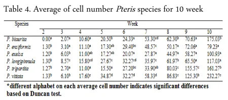
The growth rate of Pteris gametophyte is varied among observed species in every week (Figure 4) and were significantly different among species based on ANOVA and Duncan test (Table 4). The growth rate of Pteris gametophyte began to differ among species on the second week, and then the gametophyte growth was continuously differed by the gametophyte age. Pteris biaurita did not grow until the second week. Pteris vittata showed the fastest growth rate during the fourth until the eighth week. On the eight-week, the growth of P. ensiformis gametophyte showed the slow-est rate while that of P. tripartita showed the fastslow-est. On the ninth week, the growth of P. longipinnula ga-metophyte showed the slowest rate while that of P. tri-partita showed the fastest. On the tenth week, P. vit-tata grew the fastest while P. ensiformis grew the slowest.
Figure 3. Comparison of gametophyte development time among Pteris spesies. Germinated time ( ), formation time of laminar ( ), formation time of gametangia ( )
Table 3. Gametophyte development of observed Pteris and a previous study [10]
Table 4. Average of cell number Pteris species for 10 week
Laminar phase
The type of gametophyte development varies within the genus Pteris, type Ceratopteris, Adiantum, and modifications of the two kinds (Figure 5). However, previous studies had reported that the gametophyte de-velopment in Pteris genus has Ceratopteris-type [20].
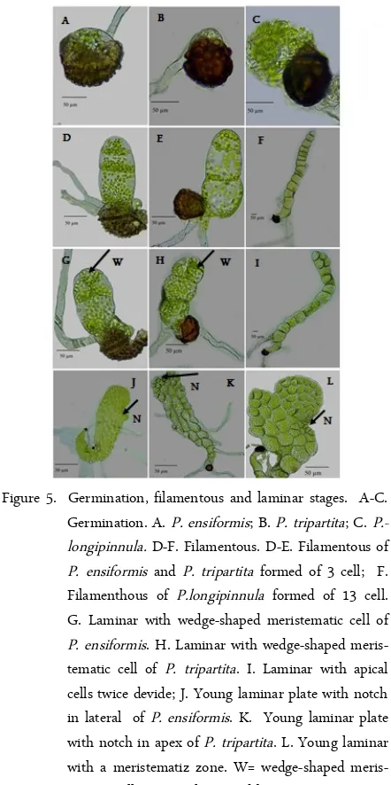
The gametophyte development of all Pteris species is the Ceratopteris-type, except P. ensiformis and P. tri-partita. This pattern is similar to that of P. deflexa, P. tristicula, P. cretica, P. berteroana, P. fauriei, P. incom-pleta, P. multifida, P. vittata, and P. wallichiana [7-12]. In this type, the first cell division is in apical and sometime in subapical, following by repeated division (Figure 5I). Since the meristem is in lateral position, the cell differentiation in the laminar stage is asymmetric. By the time, the activity of meristematic cells gives rise to form a cordate stage after 15-25 days. P. ensiformis has two types of gametophyte devel-opment, the Adiantum-type in the early stage, and the Ceratopteris-type in the next phase of its prothallia de-velopment. During the early phase of this type, the oblique wall division occurs in a terminal cell of the filament and was followed by a second oblique wall di-vision at the right angles. A wedge-shaped then was formed in an apical region (Figure 5G). By the time, the activity of meristem cell in lateral position develops a young asymmetric gametophyte. Previous studies re-ported that the type of development gametophyte of P. ensiformis follow Ceratopteris-type [7,10].
The gametophyte development of P. tripartita fol-lows the Adiantum-type as described by Nayar and Kaur (1971) [20]. In this type, the first division in the terminal cell is parallel to the long axis of the filament. The second division is oblique, then was followed by all cell divisions in the meristematic cells (Figure 5H). An expanded one-cell-thick of the obovate prothallia plate is formed by repeated transverse and longitudinal
divisions. The location of the meristematic cell turned into the notch is in the thallus apex. Among these six species, the apical notches of P. tripartita were formed earlier than that of the others. Rhizoids were usually formed on the ventral surface of the prothallus, but they were sometimes found on the dorsal surface of the cushion or the wing margins. Results of this study dif-fer from Ravi et al. (2014) [13] who reported that ga-metophyte development of P. tripartita is Ceratopteris-type.
Mature Gametophytes and Sexual Expression
The shapes of the mature cordate prothalli are var-ied among all Pteris species (Figure 6 D-I). The mature gametophytes of P. biaurita, P. longipinnula, P. vittata,
Figure 5. Germination, filamentous and laminar stages. A-C. Germination. A. P. ensiformis; B. P. tripartita; C. P.-longipinnula. D-F. Filamentous. D-E. Filamentous of P. ensiformis and P. tripartita formed of 3 cell; F. Filamenthous of P.longipinnula formed of 13 cell. G. Laminar with wedge-shaped meristematic cell of P. ensiformis. H. Laminar with wedge-shaped meris-tematic cell of P. tripartita. I. Laminar with apical cells twice devide; J. Young laminar plate with notch in lateral of P. ensiformis. K. Young laminar plate with notch in apex of P. tripartita. L. Young laminar with a meristematiz zone. W= wedge-shaped meris-tematic cell, N= Notch pointed by an arrow. Figure 4. The cell growth of gametophyte Pteris, P. biaurita
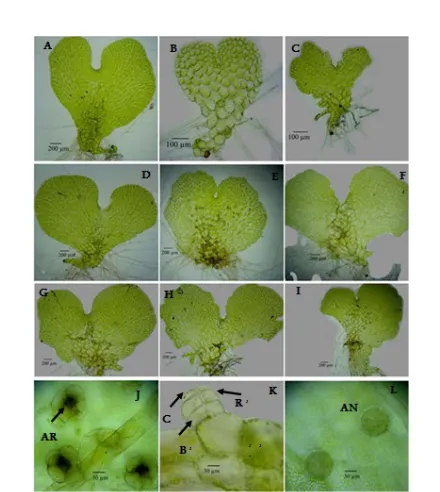
and P. exelsa are asymetric cordiform, whereas P. tri-partita and P. ensiformis are nearly symetric cordiform. Formation time of sex organ is varied among species. Pteris gametophytes began to produce an-theridia 40–92 DAS. Pteris vittata produced sex organ (antheridia) the earliest, 40 DAS, while P. longipinnula is the last producing sex organ, on 92 DAS (Figure 3). The hermaphrodite gametophyte produced antheridia earlier than archegonia. The archegonia were produced on the ventral surface or notches of the prothalli, 74– 102 DAS. Typically, antheridia were produced on the ventral surface of the wings, distributed over the dorsal surface, along the wing margins, or both sides of the gametophytes. Antheridia consist of a cap cell, a ring cell, and a basal cell (Figure 6K).
Sex organs of P. exelsa, P. longipinnula, P. vittata, and P. biaurita give rise to monosexual/male gameto-phyte. P. ensiformis spores produced 17% of hermaph-rodite gametophyte and 83% of the male gametophyte
while P. tripartita produced 27% of hermaprodite and 73% of the male gametophyte (Figure 7).
Apogamous types of P. exelsa, P. biaurita, P. longipinnula, and P. vittata produced only the func-tional antheridium. The same evident also found in P. cretica, P. pellucidifolia, and P. wulaensis [30]. How-ever, P. wulaensis produced only archegonium, and the embryo appeared from the gametophyte cells indicat-ing that it has apogamous reproduction type. Laird and Sheffield (1986) [31] reported that apogamous P. cret-ica is hermaprodite, but the archegonia had lost func-tion because of the failure of the neck canal to open.
Results of this study differ from the previous study on P. exelsa, P. ensiformis, and P. vittata that produces 100% gametophyte hermaphrodites [10]. The differ-ences of this result may be due to physical factors such as nutrition, density gametophyte, the influence of light, and the interaction between the gametophyte [32, 33].
Spore germination of Pteris genus is the Vittaria type. Pteris spores were germinated in 10 to 18 DAS. The spores of P. ensiformis were germinated faster, 10 DAS while the spores of P. longipinnula were germi-nated slower, 18 DAS. The number of cells in each week of gametophyte growth were significantly differ-ent among species based on ANOVA and Duncan test. The gametophytes among Pteris species are differed in the number of filament cell, germinated time, the for-mation time of notch and gametangia, margin shape, and the development type. Gametophyte morphologi-cal characters can be used as a genus characteristic.
The author expresses the deepest appreciation to Indonesian Government through the Directorate Gen-eral of Higher Education for providing financial sup-port for her research, Dr. Wen-Liang Chiou, Cibodas
ACKNOWLEDGMENT CONCLUSIONS Figure 6. Development stages of the gametophyte and sex
or-gans of Pteris. A-C Young gametophyte A. P. ensi-formis; B. P. tripartita; C. P.longipinnula. D-I Ma-ture Gametophyte, D. Asymetric cordate of P. ensi-formis; E. Symetric cordate of P. tripartita; F. Asy-metric cordate of P.longipinnula; G. AsyAsy-metric cor-date of P. biaurita; H-I Asymetric corcor-date of P. ex-elsa and P. vittata; J. Top view of the archegonia P. tripartita; K. Side view of antheridium P. exelsa with a cap cell (C), a ring cell (R), and a basal cell (B); L. Top view of Antheridia. AR= Archegonium, AN= Antheridium. B, C, R, AR are pointed by an arrow.
Botanical Gargen, and Bogor Botanical Garden.
1. Holttum RE (1966) A Revised Flora of Malaya. Vol II. Singapura (SG): Government Printing Off.
2. Tryon R M; Tryon A F (1982) Ferns and allied plants. New York: Springer Verlag.
3. Backer CA, Posthumus O. Varenflora voor Java (1939) Buitenzorg (NL): Archipel Drukkerij.
4. Tryon RM, Tryon AF, Kramer KU (1990) Pteridaceae. In Kramer KU, Green PS, editor. The Families and Genera of Vascular Plants: Pteridophytes and Gymnosperms. Vol-ume I. Berlin (DE): Springer Verlag.
5. Ma LQ, Komar KM, Tu C, Zhang WH, Cai Y, Kennelley ED (2001) A fern that hyperaccumulates arsenic: A hardy, versatile, fast-growing plant helps to remove arsenic from contaminated soils. Nature. 409: 578-579.
6. Copeland EB (1958) Fern Flora of the Philippines. Vol I. Manila (PH): Bureau of printing.
7. Chiou WL (1992) The gametophyte of Pteris ensiformis Burm. In Proceeding of the Second Seminar Asian Pteri-dology. Taiwan. Edited by Tsai & Shieh WC: National Chung Hsing University and National Science Council. 49-54.
8. Mendoza A, Pérez-García B, Reyes Jaramillo I, Ricci M (1997) Desarrollo del gametófito de Pteris berteroana (Pteridaceae: Pterideae). Rev Biol Trop 44(45): 51-57. 9. Prada C, Moreno V, Gabriel y Galán JM (2008)
Gameto-phyte development, sex expression and antheridiogen sys-tem in Pteris incompleta Cav. (Pteridaceae). Am Fern J. 98(1): 14–25.
10. Zhang KM, Shi L, Zhang XC, Jiang CD (2008) Gameto-phyte morphology and development of six Chinese species of Pteris (Pteridaceae). Am Fern J. 98(1): 33-41.
11. Martinez OG (2010) Gametophytes and young sporo-phytes of four species of the fern genus Pteris (Pteri-daceae) naturalized in the American continent. Rev Biol Trop. 1: 89-102.
12. Martinez OG, Prada C, Tanco ME, Bonomo MC (2013) Sexual phase of three spesies (Pteridaceae). Trop Plant Biol. 6: 46-52.
13. Ravi BX, Robert J, Gabriel M (2014) Invitro spore germi-nation and gametophytic growth development of the criti-cally endangered Pteris tripartita SW. Acad J. 13(23): 2350-2358.
14. Walker TG (1962) Cytology and evolution in the fern genus Pteris L. Evolution. 16: 27-43.
15. Zubaidah S (1998) Kajian sitologi, tipe reproduksi dan ciri-ciri morfologi Pteris biaurita L. di daerah berketing-gian berbeda [Tesis]. Malang (ID), UNM.
16. Praptosuwiryo Tng, Darnaedi D (2008) Cytological
obser-vation on fern genus Pteris in Bogor Botanic Garden. Bul Kebun Raya. 11: 15-24.
17. Hastuti DV, Praptosuwiryo TN, Djuita NR (2011) Sitologi dan tipe reproduksi Pteris multifida Poir. (Pteridaceae). Bul Kebun Raya. 14(1): 8-18.
18. Efendi M, Chikmawati T, Darnaedi D (2014) New cy-totipes of Pteris ensiformis var. victoria from Indonesia. Reinwardtia. 14(1): 133.
19. Mumpuni M (2014) Sitotaksonomi Pteris vittata (Pteridaceae) di Pulau Jawa. [Tesis]. Bogor (ID), Sekolah Pascasarjana IPB.
20. Nayar BK, Kaur S (1971) Gametophytes of homosporous ferns. Bot Rev. 37: 298-333.
21. Chiou WL, Farrar DR (1997) Comparative gametophyte morphology of selected species of the family the Polypodi-aceae. Amer Fern J. 87: 77-86.
22. Chiou WL, Farrar DR (2001) Separating spores from spo-rangia. Bull Am fern Soc. 21(3): 22.
23. Knobloch IW (1966) A preliminary review of spore num-ber and apogamy within the genus Cheilanthes. Am Fern J. 56: 163-167.
24. Huang YM, Hsu SY, Huang MH, Chiou WL (2009) Re-productive biology of three Cheilanthoid Ferns in Taiwan. J Plant Reprod Biol. 1(2): 109-116.
25. Klekowski EJ Jr (1973) Sexual and subsexual systems in homosporous pterydophytes a new hypothesis. Am J Bot. 60: 535-544.
26. Chang YJ, Shieh WC, Tsai JL (1992) Studies on the kary-otypes of the fern genus Pteris in Taiwan. In Proceeding of the Second Seminar Asian Pteridology. Taiwan. Edited by Tsai & Shieh WC: National Chung Hsing University and National Science Council. 93–109.
27. Darnaedi D (1992) A preliminary cytological study of fern flora of Gede-Pangrango National Park (West Java). In Proceeding of the Second Seminar Asian Pteridology. Tai-wan. Edited by Tsai & Shieh WC: National Chung Hsing University and National Science Council. 73 –78. 28. Kato, M. and K. Iwatsuki (1986) Variation in ecology,
morphology and reproduction of AspleniumSec. Hy-menasplenium (Aspleniaceae) in Seram, Indonesia. J Fac SciTokyo Univ. 3: 37-48.
29. Gifford E, Foster AS (1989) Morphology and Evolution of Vascular Plants. New York (US): WH Freeman and Co. 30. Huang YM, Hsu SY, Hsieh TH, Chou HM, Chiou WL
(2011) Three Pteris species (Pteridaceae: Pteridophyta) re-produce by apogamy. Bot Stud. 52: 79-87.
31. Laird S, Sheffield E (1986) Antheridia and archegonia of the apogamous fern Pteris cretica. Ann Bot. 57: 139-143. 32. De Soto L, Quintanilla G, Méndez M (2008)
Ecol. 96: 1319–1327.
33. Huang YM, Chou HM, Chiou WL (2004) Density affects