The SeparaTion of CelluloSe-Binding domain from
endogluCanaSe egl-ii By proTeolySiS meThod
rina masriani a 1, Taufan hidayat a, dewi Christanti Trisulo b
a Center of Pulp and Paper, Jl. Raya Dayeuhkolot No. 132, Bandung 40258, Indonesia b University of Surabaya, Jl. Raya Kalirungkut-Tenggilis, Surabaya, Indonesia
1 email: [email protected]
Diterima : 04 Maret 2013, Revisi akhir : 16 Mei 2013, Disetujui terbit : 30 Mei 2013
Pemisahan Cellulose-Binding domain dari endoglukanase
egl-ii
dengan metode Proteolisis
aBSTrak
Molekul protein dari endoglukanase Egl-II terdiri dari dua domain, yaitu cellulose-binding domain (CBD) yang berfungsi untuk mempromosikan adsorpsi enzim ke selulosa kristalin dan domain katalitik yang bertanggung jawab dalam reaksi hidrolisis. Dalam studi ini, CBD dari endoglukanase Egl-II telah dipisahkan dari protein utuhnya setelah didegradasi dengan papain. Pemisahan CBD dari campuran
hasil degradasi dilakukan dengan metode ultrafiltrasi. CBD yang dihasilkan dapat digunakan untuk memodifikasi serat kertas bekas. Hasil elektroforesis sebelum degradasi menunjukkan endoglukanase
Egl-II memiliki massa molekul sekitar 57,5 kDa. Dari elektroforegram setelah degradasi protein dan pemisahan CBD dari endoglukanase Egl-II memperlihatkan bahwa CBD telah terpisah dari protein utuhnya dan memiliki massa molekul sekitar 21 kDa. Rendemen CBD adalah 59,51%. CBD dari endoglukanase Egl-II dapat dipisahkan dengan metode ini.
Kata kunci: endoglukanase Egl-II, cellulose-binding domain, papain, ultrafiltrasi.
aBSTraCT
Protein molecule of endoglucanase Egl-II is consisted of two domains, namely cellulose-binding domain (CBD) which serves in promoting the adsorption of the enzyme to the insoluble crystalline cellulose and cellulase catalytic domain which is responsible for the hydrolysis reaction. In this study, CBD of endoglucanase Egl-II was separated from the intact protein by degradation using the papain
and then separation by ultrafiltration methods. The CBD resulted can be used to modify the waste paper fibers. The results of electrophoresis before degradation showed that endoglucanase Egl-II has
a molecular weight about 57.5 kDa. The electrophoregram after protein degradation and separation of CBD from the endoglucanase Egl-II showed that CBD was separated from the intact protein with a molecular weight about 21 kDa. The yield of CBDs were 59.51%. It can be concluded that CBD of endoglucanase Egl-II can be separated from the intact protein.
Keywords: endoglucanase Egl-II, cellulose-binding domain, papain, ultrafiltration.
inTroduCTion
Recycling of waste paper can reduce deforestation, energy consumption, pollutant emissions and waste treatment problems. One
ton of paper made from recycled fiber can save
25-30 m3 of water, 20-30 trees, 4000 kWh of
electricity and reduce environmental pollution (Dienes, 2006). Disadvantages of using waste
paper fiber as raw material in paper making
were high energy cost on the drying process and low strength on paper products (Pala et. al., 2001; Dienes, 2006). An enzymatic treatment of
once-dried fiber by commercial cellulases would
increase the freeness and drainage. The
enzyme-treated once-dried fiber is slightly stronger in
wet-web tensile strength than is the untreated
presses (Abubakr et al., 1994). We suggested to use cellulose binding domain (CBD) for
improving the waste paper fiber.
Protein molecule of endoglucanase Egl-II is consisted of two domains (Nurachman et. al., 2010), namely cellulose-binding domain (CBD) and cellulase catalytic domain (CD). The CBD serves to promote the adsorption of the enzyme to the insoluble crystalline cellulose, CD is responsible for the hydrolysis reaction (Pala et al., 2001). CBD has an important role in mediating the binding of cellulolytic enzymes to surface of cellulose doing non-hydrolytic disruption on
cellulose fibers. This stage is initiation point of
cellulose degradation process (Tormo et al.,
1996). These proteins will bind to surface of fiber,
then modifying surface or interface properties of
fibers (Henrissat and Davies, 2000).
CBDs may have potential applications
in the modification of polysaccharide fibres,
for instance in wood or paper (Lemos et al., 2000). According to Pala et al. (2001), when CBD was applied, it was possible to achieve the simultaneous increase of pulp drainage rate and paper resistance indexes (specially tensile and burst) as compared with the control. The peptide concentration seems to be critical, as the lower dosages (0.4–1.4 mg protein/g o.d. pulp) were quite favorable to the process (maximum increase in drainage of 14%, together with an increase of 9% in burst and 7% in tensile). It
seems that the CBD binding to the fiber surface,
by modifying the surface/interfacial properties
of the fibers, affects the technical properties
of the pulp and paper in a very positive way. Although it is still not possible to establish the kind and the importance of these changes to
the modification of the properties of pulp and
paper, it could be hypothesized that an excess of CBD reduces the mechanical peeling effect of
fines from the surface of fibers and consequently worsens the final pulp characteristics. According
to Machado et al. (2009), the application of carbohydrate binding module (CBM3) from the Clostridium thermocellum scaffolding protein (CipA) on E. globulus pulp was increase bursting index, tensile strength index, decrease permeability, and no effect for tear index.
Endoglucanase is hidrolyzed glucosidic linkage from internal chain of cellulose. Amino acid sequence of Endoglucanase Egl-II is composed by signal peptida (1-29), catalytic
domain (48-301) from Glycosyl Hydrolase
family 5 (GH5) and substrate binding domain
(356-437) from Cellulose Binding Module 3 (CBM 3) (Nurachman et al., 2010).
According to Tormo et al. (1996), over 100 different CBD sequences have already been
identified, which range in size from only 33 to
over 170 amino acid residues. These CBDs can be grouped into distinctive families on the basis of amino acid sequence similarities, CBD divided in 3 families. The smallest and simplest type of CBD, comprising family I, is found only in fungal cellulases and contains between 33 to 36 residues. Family II contains 110 residues amino acid, and Family III contains 150 residues amino acid. CBD from Trichoderma reesei has molecular weight of 9 kDa (Lemos et. al., 2000), belonging to family I of CBD. This CBD has increasing 14% drainage, 9% burst index, and 7% tensile index (Pala et. al., 2001).
In this research, the CBD of endoglukanase Egl-II was separated from the intact protein by degradation using the papain enzyme and
separation by ultrafiltration methods. According
to Lemos et al. (2000), separation of the two domains has also been achieved using proteolysis. Separation was carried out with the aim of cellulase catalytic core or cellulase binding domain isolation of cellulase. Proteolysis was done by papain. The result of the proteolysis process was checked for enzymatic activity and analyzed using capillary electrophoresis
(CE). The digested mixture was ultrafiltrated
through both 10 kDa and 30 kDa nominal cut-off membranes to assure the separation of the cellulose-binding domains (CBDs) from the
digested mixture. The filtrates obtained by PM10
membrane did not show any hydrolytic activity,
but CMCase activity was detected in the filtrates
obtained by PM30 membrane showing that some catalytic core managed to pass through PM30 membrane.
The purity of the filtrate was checked using
CE. The results obtained displayed one peak corresponding to a protein with a molecular weight close to 9 kDa, refer to CBD of cellulases from Trichoderma reseei. The molecular weight of the core-binding domain of fungal cellulases is close to 5 kDa, including the CBD family I, which is composed of 33-36 amino acid residues (Tormo et al., 1996).
β-mercaptoethanol and measurement activities.
The bound fraction was estimated by deducting
the fluorescence intensity of the soluble fraction from the total fluorescence of the proteins in
absence of cellulose. In these cases the interest in proteolysis was more directed to the isolation of catalytic cores of cellulases. Another technique for separated of CBD by engineering genetic (Carrad et al., 2000). The steps of this work were construction of recombinant proteins, protein
expression and purification, complex formation,
and activity measurements.
The purpose of this research is to separate CBD of endoglucanase Egl-II from the intact protein by degradation using the papain and
separation by ultrafiltration methods.
maTerialS and meThod
materials
Endoglucanase Egl-II was expressed extracellular by Bacillus megaterium that carrying the plasmid PMM1525-egII. B. megaterium was obtained from the Laboratory of Biochemistry, Institute of Technology Bandung. Chemicals such as trypton, yeast extract, NaCl, tetracycline, peptone, xylose, bovine serum albumin, carboxymethyl cellulose, dinitrosalicylic acid (DNS), cellobiose, Bradford reagent, and
NaOH were used. It was also used papain from
papaya latex (Sigma Co. Ltd) and distilled water. Polysulfone membrane with size of 10 kDa, 30
kDa and 50 kDa was used for ultrafiltration. Chemicals for electrophoresis such as ddH2O, acrylamide/bis-acrylamide solution, Tris pH 8.8, sodium dodecyl sulfate (SDS), thermo scientific™
Pierce tetramethylethylenediamine (TEMED),
thermo scientific™ Pierce ammonium persulfate (APS), thermo scientific™ Pierce LDS sample loading buffer, spectra™ Multicolor broad range protein ladder and spectra™ multicolor low
range protein ladder.
method
production of endoglucanase egl-ii
For cell multiplication, B. megaterium was grown in the liquid Luria-Bertani medium (LB) containing trypton 1% (w/v); yeast extracts 0.5%, NaCl 1%, and tetracycline 10 mg/mL (Sambrook and Russel, 2001). For the production of
endoglucanase Egl-II, B. megaterium was grown in liquid LB medium-P by replacing trypton 1% with peptone 1%. Culture B. megaterium was
firstly grown in LB medium-P and incubated at the
temperature of 37°C at 200 rpm. The growth was monitored by its optical density (OD) at 600 nm from 0.152 to reach the value of 0.982 (Masriani and Nurachman, 2012). To induce the expression of endoglucanase Egl-II, xylose was added into the LB medium-P with concentration of 0.03 M and then the incubation process was continued for 5 hours (MobiTec, 2008). Endoglucanase Egl-II was harvested by centrifugation speed of 7000 g at 4°C for 30 minutes. Supernatant obtained (Endoglucanase Egl-II) was stored at 4°C before using for further research.
determination of protein Content
Enzyme protein content was determined by Bradford method using bovine serum albumin (BSA) as a standard (Bradford, 1976). Each absorbance measurements were performed three times (triplo).
activity measurement
Activity of endoglucanase Egl-II in hydrolyze carboxymethyl cellulose (CMC) was determined from the amount of reducing sugars released.
Briefly, the mixture of enzyme reaction
containing endoglucanase Egl-II of 0.02 mg /mL and CMC of 2.5 mg/mL was incubated for 30 minutes at 50°C. Reaction was stopped by adding 50 mL DNS reagent and boiled for 10 minutes. Reducing sugar released was determined by DNS (Miller, 1959) using cellobiose as a standard. Each absorbance measurements were performed three times (triplo). One Unit Egl-II
endoglucanase activity is defined as the amount of enzyme capable of releasing 1 μmol reducing
sugar per minute under reaction conditions.
Specific activity of enzyme activity is defined as
units per mg protein.
removal of The Small protein of The Crude endoglucanase egl-ii and Concentrated The protein
Endoglucanase Egl-II filtered using membrane PM 10 kDa by ultrafiltration techniques, using
nitrogen gas at a pressure of 20 psi. Feed in
PM 50 kDa by ultrafiltration process, with
variation of the pressure of 10, 20, and 30 psi.
digestion of endoglucanase egl-ii
Digestion of endoglucanase Egl-II was performed by papain. A portion of the papain solution (1 mg/mL) was added to 100 mL crude endoglucanase Egl-II (0.0533 mg/mL) to give an endoglucanase Egl-II and papain ratio of 50:1 (w/w). The mixture was agitated for 4 hours at the temperature of ± 23°C (Lemos et. al., 2000; Yokota et al., 2008). Protein digestion results
were separated by ultrafiltration.
Separation of Cellulose Binding domain of Endoglucanase Egl-II by Ultrafiltration
Protein digestion results was filtered using ultrafiltration membrane PM 30 kDa. Permeate
and feed were checked the enzyme activity and protein content. The separation of CBD
was confirmed by sodium dodecyl sulfate
polyacrylamide gel electrophoresis (SDS
PAGE), based on protein identification method
(Boyer, 2000).
Identification of Proteins
The molecular weight of endoglucanase Egl-II and CBD protein was calculate from the graph of a linear relationship between electrophoretic mobility of a protein marker and the log of its molecular weight (Weber and Osborn in Boyer, 2000).
SDS PAGE gel concentration used in this experiment is 20%. Recipes for Polyacrylamide Gel Mix (Separating and Stacking Gels) obtained from SDS PAGE calculator (www.cytographica. com/lab/acryl2.html). For the stacking gel, the gel concentration used 6% with a total volume of 5 mL, the gels divided in two chambers, because
the 6% stacking gel solidifies slower than 20%
separating gel.
A total of 200 μL protein samples concentrated
by the TCA method (Folin and Ciocalteu, 1929). Then the sample treat by boiling method results for denaturation and linearization of protein. In the boiling method, 30 mL loading buffer 1x added to the sample, and heated for 10 minutes. Number of samples are loaded into well of SDS PAGE was 12 µL.
reSulTS and diSCuSSion
Identification of Endoglucanase Egl - II
To verify the purity and molecular weight of crude endoglucanase Egl-II, supernatant was analyzed by the SDS PAGE. Electrophoresis is an analytical tool that can examine the movement
of charged molecules in an electric field.
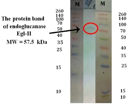
Electrophoresis has been applied to analyze the purity and size of macromolecules. The technique is especially useful for the analysis of amino acids, peptides, proteins, nucleotides, nucleic acids, and other charged molecules (Boyer, 2000). The SDS PAGE separate molecules base on molecular weight (MW). Electrophoretic mobility of a protein and the log of its molecular weight have a linear relationship (Weber and Osborn in Boyer, 2000). The band pattern on the SDS PAGE result of crude endoglucanase Egl-II and marker is showed in Figure 1.
Figure 1. SDS PAGE of Crude Endoglucanase Egl-II and Marker (M)
Figure 1 shows the crude endoglucanase Egl-II has only one protein band with a molecular weight close to 57 kDa (the graph of a linear relationship between electrophoretic mobility of a protein marker and the log of its molecular weight was not shown). According to Kurniasih (2009), ProtParam analysis result of gen eg-II showed that the enzyme of this gen will have molecular weight of 55359.9 Da. This crude enzyme was pure because only one protein band. The advantages of pure endoglucanase is simple process in the separation of CBD by
to get pure CBD and controlled hydrolysis in application of upgrading waste paper. Controlled hydrolysis is important to avoid uncontrolled degradation of cellulose that can make paper dissolved.
From Table 1, it can be seen that crude endoglucanase Egl-II has protein content only
53.3 μg/mL, because this protein only has one
band protein on the SDS PAGE (Fig. 1). One band protein means crude endoglucanase Egl-II only contain one protein i.e. endoglucanase Egl-II. The recombinant form usually has protein content lower than native form. For example, the recombinant form of the crude cellulase CelF of Clostridium cellulolyticum was overproduced in Escherichia coli, had protein content only 46.2
μg/mL (Reverbel-Leroy et. al.,1997). Compared
to crude endoglucanase from an anaerobic sulphidogenic bioreactor has protein content
1210 μg/mL (Oyekola et al., 2007).
Crude endoglucanase Egl-II has specific
activity of 3.19 U/mg, it is higher than endoglucanase Egl-II which expressed in Escherichia coli i.e. 0.01 U/mg at 50°C
pH 6.0 (Nurachman et al., 2010) and the
recombinant form of the crude cellulase CelF of Clostridium cellulolyticum was overproduced in Escherichia coli (0.0116 U/mg) (Reverbel-Leroy et. al.,1997). One unit of cellulase activity
(international unit) corresponds to 1 μmol of
reducing sugar equivalent released per min. This enzyme was better too than crude endoglucanase from an anaerobic sulphidogenic bioreactor that
has specific activity 0.5 U/mg (Oyekola et al., 2007).
Purification of Crude Endoglucanase Egl-II
Endoglucanase Egl-II was filtered using membrane PM 10 kDa by ultrafiltration method,
using nitrogen gas at a pressure of 20 psi. This
filtration has done for removing the small protein.
The Endoglucanase Egl-II has molecular weight
about 57.5 kDa, ultrafiltration process using
membrane PM 50 kDa has done to concentrate this enzyme, with variation of the pressure of 10
(flow rate of 5.69 mL/minute), 20 (flow rate of 7.75 mL/minute), and 30 psi (flow rate of 8.59
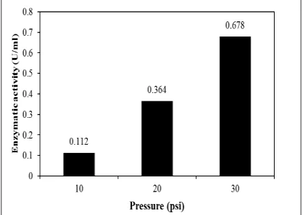
mL/minute). The feeds of this process were measured. The CMCase activity of the crude endoglucanase Egl-II after concentrate is shown in Figure 2.
The highest CMCase activity is endoglucanase Egl-II concentrated by PM 50
kDa with the pressure of 30 psi (flow rate of
7.75 mL/minute). From Figure 1, it has been known that crude endoglucanase Egl-II has the molecular weight close to 57.5 kDa so that
the feed of ultrafiltration process contain this
protein. According to Lestari et al. (2000), the
optimum conditions of α-amylase of Bacillus
slearotherinophilus purification by ultrafiltration system membrane with cut off 30 kDa was using
flow rate of 30 mL/minute and concentrated by about 10 times with 2.3 fold purification factor. By using flow rate higher than 30 mL/minute will decreased the total enzyme activity, specific
activity, and yield.
Figure 2. Enzymatic Activity of Feed from
Endoglucanase Egl-II After Ultrafiltration
Process by PM 50 kDa at Variation of The Pressure of 10, 20, and 30 psi Table 1. Enzymatic Activity of Endoglucanase Egl-II
Sample
Enzymatic activity
(U/mL)
Protein Content
(μg/mL)
Specific Aktivity
(U/mg) Crude endoglukanase
Separation of the CBd from endoglucanase egl-ii.
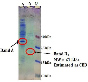
From Figure 3, it can be seen that after
digestion with papain (50:1) and ultrafiltration by PM 30kDa, with pressure of 20 psi (flow rate of
8,59 mL/minute), permeate crude endoglucanase Egl-II has protein band with the molecular weight close to 21 kDa (band B1, the graph of a linear relationship between electrophoretic mobility of a protein marker and the log of its molecular weight not showed). Refer to Nurachman et al. (2010), amino acid sequence of Endoglucanase Egl-II is composed by peptide signal (1-29), catalytic domain (48-301) from Glycosyl
Hydrolase family 5 (GH5) and substrate binding
domain (356-437) from Cellulose Binding Module 3 (CBM 3). ProtParam analysis result of amino acid sequence of substrate binding domain (356-437) have molecular weight of 22 kDa (http://au.expasy.org/tools/protparam. html). Carbohydrate binding module (CBM3) from the Clostridium thermocellum scaffolding protein (CipA) have molecular weight of 22 kDa (Machado et al., 2009). So that protein band with the molecular weight close to 21 kDa in permeate is predicted as CBD. Because in the permeate only has one band protein i.e. CBD, it can be concluded that CBD resulted was pure.
Feed of this process have several bands of protein with molecular weight of 71 kDa to 15 kDa (Figure 3). The protein band with clearlest
Figure 3. SDS PAGE of Endoglucanase Egl-II After Digestion with Papain (50:1) and Ultrafiltration
by PM 30 kDa. A. Feed of Digestion Result. B. Permeate of Digestion Result. M. Marker.
Figure 4. Protein Content of: A. Endoglucanase Egl-II
band in the electrophoregram have molecular weight of 38 kDa (the graph of a linear relationship between electrophoretic mobility of a protein marker and the log of its molecular weight was not shown). ProtParam analysis result of amino acid sequence of catalytic domain (48-301) have molecular weight of 33 kDa (http://au.expasy. org/tools/protparam.html). So that protein band with the molecular weight close to 38 kDa (band A) in feed is predicted as catalytic domain but not pure.
To determine the yield of this process, the protein content of endoglucanase Egl-II (A), feed (B) and permeate (C) of digestion result and
ultrafiltration by PM 30 kDa at 20 psi has been
checked. The result has been shown in Figure 4. It is predicted that protein in permeate as CBD having the protein content of 6.63 µg/mL and the yield of 59.51%. This CBD can apply in papermaking because paper products composed
of cellulosic fibers. Utilizing cellulose binding
module (CBM) proteins in the design of cellulose
materials is interesting. CBM has specific, high affinity for solid cellulose fibers without catalytic
activity (Yokota et al., 2009).
ConCluSionS
The results of electrophoresis before degradation showed that endoglucanase Egl-II has a molecular weight about 57.5 kDa. The electrophoregram of SDS PAGE analysis after protein degradation using the papain
(50:1) and separation by ultrafiltration methods
showed that CBD was separated from the intact protein with a molecular weight about 21 kDa. The yield of CBDs were 59.51%. It can be concluded that CBD of endoglucanase Egl-II can be-separated from the intact protein by this method.
referenCeS
Abubakr, S., K. Rutledge-Cropsey, and J.H.
Klungness, 1994. Papermachine runnability of never dried, dried, and enzymatically treated dried pulp. Proceedings of the 6th International Conference on Biotechnology in the Pulp and Paper Industry: Advances in Applied and Fundamental Research. 151-156. Australian Proteome Analysis Facility, 2003.
ProtParam Tool. http://au.expasy.org/tools/ protparam.html.
Bradford, M.M., 1976. A rapid and sensitive method for the quantitation of microgram quantities of protein utilizing the principle of protein-dye binding. Anal. Biochem., Vol. 72, 248-254. DOI: 10.1016/0003-2697(76)90527-3
Boyer, R. 2000. Modern Experimental Biochemistry. 3th edition. Addison Wesley
Longman.
Carrard, G., Koivula, A., Soderlund, H., Beguin,
P., 2000. Cellulose-Binding Domains
Promote Hydrolysis of Different Sites on
Crystalline Cellulose. PNAS, Vol. 97, No. 19, 10342–10347.
Dienes, D., 2006. “Effect of cellulase enzymes on secondary fiber properties”. Ph. D. Thesis, Budapest University of Technology and Economics. Budapest,
Hungaria.
Folin, O., and Ciocalteu, V. 1929. J. Biol. Chem. 73, 627
Henrissat, B. and Davies, G. J., 2000. Glycoside
Hydrolases and Glycosyltransferases.
Families, Modules, and Implications for Genomics. Plant Physiology, Vol. 124, 1515–1519.
Kurniasih, S. D., 2009. Isolation and Cloning Gene Encoding Endoglucanase from Marine Bacterium Bacillus amyloliquefaciens. Skripsi, Jurusan Kimia, Fakultas Matematika dan Ilmu Pengetahuan Alam, Institut Teknologi Bandung.
Lemos, M. A., Teixeira, J. A., Mota, M., and Gama, F. M., 2000. A Simple Method to Separate Cellulose-Binding Domain of Fungal Cellulases after Digestion by a Protease. Biotechnology letters, Vol. 22, 703-707.
Lestari, P., Richana, N., and Murdiyatmo,
U., 2000. Pemurnian α-Amilase Bacillus
stearothermophilus dengan Membran
Ultrafiltrasi. Jurnal Mikrobiologi Indonesia. Vol. 5, No. 1, 10-14.
Machado, J., Arau´jo, A., Pinto, R. and Gama, F. M., 2009. Studies on the interaction of the carbohydrate binding module 3 from the Clostridium thermocellum CipA scaffolding
protein with cellulose and paper fibres.
Cellulose. Vol. 16, 817–824.
Masriani, R., Nurachman, Z., 2012. Modifikasi
serat kertas bekas menggunakan endoglukanase Egl-II. Journal Selulosa. Vol. 2, No. 2, 53-60
MoBiTec, 2008. Bacillus megaterium Protein Expression System, Molecular Biologische Technologie. Jerman.
Nurachman, Z., Kurniasih, S. D., Puspitawati,
F., Hadi, S., Radjasa, O. K., and Natalia,
D., 2010. Cloning of Endoglucanase Gene from a Bacillus amyloliquefaciens PSM 3.1 in Escherichia coli Revealed Catalytic Triad
Residues Thr-His-Glu. American Journal of
Biochemistry and Biotechnology. Vol. 6, No. 4, 268-274.
Oyekola, O. O., Ngesi, N., and Whitelet,
C. G. 2007. Isolation, Purification and
Characterisation of An Endoglucanase
and β-Glucosidase from An Anaerobic
Sulphidogenic Bioreactor. Enzyme and Microbial Technology. Vol. 40, 637-644.
Pala, H. , Lemos, M. A., Mota, M. , and
Gama, F. M., 2001. Enzymatic upgrade of old paperboard containers. Enzyme and Microbial Technology. Vol. 29, 274-279. Reverbel-Leroy, C., Pages, S., Belaich, A., Belaich,
J., and Tardif, C., 1997. The Processive Endocellulase CelF, a Major Component of the Clostridium cellulolyticum Cellulosome:
Purification and Characterization of the
Recombinant Form. Journal of Bacteriology. Vol. 179, No. 1, 46–52.
Sambrook, J and D. W Russel, 2001. Molecular Cloning, Third edition. Gpld Spring Harbor Laboratory Press. United State of America. www.MolecularCloning.com
Tormo, T., Lamed, R., Chirinol, A. J., Morag, E., Bayer, E. A, Shoham, Y., Steitz, T. A., 1996. Crystal Structure of a Bacterial Family-III Cellulose-Binding Domain: A General Mechanism for Attachment to Cellulose. The EMBO Journal,, Vol.15, No.21, 5739–5751.
Yokota, S., K. Matsuo, T. Kitaoka, H. Wariishi, 2008. Specific interaction acting at a
cellulose-binding domain/cellulose interface for papermaking application. BioResources, Vol.3, No.4, 1030–1041.
Yokota, S., K. Matsuo, T. Kitaoka, H. Wariishi,